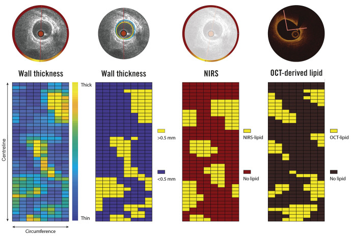
Central illustration. Methodology of sectorial wall thickness quantification.
A division into paired 1.5 mm segments of 45° sectors has been selected to ensure maximal overlap of the baseline and follow-up IVUS data, even in the presence of small registration errors (max ±1 IVUS frame), thereby maximally accounting for spatial heterogeneity in WT changes. NIRS: near-infrared spectroscopy; IVUS: intravascular ultrasound; OCT: optical coherence tomography; WT: wall thickness

