Abstract
The vascular endothelium plays a vital role during embryogenesis and aging and is a cell monolayer that lines the blood vessels. The immune system recognizes the endothelium as its own. Therefore, an abnormality of the endothelium exposes the tissues to the immune system and provokes inflammation and vascular diseases such as atherosclerosis. Its secretory role allows it to release vasoconstrictors and vasorelaxants as well as cardio-modulatory factors that maintain the proper functioning of the circulatory system. The sealing of the monolayer provided by adhesion molecules plays an important role in cardiovascular physiology and pathology.
Keywords: endothelium, endothelium physiology, endothelium pathology, endothelium dysfunction, hypertension, endothelium remodeling, endothelium released factors, atherosclerosis, adhesion molecules, calcium, ion transporters
1. Introduction
The vascular endothelium is much more than just a physical barrier at the interface between the circulating blood and the vascular wall [1,2,3]. In the 1960s, the vascular endothelium was considered a passive barrier protecting the vascular wall from circulating blood [2,3,4]. In the 1980s, it emerged as a multifunctional endocrine organ, playing an essential role in regulating cardiovascular tone [2,5,6,7,8,9].
2. The Vascular Endothelium
The vascular endothelium is a simple tissue in its morphology but complex in its function. Although it is formed as a single monolayer, it is capable of sensing hemodynamic and rheologic changes as well as responding to these modifications of its environment. Vascular tone is maintained by balancing vasodilating and vasoconstricting factors released by the endothelium. In addition, the endothelium plays a key role in controlling the migration and proliferation of VSMCs [2].
The integrity of the endothelial monolayer is essential to regulate vascular permeability and protect the vessel against platelet deposition and thrombus formation. Furthermore, the integrity of this monolayer requires that the morphology and the contacts between ECs do not change.
3. Origin and Differentiation of the Vascular Endothelium
The endothelium is the first cell type to constitute blood vessels. The formation of blood vessels and vasculogenesis result from the differentiation of the mesodermal cells into angioblasts due to the presence of specific proteins. These angioblasts are the precursors of endothelial and blood cells. The physiological primitive angiogenesis takes place to form the vascular tree, which gives birth to buds of the branches that give birth to the heart, including its endocardial endothelial cells. Then, remodeling of the vascular tree takes place to form capillaries and veins, including large arteries [2,10,11]. The endothelium of these newly formed blood vessels differs depending on the type of vessels. For example, fibroblast growth factor (FGF) receptors seem to be expressed only in large vessels [12]. Several ligands and their corresponding receptors are implicated in the differentiation and formation of the endothelium, including vascular endothelial growth factor (VEGF) and its receptors 1 and 2 [10].
The heart’s formation is more than a deformation of blood vessels, and differentiation of vascular endothelium into endocardial endothelium forms the left (arterial) and right (venous) endocardial endothelium layers. Both endocardial and vascular endothelium are separated from their muscle cells by a basal lamina membrane [2]. Vasculogenesis occurs during early embryonic development, whereas angiogenesis happens during adulthood. Angiogenesis is usually associated with diseases [11]. Several endothelial markers exist, such as VE-cadherin, PECAM-1, Tie-1 and 2, and flk1. Notch family activation plays an essential role in defining the characteristics and identities of arterial endothelial cells [13]. Although the molecular aspect of the arterial specification is more precise, little is known concerning the venous specification [13]. For example, the vascular endothelium can adapt its function depending on the environment. Still, it does not change its phenotype, such as in transplanted arterial and venous vessel grafts, where the graft vessel’s endothelium matches the host vessels’ characteristics [14,15].
4. Role of the Endothelium in Vascular Physiology
All blood vessels contain endothelial cells that form the intima. Two types of blood vessels only have endothelial cells: capillaries and venules. The intima is formed by continuous and discontinuous (fenestrated) layers of endothelial cells. Examples of continuous endothelium are arteries and veins. Tight, adherent junctions connect the continuous layer of endothelium side by side. Transport molecules go through this sealed monolayer of the endothelium via a transcytosis mechanism, such as caveolae (caveolin-1) and vesiculo-vacuolar organelles [16]. A fenestrated, discontinuous layer of endothelium permits extensive transport of molecules toward tissues such as the liver.
Several physiological functions are attributed to vascular endothelium, independent of their localization in the vascular tree, such as tuning the level of vascular endothelial cells (VECs) vasoconstrictors and vasorelaxers [2,17], regulation of coagulation and inflammation [3,18], and playing an essential role as a gatekeeper of fatty acids transport [19,20,21,22,23,24].
Among the vasoconstrictive factors released by the vascular endothelium are endothelin-1 (ET-1), thromboxane A2 (TxA2), as well as angiotensin-II (AngII) [2,16,17,25,26]. On the other hand, the most vasorelaxant factors released by the endothelium are nitric oxide (NO), prostacyclin (PGI2), and endothelium-derived hyperpolarizing factor (EDHF) [2,27].
The blood vascular system consists of a circuit of vessels in which the continuous movement of the heart pump maintains the blood flow. Blood vessels distribute nutrients, oxygen, and hormones to all organs and tissues and transport the products of cellular metabolism. The walls of arteries and veins, such as the thoracic aorta, used commonly in the literature, consist of three concentric tunics that are firmly joined from the inside out [2] (Figure 1): (1) The intima is the thin innermost layer that lines the various vascular walls, including those of the capillaries and venules. It is composed of a monolayer of endothelial cells (ECs) in direct contact with the blood and forming the vascular endothelium. The ECs provide a smooth inner surface that minimizes friction, which facilitates blood flow. The vascular endothelium is supported by a basal lamina and a thin connective tissue formed by collagen and some elastic fibers;
Figure 1.
Structure of the vascular wall. Schematic representation showing the three layers of the vascular wall: tunica intima, tunica media, and tunica adventitia, as well as the components of each layer. VSMC: vascular smooth muscle cells (from Bkaily et al., 2021 [2]).
(2) The media is the thickest intermediate layer of the vascular wall. It consists of vascular smooth muscle cells (VSMCs), collagen, and elastin. This layer is absent in the capillaries and venules; (3) The adventitia is the outermost layer of the vascular wall. It is absent in capillaries and venules. This layer is formed of supporting connective tissue consisting mainly of collagen. It is also crossed by numerous nerve endings controlling the activity of the muscle fibers as well as the blood vessels feeding the vascular wall, called vasa vasorum (vessels of the vessels). The relative importance of these three layers varies according to the type of blood vessel [2]. In conclusion, all blood vessels have an endothelium but not necessarily adventitia or VSMCs, hence the importance of studying and learning more about the vascular endothelium.
5. Structure of the Vascular Endothelium of Arteries and Veins
A monolayer of flat cells forms the vascular endothelium of arteries and veins, with a central nucleus measuring 10–20 µm in diameter. VECs are characterized by extensive intercellular overlap and long, deep slits that contribute to the integrity of the vascular endothelium [2]. The integrity of this monolayer is ensured by a dynamic cytoskeleton [2,28,29,30] as well as by contacts between cells and between these cells and the extracellular matrix [2,31,32]. In vivo and in situ morphology studies have shown the presence of tight junctions, adhesion junctions, and gap junctions between adjacent VECs (including aortic VECs) [32,33,34]. In addition, several roles have been attributed to junctional communication at the vascular endothelium level, including intercellular nutrient exchange, regulation of growth and differentiation, coordination of cellular response to exogenous and endogenous stimuli, and maintenance of vascular tissue homeostasis [35,36,37,38].
The cytoskeleton is well-developed in ECs. It contributes to vascular homeostasis and seems to play an essential role in the repair and integrity of these cells [28,29,30]. VECs contain the actin protein in its filamentous polymeric form, called F-actin, and in its globular monomeric form, called G-actin [39,40]. Therefore, F- and G-actin play a role in the shape of ECs. The balance between the monomeric and polymeric forms could be altered during stimulation of ECs and contribute to the modulation of intercellular junctions that affect the vascular permeability of the endothelial layer. Indeed, during the migration of VECs, G-actins increase compared to F-actins [41]. The migration of these cells also involves the redistribution of centrosomes [28]. Actin microfilaments are localized within the cell as short, thin stress fibers and form a continuous band at the periphery [28,39]. In situ studies have also demonstrated the presence of the protein myosin at the level of these microfilaments [42,43], which plays an essential role in cell adhesion, and facilitates the adaptation of the vascular wall to variations in blood flow pressure [28]. The presence or absence of an actin isoform allows the identification of ECs. Therefore, the presence of α-actin in VECs is considered to be a marker for this type of cell [44].
6. Role of the Endothelium in Vascular Activity
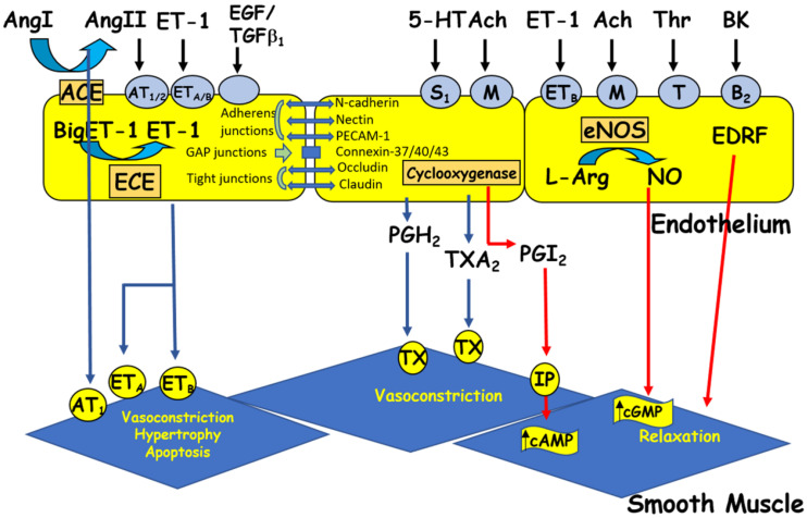
ECs respond to chemical and physical stimuli by synthesizing and releasing various vasoactive and growth factors [2] (Figure 2). The endothelium possesses anti-adhesive substances that prevent blood from clotting. The anticoagulant and antithrombotic properties of the vascular endothelium, which are essential for vascular homeostasis, are due to the synthesis of vasodilatory factors such as nitric oxide (NO) and prostacyclin [5,16,42,45,46] (Figure 2). On the other hand, the vascular endothelium secretes several vasoconstrictor substances (Figure 2), including endothelin-1 (ET-1), prostaglandins, and several components of the renin-angiotensin system (RAS), such as angiotensin II (Ang II). Ang II [25,26,47,48] and ET-1 [49,50,51] act at the plasma and nuclear membranes of ECs and induce an increase in the intracellular calcium level via activation of their respective receptors, AT1/AT2 and ETA /ETB receptors. This increase in [Ca2+]i may, in turn, modulate the secretory function of ECs [2,52] and survival [26,50]. Furthermore, a balance between the different factors secreted by the EV is essential for maintaining intracellular homeostasis and wall integrity. Any disturbance in this balance leads to endothelial dysfunction, characterized by a decreased capacity for relaxation of the vessel, an increase in the adhesion of blood cells to the vascular wall, and a disturbance in the tunica medial [1,5,9,20,53,54,55,56]. This endothelial dysfunction is generally observed during aging and in several vascular pathologies, such as hypertension, hypotension, atherosclerosis, and heart failure [26,57,58]. All VECs synthesize and secrete von Willebrand factor (vWF), a multifunctional protein involved in the typical arrest of hemorrhage [59]. Indeed, through its interaction with extracellular matrix proteins and membrane receptors, vWF plays a prominent role in blood coagulation, platelet aggregation, and platelet adhesion to the extracellular matrix [60,61]. vWF can also bind to the pro-coagulant co-enzyme, factor VIII, contributing to its stability and, indirectly, to the production of fibrin [60,61]. vWF is stored in small vesicles characteristic of endothelial cells, the Weibel–Palade bodies [60,61,62]. The latter contain other proteins, such as ET-1 [62,63] and interleukin-8 [64]. In addition, vWF is used as a marker of ECs in vitro [65].
Figure 2.
The endothelium produces vasoactive factors that cause either relaxation or contraction of the vascular smooth muscle. Ang I and II: angiotensin I and II, ACE: angiotensin-converting enzyme, Ach: acetylcholine, BK: bradykinin, cAMP/cGMP: cyclic adenosine/guanosine monophosphate, ECE: endothelin-converting enzyme, EDRF: endothelium-derived relaxing factor, ET-1: endothelin-1, 5HT: 5-hydroxytryptamine (serotonin), L-Arg: L-arginine, NO: nitric oxide, NOS: nitric oxide synthase, PGH2: prostaglandin H2, PGI2: prostacyclin, TGFβ1: transforming growth factor beta 1, Thr: thrombin, and TXA2: thromboxane A2. Circles represent receptors (AT: angiotensin receptor, B: bradykinin receptor, ET: endothelin receptor, M: muscarinic receptor, IP: purinergic receptor, S: serotonin receptor, T: thrombin receptor, and TX: thromboxane receptor).
7. Inter-Endothelial Junctions
Junctions between ECs are formed by a transmembrane protein called occludin, which connects to a group of intracellular proteins such as zonula occludin-1 (ZO-1), ZO-2, cingulin, and a new protein linked to GTP, rab13 [66,67]. Their main biological functions are: (1) to form a barrier to paracellular permeability, (2) to maintain the apical-basal polarity of cells, and (3) to assist in intercellular adhesion [68]. In vascular tissue, tight junctions are distributed according to the permeability of the endothelial monolayer in different vascular beds [69]. Figure 2 shows three different types of endothelial junctions.
The endothelium of the cerebral microcirculation is well sealed (continuous endothelium) with an extensive network of tight junctions between cells to form the blood-brain barrier [70]. ECs are more widely spaced, with relatively fewer tight intercellular junctions in muscle capillaries (fenestrated endothelium) [71,72]. On the other hand, the endothelium in postcapillary venules is highly discontinuous, with very few tight intercellular junctions, to allow more interactions between blood and interstitial tissue (discontinuous endothelium) [69].
Adhesion junctions allow ECs to act as strong structural units by linking the cytoskeletal elements of one cell to another [73,74]. These junctions are composed of two categories of proteins: (1) intracellular attachment proteins that connect the junctional complex to the actin filaments and (2) cadherins bind one or more intracellular attachment proteins, and their extracellular domains containing Ca2+ binding motifs interact with the extracellular domains of cadherins from another cell. Cadherins allow homophilic and Ca2+-dependent adhesion between cells [73,74].
The adhesion molecule PECAM is essential for EC activities, such as angiogenesis and control of leukocyte extravasation [75]. Integrins are receptor proteins of importance because they play an essential role in the interaction of ECs with leukocytes during inflammation [76]. The gap junction of ECs consists of connexons in the contact plasma membranes of two cells and forms a microchannel that allows intracellular ions and molecules of low molecular weight (<1000 Daltons) to be exchanged between ECs. One crucial role of gap junctions between ECs is that this microchannel permits the propagation of Ca2+ waves between cells and allows electrical coupling between ECs [77,78]. Connexin types include Cx43, Cx37, and Cx40 [79,80,81,82]. In addition, specific adhesion molecules such as N-cadherin and E-cadherin are essential in establishing coupling between contacting ECs.
8. Ionic Transporters in Vascular Endothelium
In addition to acting as a barrier between circulating blood and vascular smooth muscle cells, the main role of VECs is the secretion of vasoconstrictors and relaxing factors. In turn, these released endothelial vasoactive factors regulate endothelial excitation-secretion coupling. Secretion generally depends on the increase of intracellular Ca2+ (Figure 2) [2,26,48]. Calcium plays a significant role in all cell types, including VECs. Regulation of Ca2+ homeostasis in vascular endothelium relies on Ca2+ influx through Ca2+ channels, the ER, mitochondria, and the nucleus, as well as indirectly through the Na+/Ca2+ exchanger (NCX) [2,27]. In addition, the level of Ca2+ release via the ER IP3 and ryanodine-sensitive pools highly contributes to the excitation-secretion coupling of VECs [2,27,44]. The level of intracellular Ca2+ also depends on the density of the plasma membrane and ER Ca2+ pumps [9,83].
In the early 1980s, it became apparent that different calcium currents coexisted in several excitable cell types [27,52,83,84]. In VSMCs, two types of calcium currents are present: L-type (high threshold) and T-type (low threshold) [27]. Compared with type L, the T current activates and inactivates relatively quickly from the more negative membrane potentials. Furthermore, the inactivation of the L-type calcium channel is calcium-dependent, whereas the T-type channel is not [27]. There do not appear to be any L- or T-type calcium channels in endothelial cells [27,44,52,85]. In contrast, a voltage-dependent and G-protein coupled calcium channel called the R-type calcium channel has been identified in human VECs, rabbit and human aortic smooth muscle cells, the renal artery, and cardiac ventricular cells [27,44,86,87]. The existence of this calcium channel was initially suggested by Baker et al. (1971) [88]. They showed that this type of calcium channel allows the passive entry of Ca2+ into the cell during a long-term depolarization of the membrane. It was named the resting potential calcium channel by DiPolo in 1979 [89]. Unlike the L-type calcium channel, the resting-type (R) calcium channel has no inactivation gates [44,90]. The latter is activated during sustained depolarization of the cell membrane of VSMCs and has a conductance of 24 pS [27,44]. By measuring free intracellular calcium levels using techniques such as microfluorometry and imaging, sustained membrane depolarization produces a rapid, transient increase in intracellular calcium that is followed by a sustained phase [91]. The first phase is abolished by L-type calcium channel blockers such as nifedipine and L- and R-type blockers such as isradipine [87,91]. However, the sustained phase is insensitive to nifedipine, caffeine, and other blockers but sensitive to isradipine and the calcium chelator EGTA [91]. In addition, the R-type calcium channel is responsible for sustained increases in calcium during sustained contraction of vascular smooth muscle in response to various vasoactive and proinflammatory agents such as PAF (Platelet Activating Factor), endothelin-1, as well as insulin [44,90,91]. In addition, calcium influx via the R (resting) channel is primarily responsible for maintaining normal physiological intracellular calcium concentration and the basic tone of vascular smooth muscle [27]. Finally, this channel seems to be involved in basal secretion phenomena of the vascular endothelium [44].
Several calcium channels were suggested to be present in vascular endothelium, such as transient receptor potential channels 1–7 (TRPC1-7), store-operated channels, receptor-operated channels (SOCs), receptor-operated calcium channels (ROC), ryanodine calcium release receptors (RyR), IP3 receptor calcium release channels (IP3R), and storage-operated calcium entry (SOCE) [89,92,93,94,95,96]. Except for RYR and IP3R, all other reported calcium transporters were indirectly suggested to be present in vascular endothelium because it is difficult to prove their presence and contribution to Ca2+ homeostasis due to the absence of specific pharmacological blockers and because they cannot be recorded using biophysical techniques. Thus, their presence and contribution to intracellular Ca2+ homeostasis are questionable.
9. Morphological and Functional Remodeling of Vascular Endothelium
As mentioned in the previous section, the vascular endothelium secretes substances that cause relaxation and others that cause contraction [6,9,18,21,24,25,97,98] (Figure 2), and a balance between these substances is a significant determinant of vascular homeostasis. Therefore, any activation or damage to endothelial cells, as in cardiovascular diseases such as atherosclerosis, hypertension, and cardiac failure, may cause disequilibrium in their secretory functions and hence contribute to the different symptoms associated with these diseases [6,9,19,20,23,30,92,94,98].
In the last decade, several studies investigating endothelial function have provided strong evidence for the role of endothelial dysfunction in both large conduit and small resistance vessels in patients with heart failure [99]. These studies have shown attenuated endothelium-dependent vasodilation in patients with chronic heart failure [99]. Furthermore, a study has shown coronary endothelial dysfunction in patients with new-onset idiopathic dilated cardiomyopathy, suggesting that changes in endothelial function occur early in the course of the disease [100].
Recent work showed that high sodium salt-induced hypertension induced glycocalyx destruction and morphological remodeling in human VSMCs [101]. Since the glycocalyx plays an essential role in VE function, its collapse will cause morphological and functional remodeling, thus affecting VSM function and promoting the remodeling of VSMC [102]. In addition, it was reported in hereditary cardiomyopathy that morphological remodeling takes place early in life before the development of cardiac hypertrophy [103] (Figure 3). Thus, remodeling of the endothelium could be considered a marker of hereditary cardiomyopathy.
Figure 3.
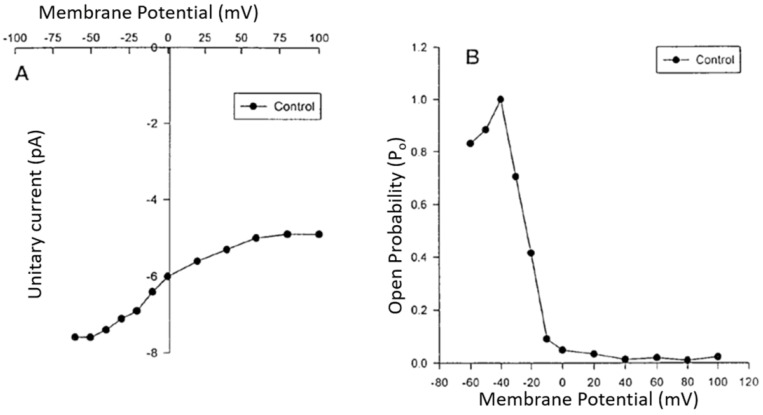
Current to voltage (I/V) relationship curve (A) and open probability/voltage relationship (B) of the voltage-dependent steady-state R-type Ca2+ channel in human aortic VSMCs recorded using the patch clamp technique (modified from Bkaily et al. 1997) [44].
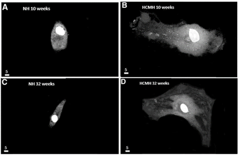
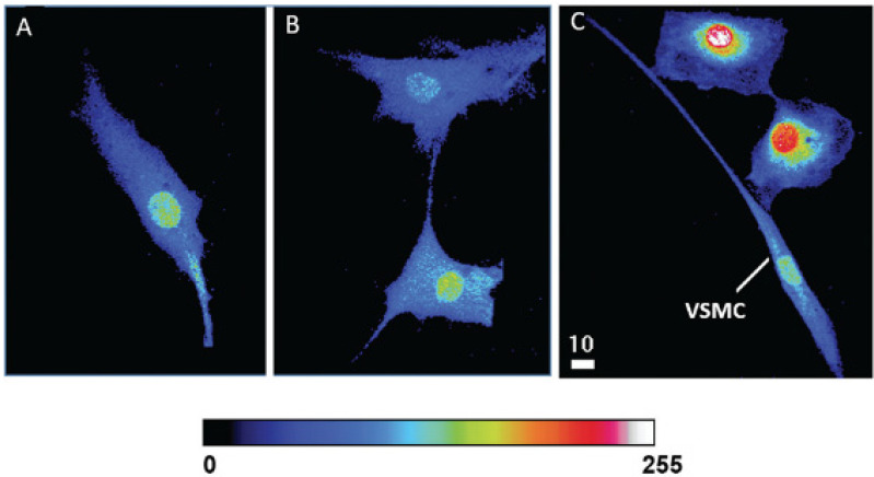
As mentioned, the VECs are separated from the VSMCs by a basal membrane and an internal elastic lamina. However, VECs develop a finger-like projection into the basal membrane, and hypertrophic cell remodeling will promote physical contact between VECs and VSMCs, which promotes cytosolic and nuclear calcium increase. Figure 4 and Figure 5 show examples [104,105].
Figure 4.
Increase in the volume of endocardial endothelial cells (EECs) in young and old hereditary cardiomyopathic hamsters (HCMHs). (A–D) Freshly isolated and cultured EECs from 10-week-old normal hamsters (NH) (A), 10-week-old HCMH (B), 32-week-old NH (C), 32-week-old HCMH, and (D) show an increase in the volume (in µm3) of EECs in HCMHs compared to those of age-matched NHs. In panels A–D, the white scale bar is in µm (modified from Jacques and Bkaily 2019) [104].
Figure 5.
Modulation of cytosolic and nuclear calcium levels of human VSMCs by physical contact with human VECs. Quantitative 3D confocal microscopy images showing the distribution of cytosolic and nuclear Ca2+ in hVSMCs in pure culture of hVSMCs without contact (A) and in contact with other hVSMCs (B), as well as in co-culture with hVECs (C). The pseudo-color scale represents the level of fluorescence intensity of the Fluo-3–Ca2+ complex from 0 (black, absence of fluorescence) to 255 (white, maximum fluorescence) in panels A, B, and C. The white scale bar value is in micrometers (modified from Hassan et al., 2018) [105].
10. Atherosclerosis
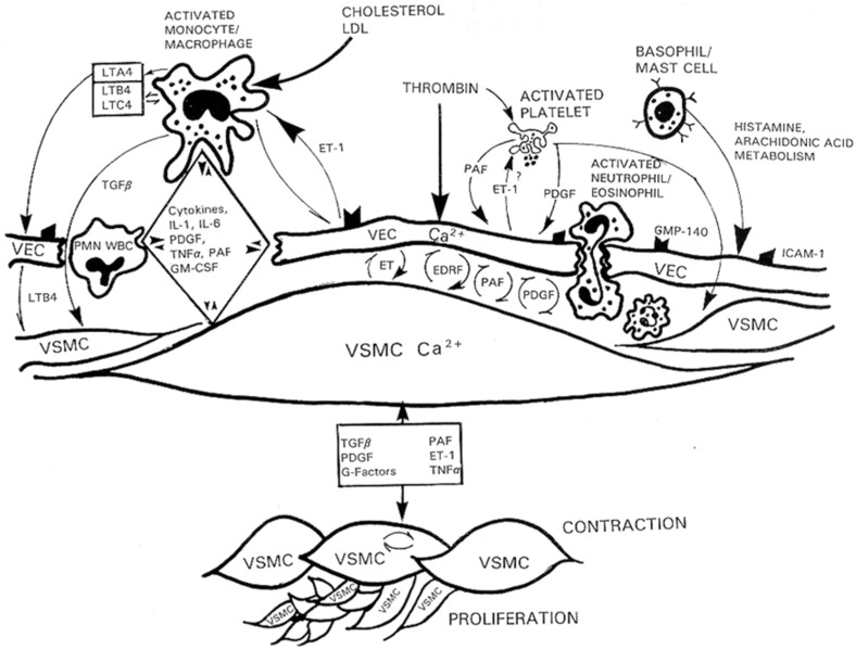
Atherosclerosis is a disease that starts at the endothelial cell level. Several major risk factors for atherosclerosis, such as hypercholesterolemia [19,22,24], angioplasty restenosis [56], hypertension, and diabetes [9,20,24], are characterized by abnormal arterial vascular endothelium function, leading to an increase in the inter endothelial cell junction. VECs injury, platelet adhesion/degranulation, invasion of the subendothelium by leucocytes from the circulating blood, and proliferation of contractile and non-contractile VSMCs from the media are critical early events in atherogenesis [9,20,30]. The leading risk factor for atherosclerosis is chronic hypercholesterolemia-induced monocyte attachment to the vascular endothelium, leading to an increase in vascular permeability, which permits entry of immune cells and blood-circulating factors and promotes VSMC proliferation (Figure 6). When immune-inflammatory cells come into contact with VSMCs, they become activated and release cytokines, platelet-activating factor (PAF), tumor necrosis factor-alpha (TNF-α), IL-1, IL6, and eicosanoids. These factors stimulate the release of endothelium factors such as endothelin-1, PAF, and PDGF. In addition, all these factors promote the proliferation of contractile VSMCs (Figure 6). All these factors can be considered pathological first messengers for atherosclerosis. In addition, most, if not all, of these factors induced an increase of intracellular Ca2+ via stimulation of R-type calcium channels, leading to secretion and activation of Ca2+-dependent intracellular signaling and hypertrophy of VECs.
Figure 6.
Activation of immune cells induces the release of factors that modulate the function and junctions between VECs, which permits the infiltration of activated neutrophils/eosinophils, and polymorphonuclear cells (PMN). The remodeling of the vascular endothelium induces the secretion of factors that induce either relaxation or contraction of VSMCs. The activated monocytes/macrophages and VECs’ released factors promote the proliferation of contractile and non-contractile VSMCs that characterize atherosclerosis (modified from Bkaily, 1994) [27].
One of the critical phenomena of atherosclerosis is endothelial denudation. Studies have suggested a loss of gap junctions between ECs in the early development of this disease [105]. Later, gap junctions increase in volume concomitantly with endothelial regeneration [106]. Thus, ECs appear to maintain their gap junctional contact under stressful conditions, even during their migration and proliferation to fill and seal the lesion [107,108].
11. Conclusions
Knowing the physiology of the endothelium helps us better understand the role of these secretory cells in cardiovascular pathology and to determine their involvement in many diseases, such as hypertension, diabetes, obesity, inflammation, atherosclerosis, and even cancer. This allows us to develop more targeted treatments for these diseases involving the endothelial system. In addition, knowing the different contact systems and their roles in the cardiovascular system’s pathophysiology deserves more studies to design treatments to preserve the endothelial layer seal.
Author Contributions
Both authors contributed equally to the paper. All authors have read and agreed to the published version of the manuscript.
Institutional Review Board Statement
This work was carried out according to the university’s ethical committee (protocol numbers 2020–2814, date of approval, 16 February 2022).
Informed Consent Statement
Not applicable.
Data Availability Statement
Not applicable.
Conflicts of Interest
The authors declare that the research was conducted in the absence of any commercial or financial relationships that could be construed as a potential conflict of interest.
Funding Statement
This work was supported by the National Sciences and Engineering Research Council of Canada (NSERC, RGPIN-2017-05508) and the Canadian Institutes of Health Research (CIHR).
Footnotes
Disclaimer/Publisher’s Note: The statements, opinions and data contained in all publications are solely those of the individual author(s) and contributor(s) and not of MDPI and/or the editor(s). MDPI and/or the editor(s) disclaim responsibility for any injury to people or property resulting from any ideas, methods, instructions or products referred to in the content.
References
- 1.Mann M.J., Gibbons G.H., Tsao P.S., von der Leyen H.E., Cooke J.P., Buitrago R., Kernoff R., Dzau V.J. Cell cycle inhibition preserves endothelial function in genetically engineered rabbit vein grafts. J. Clin. Investig. 1997;99:1295–1301. doi: 10.1172/JCI119288. [DOI] [PMC free article] [PubMed] [Google Scholar]
- 2.Bkaily G., Abou Abdallah N., Simon Y., Jazzar A., Jacques D. Vascular smooth muscle remodeling in health and disease. Can. J. Physiol. Pharmacol. 2021;99:171–178. doi: 10.1139/cjpp-2020-0399. [DOI] [PubMed] [Google Scholar]
- 3.Kotlyarov S. Immune Function of Endothelial Cells: Evolutionary Aspects, Molecular Biology and Role in Atherogenesis. Int. J. Mol. Sci. 2022;23:9770. doi: 10.3390/ijms23179770. [DOI] [PMC free article] [PubMed] [Google Scholar]
- 4.Florey L. The endothelial cell. Br. Med. J. 1966;2:487–490. doi: 10.1136/bmj.2.5512.487. [DOI] [PMC free article] [PubMed] [Google Scholar]
- 5.Burnett J.C., Jr. Coronary endothelial dysfunction in the hypertensive patient: From myocardial ischemia to heart failure. J. Hum. Hypertens. 1997;11:45–49. doi: 10.1038/sj.jhh.1000400. [DOI] [PubMed] [Google Scholar]
- 6.Triggle C.R., Samuesl S.M., Ravishankar S., Marei I., Arunachalam G., Ding H. The endothelium: Influencing vascular smooth muscle in many ways. Can. J. Physiol. Pharmacol. 2012;90:713–738. doi: 10.1139/y2012-073. [DOI] [PubMed] [Google Scholar]
- 7.Gimbrone M.A., Jr., García-Cardeña G. Endothelial cell dysfunction and the pathobiology of atherosclerosis. Circ. Res. 2016;118:620–636. doi: 10.1161/CIRCRESAHA.115.306301. [DOI] [PMC free article] [PubMed] [Google Scholar]
- 8.Chia P.Y., Teo A., Yeo T.W. Overview of the assessment of endothelial function in humans. Front. Med. 2020;7:542567. doi: 10.3389/fmed.2020.542567. [DOI] [PMC free article] [PubMed] [Google Scholar]
- 9.Santos-Gomes J., Le Ribeuz H., Brás-Silva C., Antigny F., Adão R. Role of Ion Channel Remodeling in Endothelial Dysfunction Induced by Pulmonary Arterial Hypertension. Biomolecules. 2022;12:484. doi: 10.3390/biom12040484. [DOI] [PMC free article] [PubMed] [Google Scholar]
- 10.Ratajska A., Jankowska-Steifer E., Czarnowska E., Olkowski R., Gula G., Niderla-Bielińska J., Flaht-Zabost A., Jasińska A. Vasculogenesis and Its Cellular Therapeutic Applications. Cells Tissues Organs. 2017;203:141–152. doi: 10.1159/000448551. [DOI] [PubMed] [Google Scholar]
- 11.Ferguson J.E., 3rd, Kelley R.W., Patterson C. Mechanisms of endothelial differentiation in embryonic vasculogenesis. Arterioscler. Thromb. Vasc. Biol. 2005;25:2246–2254. doi: 10.1161/01.ATV.0000183609.55154.44. [DOI] [PubMed] [Google Scholar]
- 12.Risau W. Differentiation of endothelium. FASEB J. 1995;9:926–933. doi: 10.1096/fasebj.9.10.7615161. [DOI] [PubMed] [Google Scholar]
- 13.Atkins G.B., Jain M.K. Role of Krüppel-like transcription factors in endothelial biology. Circ. Res. 2007;100:1686–1695. doi: 10.1161/01.RES.0000267856.00713.0a. [DOI] [PubMed] [Google Scholar]
- 14.Dyer L.A., Patterson C. Development of the endothelium: An emphasis on heterogeneity. Semin. Thromb. Hemost. 2010;36:227–235. doi: 10.1055/s-0030-1253446. [DOI] [PMC free article] [PubMed] [Google Scholar]
- 15.Ladak S.S., McQueen L.W., Layton G.R., Aujla H., Adebayo A., Zakkar M. The Role of Endothelial Cells in the Onset, Development and Modulation of Vein Graft Disease. Cells. 2022;11:3066. doi: 10.3390/cells11193066. [DOI] [PMC free article] [PubMed] [Google Scholar]
- 16.Gratton J.P., Bernatchez P., Sessa W.C. Caveolae and caveolins in the cardiovascular system. Circ. Res. 2004;94:1408–1417. doi: 10.1161/01.RES.0000129178.56294.17. [DOI] [PubMed] [Google Scholar]
- 17.Sandoo A., Veldhuijzen van Zanten J.J., Metsios G.S., Carroll D., Kitas G.D. The endothelium and its role in regulating vascular tone. Open Cardiovasc. Med. J. 2010;4:302–312. doi: 10.2174/1874192401004010302. [DOI] [PMC free article] [PubMed] [Google Scholar]
- 18.Van Hinsbergh V.W. Endothelium—Role in Regulation of Coagulation and Inflammation. Springer; Amsterdam, The Netherlands: 2012. pp. 93–106. [DOI] [PMC free article] [PubMed] [Google Scholar]
- 19.Mehrotra D., Wu J., Papangeli I., Chun H.J. Endothelium as a gatekeeper of fatty acid transport. Trends Endocrinol. Metab. 2014;25:99–106. doi: 10.1016/j.tem.2013.11.001. [DOI] [PMC free article] [PubMed] [Google Scholar]
- 20.Ghosh A., Gao L., Thakur A., Siu P.M., Lai C.W. Role of free fatty acids in endothelial dysfunction. J. Biomed. Sci. 2017;24:50. doi: 10.1186/s12929-017-0357-5. [DOI] [PMC free article] [PubMed] [Google Scholar]
- 21.Northcott J.M., Czubryt M.P., Wigle J.T. Vascular senescence and ageing: A role for the MEOX proteins in promoting endothelial dysfunction. Can. J. Physiol. Pharmacol. 2017;95:1067–1077. doi: 10.1139/cjpp-2017-0149. [DOI] [PubMed] [Google Scholar]
- 22.Mallick R., Duttaroy A.K. Modulation of endothelium function by fatty acids. Mol. Cell. Biochem. 2022;477:15–38. doi: 10.1007/s11010-021-04260-9. [DOI] [PMC free article] [PubMed] [Google Scholar]
- 23.Stanek A., Fazeli B., Bartuś S., Sutkowska E. The role of endothelium in physiological and pathological states: New data. BioMed. Res. Int. 2018;2018:1–3. doi: 10.1155/2018/1098039. [DOI] [PMC free article] [PubMed] [Google Scholar]
- 24.Pi X., Xie L., Patterson C. Emerging roles of vascular endothelium in metabolic homeostasis. Circ. Res. 2018;123:477–494. doi: 10.1161/CIRCRESAHA.118.313237. [DOI] [PMC free article] [PubMed] [Google Scholar]
- 25.Kamal M., Jacques D., Bkaily G. Angiotensin II receptors’ modulation of calcium homeostasis in human vascular endothelial cells. Can. J. Physiol. Pharmacol. 2017;95:1289–1297. doi: 10.1139/cjpp-2017-0416. [DOI] [PubMed] [Google Scholar]
- 26.Jacques D., Abdel-Karim Abdel-Malak N., Abou Abdallah N., Al-Khoury J., Bkaily G. Difference in response to angiotensin II between left and right ventricular endocardial endothelial cells. Can. J. Physiol. Pharmacol. 2017;95:1271–1282. doi: 10.1139/cjpp-2017-0280. [DOI] [PubMed] [Google Scholar]
- 27.Bkaily G. Ionic Channels in Vascular Smooth Muscle. R.G. Landers Company; Georgetown, TX, USA: 1994. Biophysical and pharmacological properties of T-, L- and R-type Ca2+ channels. [Google Scholar]
- 28.Gottlieb A.I., Langille B.L., Wong M.K., Kim D.W. Structure and function of the endothelial cytoskeleton. Lab. Investig. 1991;65:123–137. [PubMed] [Google Scholar]
- 29.Gaudette S., Hughes D., Boller M. The endothelial glycocalyx: Structure and function in health and critical illness. J. Vet. Emerg. Crit. Care. 2020;30:117–134. doi: 10.1111/vec.12925. [DOI] [PubMed] [Google Scholar]
- 30.Rajendran P., Rengarajan T., Thangavel J., Nishigaki Y., Sakthisekaran D., Sethi G., Nishigaki I. The vascular endothelium and human diseases. Int. J. Biol. Sci. 2013;9:1057–1069. doi: 10.7150/ijbs.7502. [DOI] [PMC free article] [PubMed] [Google Scholar]
- 31.Koçer G., Albino I.M.C., Verheijden M.L., Jonkheijm P. Endothelial cell spreading on lipid bilayers with combined integrin and cadherin binding ligands. Bioorg. Med. Chem. 2022;68:116850. doi: 10.1016/j.bmc.2022.116850. [DOI] [PubMed] [Google Scholar]
- 32.Lampugnan A.G., Dejana E. Interendothelial junctions: Structure, signaling and functional roles. Curr. Opin. Cell Biol. 1997;9:674–682. doi: 10.1016/S0955-0674(97)80121-4. [DOI] [PubMed] [Google Scholar]
- 33.Francke W.W., Cowin P., Grund C., Kuhn C., Kapprel H.P. The endothelium junction. The plaque and its components. In: Siminescu N., Simionescu M., editors. Endothelial Cell Biology in Health and Disease. Plenum Publishing Corp.; New York, NY, USA: 1988. [Google Scholar]
- 34.Nguyen Duong C., Vestweber D. Mechanisms ensuring endothelial junction integrity beyond Ve-cadherin. Front. Physiol. 2020;11:1–9. doi: 10.3389/fphys.2020.00519. [DOI] [PMC free article] [PubMed] [Google Scholar]
- 35.Seebach J., Klusmeier N., Schnittler H. Autoregulatory “Multitasking” at Endothelial Cell Junctions by Junction-Associated Intermittent Lamellipodia Controls Barrier Properties. Front. Physiol. 2021;11:586921. doi: 10.3389/fphys.2020.586921. [DOI] [PMC free article] [PubMed] [Google Scholar]
- 36.Charollais A., Gjinovci A., Huarte J., Bauquis J., Nadal A., Martin F., Andreu E., Sanchez-Andres J.V., Calabrese A., Bosco D., et al. Junctional communication of pancreatic beta cells contributes to the control of insulin secretion and glucose tolerance. J. Clin. Investig. 2000;106:235–243. doi: 10.1172/JCI9398. [DOI] [PMC free article] [PubMed] [Google Scholar]
- 37.Saez J.C., Branes M.C., Corvalan L.A., Eugenin E.A., Gonzalez H., Martinez A.D., Palisson F. Gap junctions in cells of the immune system: Structure, regulation and possible functional roles. Braz. J. Med. Biol. Res. 2000;33:447–455. doi: 10.1590/S0100-879X2000000400011. [DOI] [PubMed] [Google Scholar]
- 38.Morini M.F., Giampietro C., Corada M., Pisati F., Lavarone E., Cunha S.I., Conze L.L., O’Reilly N., Joshi D., Kjaer S., et al. VE-Cadherin-Mediated Epigenetic Regulation of Endothelial Gene Expression. Circ. Res. 2018;122:231–245. doi: 10.1161/CIRCRESAHA.117.312392. [DOI] [PMC free article] [PubMed] [Google Scholar]
- 39.Becker C.G., Murphy G.E. Demonstration of contractile protein in endothelium and cells of the heart valves, endocardium, intima, arteriosclerotic plaques, and Aschoff bodies of rheumatic heart disease. Am. J. Pathol. 1969;55:1–37. [PMC free article] [PubMed] [Google Scholar]
- 40.Pollard T.D. Actin and Actin-Binding Proteins. Cold Spring Harb. Perspect. Biol. 2016;8:a018226. doi: 10.1101/cshperspect.a018226. [DOI] [PMC free article] [PubMed] [Google Scholar]
- 41.Willingham M.C., Yamada S.S., Davies P.J., Rutherford A.V., Gallo M.G., Pastan I. Intracellular localization of actin in cultured fibroblasts by electron microscopic immunocytochemistry. J. Histochem. Cytochem. 1981;29:17–37. doi: 10.1177/29.1.7009728. [DOI] [PubMed] [Google Scholar]
- 42.Majolée J., Kovačević I., Hordijk P.L. Ubiquitin-based modifications in endothelial cell-cell contact and inflammation. J. Cell. Sci. 2019;132:jcs227728. doi: 10.1242/jcs.227728. [DOI] [PubMed] [Google Scholar]
- 43.Alonso F., Spuul P., Daubon T., Kramer I., Génot E. Variations on the theme of podosomes: A matter of context. Biochim. Biophys. Acta Mol. Cell. Res. 2019;1866:545–553. doi: 10.1016/j.bbamcr.2018.12.009. [DOI] [PubMed] [Google Scholar]
- 44.Bkaily G., Pothier P., D’Orléans-Juste P., Simaan M., Jacques D., Jaalouk D., Belzile F., Hassan G., Boutin C., Haddad G., et al. The use of confocal microscopy in the investigation of cell structure and function in the heart, vascular endothelium and smooth muscle cells. Mol. Cell. Biochem. 1997;172:171–194. doi: 10.1023/A:1006840228104. [DOI] [PubMed] [Google Scholar]
- 45.Cerutti C., Ridley A.J. Endothelial cell-cell adhesion and signaling. Exp. Cell. Res. 2017;358:31–38. doi: 10.1016/j.yexcr.2017.06.003. [DOI] [PMC free article] [PubMed] [Google Scholar]
- 46.Bkaily G., D’Orleans-Juste P., Jacques D. A new paradigm: Calcium independent and caveolae internalization dependent release of nitric oxide by the endothelial nitric oxide synthase. Circ. Res. 2006;99:793–794. doi: 10.1161/01.RES.0000247757.36872.d6. [DOI] [PubMed] [Google Scholar]
- 47.Bkaily G., Nader M., Avedanian L., Choufani S., Jacques D., D’Orléans-Juste P., Al-Khoury J. G-protein-coupled receptors, channels, and Na+–H+ exchanger in nuclear membranes of heart, hepatic, vascular endothelial, and smooth muscle cells. Can. J. Physiol. Pharmacol. 2006;84:431–441. doi: 10.1139/y06-002. [DOI] [PubMed] [Google Scholar]
- 48.Bkaily G., Al-Khoury J., Jacques D. Nuclear membranes GPCRs: Implication in cardiovascular health and diseases. Curr. Vasc. Pharmacol. 2014;12:15–22. doi: 10.2174/1570161112666140226120837. [DOI] [PubMed] [Google Scholar]
- 49.Mikhail M., Vachon P.H., D’Orléans-Juste P., Jacques D., Bkaily G. Role of endothelin-1 and its receptors, ETA and ETB, in the survival of human vascular endothelial cells. Can. J. Physiol. Pharmacol. 2017;95:1298–1305. doi: 10.1139/cjpp-2017-0412. [DOI] [PubMed] [Google Scholar]
- 50.Bonetti P.O., Lerman L.O., Lerman A. Endothelial dysfunction: A marker of atherosclerotic risk. Arterioscler. Thromb. Vasc. Biol. 2003;23:168–175. doi: 10.1161/01.ATV.0000051384.43104.FC. [DOI] [PubMed] [Google Scholar]
- 51.Jules F., Avedanian L., Al-Khoury J., Keita R., Normand A., Bkaily G., Jacques D. Nuclear Membranes ETB Receptors Mediate ET-1-induced Increase of Nuclear Calcium in Human Left Ventricular Endocardial Endothelial Cells. J. Cardiovasc. Pharmacol. 2015;66:50–57. doi: 10.1097/FJC.0000000000000242. [DOI] [PubMed] [Google Scholar]
- 52.Nilius B., Droogmans G. Ion channels and their functional role in vascular endothelium. Physiol. Rev. 2001;81:1415–1459. doi: 10.1152/physrev.2001.81.4.1415. [DOI] [PubMed] [Google Scholar]
- 53.Lüscher T.F., Barton M. Biology of the endothelium. Clin. Cardiol. 1997;20:1415–1459. doi: 10.1002/j.1932-8737.1997.tb00006.x. [DOI] [PubMed] [Google Scholar]
- 54.Mombouli J.V., Vanhoutte P.M. Endothelial dysfunction: From physiology to therapy. J. Mol. Cell. Cardiol. 1999;31:61–74. doi: 10.1006/jmcc.1998.0844. [DOI] [PubMed] [Google Scholar]
- 55.Mitchell C.A., Risau W., Drexler H.C. Regression of vessels in the tunica vasculosa lentis is initiated by coordinated endothelial apoptosis: A role for vascular endothelial growth factor as a survival factor for endothelium. Dev. Dyn. 1998;213:322–333. doi: 10.1002/(SICI)1097-0177(199811)213:3<322::AID-AJA8>3.0.CO;2-E. [DOI] [PubMed] [Google Scholar]
- 56.Drexler H. Endothelial dysfunction: Clinical implications. Prog. Cardiovasc. Dis. 1997;39:287–324. doi: 10.1016/S0033-0620(97)80030-8. [DOI] [PubMed] [Google Scholar]
- 57.Pacinella G., Ciaccio A.M., Tuttolomondo A. Endothelial Dysfunction and Chronic Inflammation: The Cornerstones of Vascular Alterations in Age-Related Diseases. Int. J. Mol. Sci. 2022;23:15722. doi: 10.3390/ijms232415722. [DOI] [PMC free article] [PubMed] [Google Scholar]
- 58.Al-Khoury J., Jacques D., Bkaily G. Hypotension in hereditary cardiomyopathy. Pflugers Arch. 2022;474:517–527. doi: 10.1007/s00424-022-02669-9. [DOI] [PubMed] [Google Scholar]
- 59.de Wit T.R., van Mourik J.A. Biosynthesis, processing and secretion of von Willebrand factor: Biological implications. Best Pract. Res. Clin. Haematol. 2001;14:241–255. doi: 10.1053/beha.2001.0132. [DOI] [PubMed] [Google Scholar]
- 60.Ruggeri Z.M., Landolfi R. Platelet function and arterial thrombosis. Ital. Heart J. 2001;2:809–810. [PubMed] [Google Scholar]
- 61.Agostini S., Lionetti V. New insights into the non-hemostatic role of von Willebrand factor in endothelial protection. Can. J. Physiol. Pharmacol. 2017;95:1183–1189. doi: 10.1139/cjpp-2017-0126. [DOI] [PubMed] [Google Scholar]
- 62.Wagner D.D., Marder V.J. Biosynthesis of von Willebrand protein by human endothelial cells: Processing steps and their intracellular localization. J. Cell. Biol. 1984;99:2123–2130. doi: 10.1083/jcb.99.6.2123. [DOI] [PMC free article] [PubMed] [Google Scholar]
- 63.Sporn L.A., Rubin P., Marder V.J., Wagner D.D. Irradiation induces release of von Willebrand protein from endothelial cells in culture. Blood. 1984;64:567–570. doi: 10.1182/blood.V64.2.567.567. [DOI] [PubMed] [Google Scholar]
- 64.Utgaard J.O., Jahnsen F.L., Bakka A., Brandtzaeg P., Haraldsen G. Rapid secretion of prestored interleukin 8 from Weibel-Palade bodies of microvascular endothelial cells. J. Exp. Med. 1998;188:1751–1756. doi: 10.1084/jem.188.9.1751. [DOI] [PMC free article] [PubMed] [Google Scholar]
- 65.Wagner D.D., Urban-Pickering M., Marder V.J. Von Willebrand protein binds to extracellular matrices independently of collagen. Proc. Natl. Acad. Sci. USA. 1984;81:471–475. doi: 10.1073/pnas.81.2.471. [DOI] [PMC free article] [PubMed] [Google Scholar]
- 66.DeMali K.A., Adams J.C. Cell-cell and cell-matrix interactions. Mol. Biol. Cell. 2012;23:965. doi: 10.1091/mbc.e11-12-0967. [DOI] [Google Scholar]
- 67.Dejana E. Perspectives series: Cell adhesion in vascular biology. Endothelial adherens junctions: Implications in the control of vascular permeability and angiogenesis. J. Clin. Investig. 1996;98:1949–1953. doi: 10.1172/JCI118997. [DOI] [PMC free article] [PubMed] [Google Scholar]
- 68.Liu J.P., Wang J., Zhou S.X., Huang D.C., Qi G.H., Chen G.T. Ginger polysaccharides enhance intestinal immunity by modulating gut microbiota in cyclophosphamide-induced immunosuppressed mice. Int. J. Biol. Macromol. 2022;223:1308–1319. doi: 10.1016/j.ijbiomac.2022.11.104. [DOI] [PubMed] [Google Scholar]
- 69.Schneeberger E.E., Lynch R.D. Tight junctions. Their structure, composition, and function. Circ. Res. 1984;55:723–733. doi: 10.1161/01.RES.55.6.723. [DOI] [PubMed] [Google Scholar]
- 70.Simionescu N., Simionescu M. Cellular interactions of lipoproteins with the vascular endothelium: Endocytosis and transcytosis. Targeted Diagn. Ther. 1991;5:45–95. doi: 10.1201/9780203748831-3. [DOI] [PubMed] [Google Scholar]
- 71.Claassen J.A.H.R., Thijssen D.H.J., Panerai R.B., Faraci F.M. Regulation of cerebral blood flow in humans: Physiology and clinical implications of autoregulation. Physiol. Rev. 2021;101:1487–1559. doi: 10.1152/physrev.00022.2020. [DOI] [PMC free article] [PubMed] [Google Scholar]
- 72.Pries A.R., Kuebler W.M. Normal endothelium. Handb. Exp. Pharmacol. 2006;176:1–40. doi: 10.1007/3-540-32967-6_1. [DOI] [PubMed] [Google Scholar]
- 73.Kojimahara M., Yamazaki K., Ooneda G. Ultrastructural study of hemangiomas. 1. Capillary hemangioma of the skin. Acta Pathol. Jpn. 1981;31:105–115. [PubMed] [Google Scholar]
- 74.Zhang X., Gong P., Zhao Y., Wan T., Yuan K., Xiong Y., Wu M., Zha M., Li Y., Jiang T., et al. Endothelial caveolin-1 regulates cerebral thrombo-inflammation in acute ischemia/reperfusion injury. BioMedicine. 2022;84:104275. doi: 10.1016/j.ebiom.2022.104275. [DOI] [PMC free article] [PubMed] [Google Scholar]
- 75.Ramasubramanian B., Kim J., Ke Y., Li Y., Zhang C.O., Promnares K., Tanaka K.A., Birukov K.G., Karki P., Birukova A.A. Mechanisms of pulmonary endothelial permeability and inflammation caused by extracellular histone subunits H3 and H4. FASEB J. 2022;36:e22470. doi: 10.1096/fj.202200303RR. [DOI] [PubMed] [Google Scholar]
- 76.Marcinczyk N., Misztal T., Gromotowicz-Poplawska A., Zebrowska A., Rusak T., Radziwon P., Chabielska E. Utility of Platelet Endothelial Cell Adhesion Molecule 1 in the Platelet Activity Assessment in Mouse and Human Blood. Int. J. Mol. Sci. 2021;22:9611. doi: 10.3390/ijms22179611. [DOI] [PMC free article] [PubMed] [Google Scholar]
- 77.Halder S.K., Sapkota A., Milner R. The impact of genetic manipulation of laminin and integrins at the blood-brain barrier. Fluids Barriers CNS. 2022;19:50. doi: 10.1186/s12987-022-00346-8. [DOI] [PMC free article] [PubMed] [Google Scholar]
- 78.Jackson W.F. Endothelial Ion Channels and Cell-Cell Communication in the Microcirculation. Front. Physiol. 2022;13:805149. doi: 10.3389/fphys.2022.805149. [DOI] [PMC free article] [PubMed] [Google Scholar]
- 79.Hautefort A., Pfenniger A., Kwak B.R. Endothelial connexins in vascular function. Vasc. Biol. 2019;1:H117–H124. doi: 10.1530/VB-19-0015. [DOI] [PMC free article] [PubMed] [Google Scholar]
- 80.Pogoda K., Kameritsch P., Mannell H., Pohl U. Connexins in the control of vasomotor function. Acta Physiol. 2019;225:e13108. doi: 10.1111/apha.13108. [DOI] [PubMed] [Google Scholar]
- 81.Tyml K. Role of connexins in microvascular dysfunction during inflammation. Can. J. Physiol. Pharmacol. 2011;89:1–12. doi: 10.1139/Y10-099. [DOI] [PubMed] [Google Scholar]
- 82.Johnstone S., Isakson B., Locke D. Biological and biophysical properties of vascular connexin channels. Int. Rev. Cell. Mol. Biol. 2009;278:69–118. doi: 10.1016/S1937-6448(09)78002-5. [DOI] [PMC free article] [PubMed] [Google Scholar]
- 83.Brisset A.C., Isakson B.E., Kwak B.R. Connexins in vascular physiology and pathology. Antioxid. Redox Signal. 2009;11:267–282. doi: 10.1089/ars.2008.2115. [DOI] [PMC free article] [PubMed] [Google Scholar]
- 84.Bean B.P. Two kind of Ca2+ ion canine atrial cells. Differences in kinetics selectivity and pharmacology. J. Gen. Physiol. 1985;86:1–30. doi: 10.1085/jgp.86.1.1. [DOI] [PMC free article] [PubMed] [Google Scholar]
- 85.Bkaily G., Sculptoreanu A., Wang S., Nader M., Hazzouri K.M., Jacques D., Regoli D., D’Orleans-Juste P., Avedanian L. Angiotensin II-induced increase of T-type Ca2+ current and decrease of L-type Ca2+ current in heart cells. Peptides. 2005;26:1410–1417. doi: 10.1016/j.peptides.2005.03.021. [DOI] [PubMed] [Google Scholar]
- 86.Takeda K., Klepper M. Voltage-dependent and agonist-activated ionic currents in vascular endothelial cells: A review. Blood Vessel. 1990;27:169–183. doi: 10.1159/000158808. [DOI] [PubMed] [Google Scholar]
- 87.Bkaily G., Jaalouk D., Jacques D., Economos D., Hassan G., Simaan M., Regoli D., Pothier P. Bradykinin activates R-, T-, and L-type Ca2+ channels and induces a sustained increase of nuclear Ca2+ in aortic vascular smooth muscle cells. Can. J. Physiol. Pharmacol. 1997;75:652–660. doi: 10.1139/y97-083. [DOI] [PubMed] [Google Scholar]
- 88.Baker P.F., Hodgkin A.L., Ridgway E.B. Depolarization and calcium entry in squid giant axons. J. Physiol. 1971;218:709–755. doi: 10.1113/jphysiol.1971.sp009641. [DOI] [PMC free article] [PubMed] [Google Scholar]
- 89.DiPolo R. Calcium influx in internally dialyzed squid giant axons. J. Gen. Physiol. 1979;73:91–113. doi: 10.1085/jgp.73.1.91. [DOI] [PMC free article] [PubMed] [Google Scholar]
- 90.Bkaily G., Economos D., Potvin L., Ardilouze J.L., Marriott C., Corcos J., Bonneau D., Fong C.N. Blockade of insulin sensitive steady-state R-type Ca2+ channel by PN 200-110 in heart and vascular smooth muscle. Mol. Cell. Biochem. 1992;117:93–106. doi: 10.1007/BF00230415. [DOI] [PubMed] [Google Scholar]
- 91.Bkaily G., D’Orléans-Juste P., Naik R., Pérodin J., Stankova J., Abdulnour E., Rola-Pleszczynski M. PAF activation of a voltage-gated R-type Ca2+ channel in human and canine aortic endothelial cells. Br. J. Pharmacol. 1993;110:519–520. doi: 10.1111/j.1476-5381.1993.tb13841.x. [DOI] [PMC free article] [PubMed] [Google Scholar]
- 92.Moccia F., Berra-Romani R., Tanzi F. Update on vascular endothelial Ca2+ signalling: A tale of ion channels, pumps and transporters. World J. Biol. Chem. 2012;3:127–158. doi: 10.4331/wjbc.v3.i7.127. [DOI] [PMC free article] [PubMed] [Google Scholar]
- 93.Becchetti A., Munaron L., Arcangeli A. The role of ion channels and transporters in cell proliferation and cancer. Front. Physiol. 2013;4:312. doi: 10.3389/fphys.2013.00312. [DOI] [PMC free article] [PubMed] [Google Scholar]
- 94.Kwan H.Y., Huang Y., Yao X. TRP channels in endothelial function and dysfunction. BBA Mol. Basis Dis. 2007;1772:907–914. doi: 10.1016/j.bbadis.2007.02.013. [DOI] [PubMed] [Google Scholar]
- 95.Moccia F., Tanzi F., Munaron L. Endothelial remodelling and intracellular calcium machinery. Curr. Mol. Med. 2014;14:457–480. doi: 10.2174/1566524013666131118113410. [DOI] [PubMed] [Google Scholar]
- 96.Bkaily G. Ionic Channels in Vascular Smooth Muscle. R.G. Landers Company; Georgetown, TX, USA: 1994. Regulation of Ca2+ channels in VSM by monocyte-released factors. [Google Scholar]
- 97.Munaron L. Intracellular calcium, endothelial cells and angiogenesis. Recent Pat. Anticancer Drug Discov. 2006;1:105–119. doi: 10.2174/157489206775246502. [DOI] [PubMed] [Google Scholar]
- 98.Shichiri M., Kato H., Marumo F., Hirata Y. Endothelin-1 as an autocrine/paracrine apoptosis survival factor for endothelial cells. Hypertension. 1997;30:1198–1203. doi: 10.1161/01.HYP.30.5.1198. [DOI] [PubMed] [Google Scholar]
- 99.Hu R.M., Levin E.R., Pedram A., Frank H.J. Insulin stimulates production and secretion of endothelin from bovine endothelial cells. Diabetes. 1993;42:351–358. doi: 10.2337/diab.42.2.351. [DOI] [PubMed] [Google Scholar]
- 100.Elkayam U., Khan S., Mehboob A., Ahsan N. mpaired endothelium-mediated vasodilation in heart failure: Clinical evidence and the potential for therapy. J. Card. Fail. 2002;8:15–20. doi: 10.1054/jcaf.2002.31910. [DOI] [PubMed] [Google Scholar]
- 101.Ramsey M.W., Goodfellow J., Jones C.J., Luddington L.A., Lewis M.J., Henderson A.H. Endothelial control of arterial distensibility is impaired in chronic heart failure. Circulation. 1995;92:3212–3219. doi: 10.1161/01.CIR.92.11.3212. [DOI] [PubMed] [Google Scholar]
- 102.Bkaily G., Simon Y., Jazzar A., Najibeddine H., Normand A., Jacques D. High Na+ Salt Diet and Remodeling of Vascular Smooth Muscle and Endothelial Cells. Biomedicines. 2021;9:883. doi: 10.3390/biomedicines9080883. [DOI] [PMC free article] [PubMed] [Google Scholar]
- 103.Bkaily G., Simon Y., Menkovic I., Bkaily C., Jacques D. High salt-induced hypertrophy of human vascular smooth muscle cells associated with a decrease in glycocalyx. J. Mol. Cell. Cardiol. 2018;125:1–5. doi: 10.1016/j.yjmcc.2018.10.006. [DOI] [PubMed] [Google Scholar]
- 104.Jacques D., Bkaily G. Endocardial endothelial cell hypertrophy takes place during the development of hereditary cardiomyopathy. Mol. Cell. Biochem. 2019;453:157–161. doi: 10.1007/s11010-018-3440-7. [DOI] [PubMed] [Google Scholar]
- 105.Hassan G.S., Jacques D., D’Orléans-Juste P., Magder S., Bkaily G. Physical contact between human vascular endothelial and smooth muscle cells modulates cytosolic and nuclear calcium homeostasis. Can. J. Physiol. Pharmacol. 2018;96:655–661. doi: 10.1139/cjpp-2018-0093. [DOI] [PubMed] [Google Scholar]
- 106.Spagnoli L.G., Pietra G.G., Villaschi S., Johns L.W. Morphometric analysis of gap junctions in regenerating arterial endothelium. Lab. Invest. 1982;46:139–148. [PubMed] [Google Scholar]
- 107.Spagnoli L.G., Villaschi S., Neri L., Palmieri G. Gap junctions in myo-endothelial bridges of rabbit carotid arteries. Experientia. 1982;38:124–125. doi: 10.1007/BF01944566. [DOI] [PubMed] [Google Scholar]
- 108.Glassberg M.K., Bern M.M., Coughlin S.R., Haudenschild C.C., Hoyer L.W., Antoniades H.N., Zetter B.R. Cultured endothelial cells derived from the human iliac arteries. Vitr. Plant. 1982;18:859–866. doi: 10.1007/BF02796327. [DOI] [PubMed] [Google Scholar]
Associated Data
This section collects any data citations, data availability statements, or supplementary materials included in this article.
Data Availability Statement
Not applicable.


