Abstract
Targeted therapies have come into prominence in the ongoing battle against non-small cell lung cancer (NSCLC) because of the shortcomings of traditional chemotherapy. In this context, indole-based small molecules, which were synthesized efficiently, were subjected to an in vitro colorimetric assay to evaluate their cyclooxygenase (COX) inhibitory profiles. Compounds 3b and 4a were found to be the most selective COX-1 inhibitors in this series with IC50 values of 8.90 µM and 10.00 µM, respectively. In vitro and in vivo assays were performed to evaluate their anti-NSCLC and anti-inflammatory action, respectively. 2-(1H-Indol-3-yl)-N′-(4-morpholinobenzylidene)acetohydrazide (3b) showed selective cytotoxic activity against A549 human lung adenocarcinoma cells through apoptosis induction and Akt inhibition. The in vivo experimental data revealed that compound 3b decreased the serum myeloperoxidase and nitric oxide levels, pointing out its anti-inflammatory action. Moreover, compound 3b diminished the serum aminotransferase (particularly aspartate aminotransferase) levels. Based on the in vitro and in vivo experimental data, compound 3b stands out as a lead anti-NSCLC agent endowed with in vivo anti-inflammatory action, acting as a dual COX-1 and Akt inhibitor.
Keywords: Akt, anti-inflammatory action, COX-1, hydrazones, non-small cell lung cancer, thiosemicarbazides
1. Introduction
Non-small cell lung cancer (NSCLC), which accounts for the majority (~85%) of lung cancer cases, is by far the primary cause of cancer-related death throughout the world [1]. Despite significant advances in both diagnosis and treatment, the prognosis for patients with NSCLC still remains poor and the 5-year survival rates of the patients are very low [2]. Surgery, chemotherapy, radiotherapy, immunotherapy, and targeted therapy are existing treatment modalities for NSCLC [3]. Clinical outcomes of patients with NSCLC depend on the cancer stage at the time of diagnosis [4]. The early stages of NSCLC carry the maximum potential for therapeutic intervention and, therefore, its early detection is critical for managing the disease and improving the survival rate [4]. However, there are many challenges in the diagnosis of NSCLC, as it is often asymptomatic early in its course [5,6].
The best treatment option for early stage NSCLC continues to be surgical resection. When the disease is diagnosed at an advanced stage, surgical intervention is no longer an option [7]. In this case, radiotherapy and chemotherapy (e.g., platinum-based chemotherapy) become major therapeutic approaches for unresectable NSCLC [1]. Despite their benefits in NSCLC therapy, conventional chemotherapeutic agents destroy normal cells along with cancer cells and, therefore, these drugs cause severe toxicity and adverse effects [8,9]. Two major barriers to NSCLC management are resistance to radio(chemo)therapy and metastasis [1,9], both of which are the main causes of NSCLC-related mortality [10,11].
The above-mentioned drawbacks have shifted the paradigm of cancer therapy from traditional chemotherapy to targeted therapy, a milestone approach that aims to maximize therapeutic benefits with negligible side effects [3].
The lungs are particularly prone to injury and inflammation since the lungs are continuously exposed to the external environment [12]. Mounting evidence has demonstrated the causal link between chronic inflammation and lung cancer. According to epidemiological data, approximately 20% of cancer-related deaths are associated with unabated inflammation [13]. Chronic inflammation plays a multifaceted role in carcinogenesis; conversely, cancer can also lead to inflammation [12]. Inflammation predisposes to the development of lung cancer [14] and can contribute to tumor initiation, promotion, progression, and metastasis [15]. Targeting inflammation stands out as a rational strategy not only for cancer therapy but also for cancer prevention [16]. Nonsteroidal anti-inflammatory drugs (NSAIDs) significantly diminish the risk of developing certain types of cancer (e.g., colon, lung, breast, and prostate cancer) by reducing tumor-related inflammation [13]. Long-term aspirin use has been reported to reduce the incidence and mortality associated with several cancer types. Several possible mechanisms have been suggested to explain the link between NSAID use and cancer prevention. One of those is cyclooxygenase (COX) inhibition, which reduces the production of inflammatory mediators, particularly prostaglandins (PGs) [16].
COX-1 expression has been reported to be up-regulated in tumorigenesis [17] and implicated in multiple aspects of cancer pathophysiology and, therefore, the inhibition of COX-1, by a variety of selective and nonselective inhibitors, is an emerging approach for pharmacologic intervention in cancer. However, there is only one selective COX-1 inhibitor currently prescribed as an NSAID (mofezolac), just in Japan, for the management of pain and inflammation [17,18,19,20].
Akt, also known as protein kinase B (PKB), is one of the most frequently hyperactivated protein kinases in a variety of human cancers including NSCLC [21,22,23]. Akt overactivation affects several downstream effectors and mediates multiple pathways that promote tumorigenesis (e.g., cell survival, growth, and proliferation) [21]. Furthermore, the hyperactivation of Akt intrinsically up-regulates the nuclear factor-κB (NF-κB) pathway, which transcriptionally initiates pro-inflammatory networks to build up the inflammatory tumor microenvironment [24]. Although diverse small molecule Akt inhibitors have been entered in clinical trials, none of them have been approved [25].
Hydrazides-hydrazones are not only versatile intermediates for the synthesis of various heterocyclic compounds but also commonly occurring motifs in drug-like molecules because of their unique features (e.g., serving as both H-bond donors and acceptors) and diverse pharmacological applications for the management of microbial infections, cancer, and inflammation [26,27,28,29]. Hydrazones exert pronounced antitumor action through diverse mechanisms including apoptosis induction, cell cycle arrest, angiogenesis inhibition, and inhibition of a plethora of biological targets related to the pathogenesis of cancer, including Akt [29,30,31,32,33,34,35,36]. Moreover, mounting evidence has demonstrated the anti-inflammatory and/or COX inhibitory potential of hydrazones [37,38,39,40,41].
Thiosemicarbazides are sulfur and nitrogen-containing ligands distinguished by their capability to form complexes with transition metals (e.g., iron, zinc, and copper) [42]. Thiosemicarbazides have aroused great interest not only as intermediates for the synthesis of biologically active heterocycles but also privileged motifs in many bioactive pharmaceutical products [42,43,44,45]. Thiosemicarbazides/thiosemicarbazones show a wide range of pharmacological activities ranging from anticancer activity to anti-inflammatory potency due to their unique structural features, allowing them to interact with the pivotal residues of biological targets associated with the pathogenesis of many diseases, particularly cancer and inflammation [42,43,44,45,46,47,48,49,50,51,52,53,54,55,56]. Triapine, a synthetic thiosemicarbazone, is a small molecule antineoplastic agent endowed with ribonucleotide reductase (RNR) inhibitory activity [42,43,44,45].
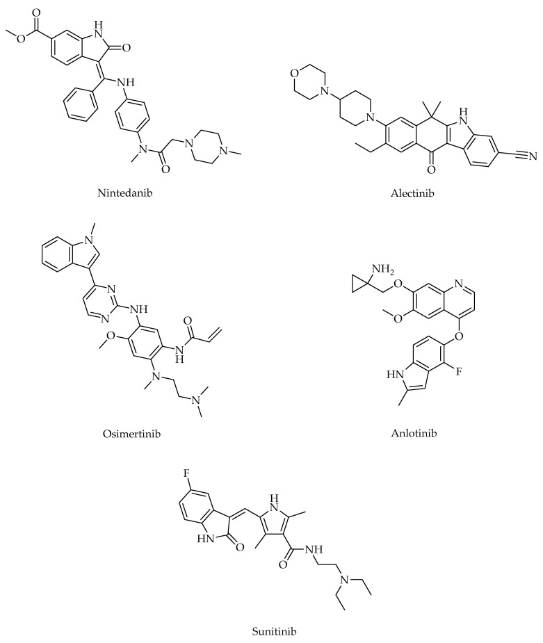
The indole ranks among the top 25 most common nitrogen heterocycles in U.S. Food and Drug Administration (FDA)-approved drugs. It is also a key structural component of an essential amino acid (tryptophan), a monoamine neurotransmitter (serotonin), and countless natural products (e.g., vinca alkaloids) [57]. The diverse applications of the indole core in challenging diseases (e.g., lung cancer, inflammatory diseases) make it one of the most privileged heterocyclic scaffolds for drug discovery [57,58]. Among vinca alkaloids, vinorelbine is the most frequently used antimitotic drug to treat lung cancer and vinblastine, in combination with cisplatin, is used in the management of NSCLC. Nintedanib (in combination with docetaxel), alectinib, osimertinib, anlotinib, and sunitinib are indole-based anti-NSCLC agents (Figure 1) [59]. In general, indole derivatives have been reported to exert marked anti-NSCLC action through diverse mechanisms including the induction of apoptosis, the inhibition of crucial biological targets such as microtubule, topoisomerases, protein kinases (e.g., Akt), and histone deacetylases (HDACs) [58,59,60,61,62]. The indole is also considered to be one of the most eligible scaffolds for anti-inflammatory drug discovery [62,63,64,65,66,67]. Indomethacin (Figure 2) is one of the most commonly prescribed NSAIDs exerting its action through the inhibition of COXs. Moreover, several experimental studies have revealed that indomethacin shows significant antiproliferative activity against a broad array of cancer (e.g., colorectal, lung) cell lines [68,69,70,71,72].
Figure 1.
Indole-based anti-NSCLC agents.
Figure 2.
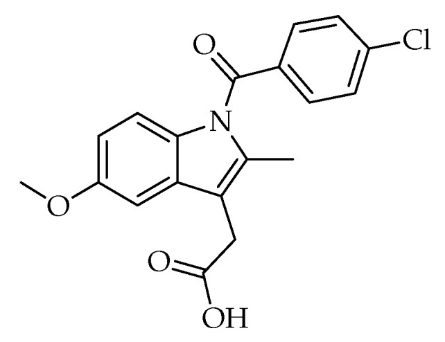
Indomethacin.
Taken together, the aforementioned data [26,27,28,29,30,31,32,33,34,35,36,37,38,39,40,41,42,43,44,45,46,47,48,49,50,51,52,53,54,55,56,57,58,59,60,61,62,63,64,65,66,67,68,69,70,71,72] prompted us to design two classes of indole-based small molecules (3a-j, 4a-g) for the targeted therapy of NSCLC. In this context, we performed the synthesis of new hydrazones (3a-j) and thiosemicarbazides (4a-g) efficiently and conducted in vitro and in vivo assays to assess their potential for the targeted therapy of NSCLC.
2. Results
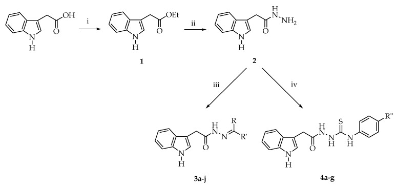
The reaction sequence for the preparation of the hitherto unreported small molecules (3a-j, 4a-g) is depicted in Figure 3, starting from 2-(1H-indol-3-yl)acetic acid. The convenient and efficient reaction of compound 2 with aromatic aldehydes or ketones and aryl isothiocyanates yielded new hydrazones (3a-j), and thiosemicarbazides (4a-g), respectively.
Figure 3.
The synthetic route for the preparation of compounds 3a-j and 4a-g. Reagents and conditions: (i) EtOH, H2SO4, reflux, 12 h; (ii) NH2NH2.H2O, EtOH, reflux, 4 h; (iii) RCHO or RCOR′, EtOH, reflux, 15 h; (iv) R″C6H4NCS, EtOH, rt, 8 h.
New hydrazones (3a-j) and thiosemicarbazides (4a-g) were subjected to in vitro assays to determine their COX inhibitory profiles. Among compounds 3a-j, compound 3a was found to be a nonselective COX inhibitor with IC50 values of 10.35 µM and 12.50 µM for COX-1 and COX-2, respectively (Table 1). On the other hand, compound 3b was the most selective COX-1 inhibitor (IC50 = 8.90 µM) in this series with a selectivity index (SI) value of 0.13.
Table 1.
COX inhibitory profiles of compounds 3a-j and positive controls.

| |||||
|---|---|---|---|---|---|
| Compound | R | R′ | IC50 (µM) | SI * | |
| COX-1 | COX-2 | ||||
| 3a | 4-(Pyrrolidin-1-yl)phenyl | H | 10.35 ± 0.35 | 12.50 ± 0.71 | 0.83 |
| 3b | 4-Morpholinophenyl | H | 8.90 ± 0.14 | 71.00 ± 1.41 | 0.13 |
| 3c | 4-(Piperidin-1-yl)phenyl | H | >100 | >100 | - |
| 3d | 4-(4-Methylpiperazin-1-yl)phenyl | H | 78.50 ± 8.50 | >100 | <0.79 |
| 3e | 4-(Methylsulfonyl)phenyl | H | 83.75 ± 6.25 | 35.00 ± 9.90 | 2.39 |
| 3f | 4-(Methylsulfonyl)phenyl | CH3 | 93.75 ± 6.25 | 51.00 ± 12.73 | 1.84 |
| 3g | 4-Morpholinophenyl | CH3 | 51.00 ± 1.00 | 52.50 ± 0.71 | 0.97 |
| 3h | 4-(2-Morpholinoethoxy)phenyl | H | 38.50 ± 1.5 | 50.50 ± 0.70 | 0.76 |
| 3i | 1-Methyl-1H-indol-3-yl | H | 31.25 ± 1.25 | >100 | <0.31 |
| 3j | 5-Methoxy-1H-indol-3-yl | H | >100 | 44.50 ± 6.36 | >2.25 |
| Indomethacin | - | - | 0.12 ± 0.01 | 0.58 ± 0.08 | 0.21 |
| Celecoxib | - | - | 8.88 ± 0.38 | 2.75 ± 0.05 | 3.23 |
* IC50 for COX-1/IC50 for COX-2.
Among compounds 4a-g, compound 4a was the most selective COX-1 inhibitor (IC50 = 10.00 µM) (Table 2). Other compounds did not show any inhibitory potency on COX-1 at the tested concentrations.
Table 2.
COX inhibitory profiles of compounds 4a-g and positive controls.
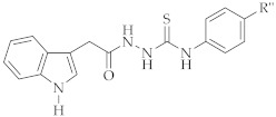
| ||||
|---|---|---|---|---|
| Compound | R″ | IC50 (µM) | SI * | |
| COX-1 | COX-2 | |||
| 4a | Br | 10.00 ± 0.13 | 76.50 ± 6.36 | 0.13 |
| 4b | CF3 | >100 | 61.50 ± 0.71 | >1.63 |
| 4c | CN | >100 | 56.50 ± 4.49 | >1.77 |
| 4d | Piperidin-1-ylsulfonyl | >100 | 59.00 ± 8.48 | >1.69 |
| 4e | 1H-Pyrazol-1-yl | >100 | 31.50 ± 2.12 | >3.17 |
| 4f | 3,4-Methylenedioxy | >100 | 51.50 ± 0.71 | >1.94 |
| 4g | Benzyloxy | >100 | 59.50 ± 2.12 | >1.68 |
| Indomethacin | - | 0.12 ± 0.01 | 0.58 ± 0.08 | 0.21 |
| Celecoxib | - | 8.88 ± 0.38 | 2.75 ± 0.05 | 3.23 |
* IC50 for COX-1/IC50 for COX-2.
All compounds were examined for their cytotoxic effects on L929 mouse fibroblast (normal) cells using the MTT test. Based on the in vitro experimental data, compound 3a, the nonselective COX inhibitor, showed cytotoxicity toward L929 cells with an IC50 value of 17.33 µM (Table 3), which is close to its IC50 values indicated in Table 1. On the other hand, compounds 3b and 4a did not show cytotoxicity against L929 cells at their effective concentrations. As a result, compounds 3b and 4a (Figure 4), the selective COX-1 inhibitors in this series, were chosen for further studies.
Table 3.
IC50 values of all compounds for L929 cells.
| Compound | IC50 (µM) |
|---|---|
| 3a | 17.33 ± 2.08 |
| 3b | 176.67 ± 5.77 |
| 3c | 21.33 ± 0.58 |
| 3d | 20.00 ± 1.73 |
| 3e | 85.00 ± 17.32 |
| 3f | <3.90 |
| 3g | <3.90 |
| 3h | <3.90 |
| 3i | 42.33 ± 0.58 |
| 3j | 43.00 ± 1.73 |
| 4a | 84.00 ± 19.70 |
| 4b | 22.67 ± 2.08 |
| 4c | 26.33 ± 3.51 |
| 4d | 45.67 ± 6.03 |
| 4e | 62.33 ± 7.51 |
| 4f | 14.00 ± 5.29 |
| 4g | 14.00 ± 3.46 |
Figure 4.
Selective COX-1 inhibitors in this series.
Compounds 3b and 4a were also subjected to the MTT assay to assess their cytotoxicity toward A549 human lung adenocarcinoma cell line. Based on the data presented in Table 4, compound 3b was found to be the most potent anticancer agent on A549 cells with an IC50 value of 89.67 µM compared to cisplatin (IC50 = 22.67 µM). On the other hand, compound 4a showed cytotoxic activity against A549 cells with an IC50 value of 179.33 µM.
Table 4.
IC50 values of compounds 3b, 4a, and cisplatin for A549 cells.
| Compound | IC50 (µM) |
|---|---|
| 3b | 89.67 ± 10.78 |
| 4a | 179.33 ± 77.59 |
| Cisplatin | 22.67 ± 4.04 |
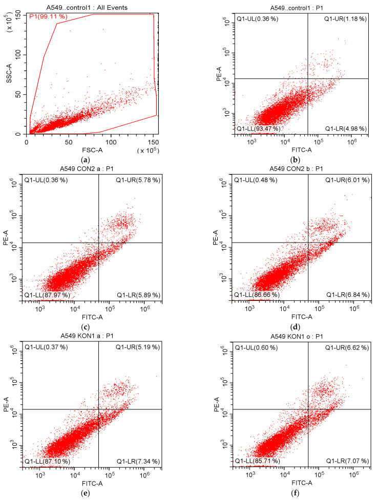
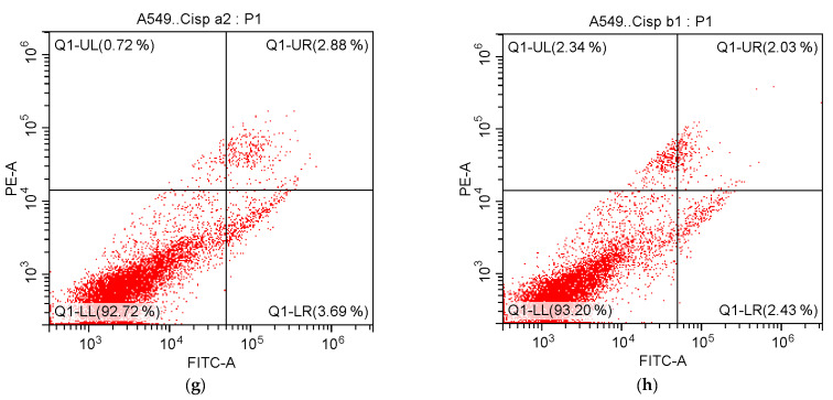
After 24 h incubation of A549 cells treated with compounds 3b and 4a in this series and cisplatin, flow cytometry-based apoptosis detection assay was performed to identify early and late apoptotic cells using Annexin V-fluorescein isothiocyanate (FITC)/propidium iodide (PI) staining. The percentages of A549 cells undergoing apoptosis (early and late) exposed to compounds 3b and cisplatin at their IC50/2 concentrations were found to be 11.67% and 6.57%, respectively. On the other hand, the percentages of A549 cells undergoing apoptosis (early and late) exposed to compounds 3b and cisplatin at their IC50 concentrations were 12.85% and 4.46%, respectively (Table 5, Figure 5). The percentages of A549 cells undergoing early and late apoptosis exposed to compound 4a at its IC50/4 concentration were 7.34% and 5.19%, respectively. On the other hand, the percentages of A549 cells undergoing early and late apoptosis exposed to compound 4a at its IC50/2 concentration were found to be 7.07% and 6.62%, respectively (Table 5, Figure 5).
Table 5.
Percentages of typical quadrant analysis of Annexin V FITC/PI flow cytometry of A549 cells treated with compounds 3b, 4a, and cisplatin.
| Compound | Early Apoptosis (%) | Late Apoptosis (%) | Necrosis (%) | Viability (%) |
|---|---|---|---|---|
| Control | 4.98 | 1.18 | 0.36 | 93.47 |
| Compound 3b at IC50/2 | 5.89 | 5.78 | 0.36 | 87.97 |
| Compound 3b at IC50 | 6.84 | 6.01 | 0.48 | 86.66 |
| Compound 4a at IC50/4 | 7.34 | 5.19 | 0.37 | 87.10 |
| Compound 4a at IC50/2 | 7.07 | 6.62 | 0.60 | 85.71 |
| Cisplatin at IC50/2 | 3.69 | 2.88 | 0.72 | 92.72 |
| Cisplatin at IC50 | 2.43 | 2.03 | 2.34 | 93.20 |
A549 cells were cultured for 24 h in medium with compound 4a (at its IC50/4 and IC50/2 concentrations), compound 3b, and cisplatin (at their IC50/2 and IC50 concentrations). At least 10,000 cells were analyzed per sample, and quadrant analysis was performed.
Figure 5.
Flow cytometric analysis of A549 cells treated with IC50/2 and IC50 concentrations of compounds 3b, 4a, and cisplatin. At least 10,000 cells were analyzed per sample, and quadrant analysis was performed. Q1-LR, Q1-UR, Q1-LL, and Q1-UL quadrants represent early apoptosis, late apoptosis, viability, and necrosis, respectively. (a) Control; (b) Control; (c) Compound 3b at IC50/2 concentration; (d) Compound 3b at IC50 concentration; (e) Compound 4a at IC50/4 concentration; (f) Compound 4a at IC50/2 concentration; (g) Cisplatin at IC50/2 concentration; (h) Cisplatin at IC50 concentration.
Akt inhibition caused by compounds 3b and 4a in A549 cells was examined using a colorimetric assay. Compounds 3b and 4a caused Akt inhibition in A549 cell line with IC50 values of 32.50 and 45.33 µM as compared to GSK690693 (IC50= 5.93 µM) (Table 6).
Table 6.
Akt inhibitory effects of compounds 3b, 4a, GSK690693, and cisplatin in A549 cells.
| Compound | IC50 (µM) |
|---|---|
| 3b | 32.50 ± 4.95 |
| 4a | 45.33 ± 6.51 |
| GSK690693 | 5.93 ± 1.20 |
| Cisplatin | 9.30 ± 2.55 |
The lipopolysaccharide (LPS)-induced sepsis model was used to assess the in vivo anti-inflammatory activities of compounds 3b and 4a. According to the data indicated in Table 7, the myeloperoxidase (MPO) activity of the LPS group increased as compared to the control group. However, this increase is not statistically significant. The LPS + compound 3b group slightly decreased the MPO activity compared to the LPS group, while the LPS + compound 4a group significantly decreased the MPO activity compared to the LPS group (p < 0.05). The decrease in the MPO activity caused by compound 4a was higher than that caused by the indomethacin therapy.
Table 7.
Effects of compounds 3b, 4a, and indomethacin on MPO levels.
| Groups | MPO (U/L) |
|---|---|
| Control | 1.03 ± 0.51 |
| LPS | 1.79 ± 0.27 |
| LPS + Compound 3b | 1.50 ± 0.81 |
| LPS + Compound 4a | 0.84 ± 0.26 # |
| LPS + Indomethacin | 1.06 ± 0.73 |
Values are given as mean ± standard deviation (SD). Significance according to LPS values, #: p < 0.05. One-way ANOVA, post-hoc Tukey test n = 8.
As presented in Table 8, there was a significant increase in the nitric oxide (NO) level after LPS administration compared to the control (p < 0.001). LPS + compound 3b, LPS + compound 4a, and LPS + indomethacin caused a significant decrease in the serum NO levels. However, this decrease in LPS + compound 3b was similar to the control. The NO level was significantly higher in the LPS group than in the control group, while it was markedly lower in the compound 4a pre-treatment group compared to the LPS group (p < 0.05).
Table 8.
Effects of compounds 3b, 4a, and indomethacin on NO levels (µmol/L).
| Groups | 25% Percentile | Median | 75% Percentile |
|---|---|---|---|
| Control | 0 | 0.07 | 0.14 |
| LPS *** | 4.482 | 6.018 | 8.386 |
| LPS + Compound 3b | 0.316 | 0.667 | 2.772 |
| LPS + Compound 4a * | 1.369 | 2.509 | 4.0 |
| LPS + Indomethacin * | 0.667 | 1.281 | 3.123 |
Significance relative to control values, *: p < 0.05, ***: p < 0.001. One-way ANOVA, Kruskal–Wallis test, n = 8.
According to the in vivo experiments, the alanine aminotransferase (ALT) level decreased in all groups compared to the LPS group (Table 9). This decrease was greater in the group treated with compound 4a compared to the group treated with compound 3b. Likewise, the aspartate aminotransferase (AST) level decreased in all groups compared to the LPS group. This decrease was greater in the group treated with compound 3b compared to the group treated with compound 4a and the group treated with indomethacin. However, the decrease caused by the compounds in the ALT and AST levels was not statistically significant compared to the LPS group.
Table 9.
Effects of compounds 3b, 4a, and indomethacin on ALT and AST levels.
| Groups | ALT (U/L) | AST (U/L) |
|---|---|---|
| Control | 45.75 ± 3.81 | 126.60 ± 24.09 |
| LPS | 57.14 ± 22.94 | 139.90 ± 26.34 |
| LPS + Compound 3b | 50.25 ± 21.86 | 114.90 ± 22.04 |
| LPS + Compound 4a | 48.29 ± 26.88 | 132.30 ± 37.06 |
| LPS + Indomethacin | 44.00 ± 14.64 | 123.50 ± 32.14 |
Values are given as mean ± SD. One-way ANOVA, post-hoc Tukey test, n = 8.
3. Discussion
Experimental studies have demonstrated that hydrazones show marked antitumor action through various mechanisms, including the inhibition of Akt [31] or the phosphatidylinositol 3-kinase (PI3K)/Akt signaling pathway [32,36]. N′-benzylidene-2-[(4-(4-methoxyphenyl)pyrimidin-2-yl)thio]acetohydrazide was previously reported to exert marked anticancer activity against the 5RP7 H-ras oncogene transformed rat embryonic fibroblast cell line via the induction of apoptosis and the inhibition of Akt (IC50 = 0.50 µg/mL) [31]. According to western blot data reported by Han et al., (S)-2-{[5-[1-(6-methoxynaphtalene-2-yl)ethyl]-4-(4-fluorophenyl)-4H-1,2,4-triazole-3-yl]thio}-N′-[(5-nitrofuran-2-yl)methylidene]acetohydrazide caused a significant decrease in the epidermal growth factor receptor (EGFR), PI3K, and Akt phosphorylation in PC3 human prostate cancer cells [32]. Bak et al. indicated that 5-hydroxy-7,4′-diacetyloxyflavanone-N-phenyl hydrazone (N101-43) induced apoptosis via the up-regulation of Fas/FasL expression, the activation of caspase cascade, and the inhibition of the PI3K/Akt signaling pathway in NSCLC cells [36].
The anti-inflammatory and/or COX inhibitory potential of hydrazones was demonstrated by in vitro and in vivo studies [37,38,39,40,41]. In our previous work, 2-[(1-methyl-1H-tetrazol-5-yl)thio]-N′-(4-(piperidin-1-yl)benzylidene)acetohydrazide and 2-[(1-methyl-1H-tetrazol-5-yl)thio]-N′-(4-(morpholin-4-yl)benzylidene)acetohydrazide caused selective COX-1 inhibition [37].
Thiosemicarbazides show pronounced antiproliferative activity toward a variety of tumor cells through diverse mechanisms [42,43,44,45,46,47,48,49,50,51,52]. Our research team reported that 4-(1,3-benzodioxol-5-yl)-1-([1,1′-biphenyl]-4-ylmethylene)thiosemicarbazide showed remarkable anticancer activity against A549 human lung adenocarcinoma and C6 rat glioma cells through apoptosis induction mediated by the disruption of ΔΨm [52].
In vitro and in vivo experimental data revealed that thiosemicarbazides exert marked anti-inflammatory action through several mechanisms including COX inhibition [53,54,55,56]. In our recent work [53], 4-[4-(piperidin-1-ylsulfonyl)phenyl]-1-[4-(4-cyanophenoxy)benzylidene]thiosemicarbazide was found to be a selective COX-1 inhibitor with an IC50 value of 1.89 µM. On the other hand, 4-[4-(piperidin-1-ylsulfonyl)phenyl]-1-[4-(4-nitrophenoxy)benzylidene]thiosemicarbazide was determined to be a nonselective COX inhibitor (COX-1 IC50 = 13.44 µM, COX-2 IC50 = 12.60 µM). Based on the LPS-induced sepsis model, these agents diminished the MPO, NO, high-sensitivity C-reactive protein (hsCRP), malondialdehyde (MDA), ALT, and AST levels. Both compounds were identified as potential anti-inflammatory agents [53].
Indole-based small molecules exert a notable anti-NSCLC action through multiple mechanisms such as the induction of apoptosis and the inhibition of crucial biological targets including protein kinases (e.g., Akt) [58,59,60,61,62]. Furthermore, mounting evidence has demonstrated that indole derivatives show marked anti-inflammatory action via COX inhibition [62,63,64,65,66,67]. In our previous study [65], 3-(5-bromo-1H-indol-3-yl)-1-(4-cyanophenyl)prop-2-en-1-one was found to be a nonselective COX inhibitor (COX-1 IC50 = 8.10 µg/mL, COX-2 IC50 = 9.50 µg/mL), while 3-(5-methoxy-1H-indol-3-yl)-1-(4-(methylsulfonyl)phenyl)prop-2-en-1-one inhibited COX-1 (IC50 = 8.60 µg/mL). According to the LPS-induced sepsis model, both compounds markedly decreased the MPO, NO, hsCRP, MDA, ALT, and AST levels. Both indole derivatives were identified as potential anti-inflammatory agents [65].
Based on the aforementioned studies [26,27,28,29,30,31,32,33,34,35,36,37,38,39,40,41,42,43,44,45,46,47,48,49,50,51,52,53,54,55,56,57,58,59,60,61,62,63,64,65,66,67,68,69,70,71,72], two classes of indole-based small molecules (3a-j, 4a-g) for the targeted therapy of NSCLC were designed. In this context, we carried out the synthesis of new hydrazones (3a-j) and thiosemicarbazides (4a-g) efficiently and performed in vitro and in vivo experiments to assess their potential for the targeted therapy of NSCLC.
Among compounds 3a-j, compound 3a was determined to be a nonselective COX inhibitor with IC50 values of 10.35 µM and 12.50 µM for COX-1 and COX-2, respectively, while compound 3b was found to be a selective COX-1 inhibitor (IC50 = 8.90 µM). Compound 3b exhibited COX-2 inhibitory activity with an IC50 value of 71.00 µM. The SI values of compounds 3a and 3b were determined to be 0.83 and 0.13, respectively. In particular, the pyrrolidine ring enhanced the inhibitory effects on both COXs, whereas the morpholine substitution caused selective COX-1 inhibitory potency. The replacement of the morpholine ring (compound 3b) with the piperidine ring (compound 3c) or the piperazine ring (compound 3d) led to a substantial drop in COX-1 inhibitory activity. Compound 3c, carrying a piperidine ring, showed the lowest COX inhibition (>100 µM) in this series.
According to the in vitro data related to the inhibitory effects of compounds 3b and 3h on COXs, it can be concluded that the ethoxy linker between the morpholine and the benzene rings diminishes COX-1 inhibition, while it enhances COX-2 inhibition. Taking into account the inhibitory effects of compounds 3b and 3g on COXs, it is important to note that the methyl branching decreases COX-1 inhibition and increases COX-2 inhibition.
The SI values of compounds 3e and 3f were found to be 2.39 and 1.84, respectively, indicating that the methylsulfonyl group significantly enhances COX-2 selectivity.
Based on the experimental results related to the inhibitory effects of compounds 3i and 3j on COXs, the methyl substituent at the 1st position of the indole scaffold enhances COX-1 selectivity, while the methoxy substituent at the 5th position of the indole core enhances COX-2 selectivity.
Among compounds 4a-g, the most selective COX-1 inhibitor was found to be compound 4a (IC50 = 10.00 µM, SI = 0.13). It can be concluded that the bromo substituent at the 4th position of the phenyl moiety significantly enhanced the COX-1 inhibitory potency. Other compounds did not exhibit any inhibitory activity towards COX-1 at the tested concentrations. Thiosemicarbazides tested in this work, except for compound 4a, were found to have a tendency to inhibit the COX-2 enzyme.
Among the indole-based small molecules (3a-j, 4a-g), compounds 3b and 4a, selective COX-1 inhibitors in this series, were chosen for further studies since both compounds did not exert cytotoxicity toward L929 (normal) cells at their effective concentrations reported for their COX-1 inhibitory activity.
To investigate their potential as anti-NSCLC agents, their cytotoxic effects on A549 cells were evaluated by means of the MTT assay protocol. Based on the experimental data, compound 3b was the most potent anticancer agent on A549 cell line with an IC50 value of 89.67 µM. It can be concluded that the anticancer activity of compound 3b against A549 cells is selective since the IC50 value of compound 3b for L929 cells is 176.67 µM. On the other hand, compound 4a showed cytotoxic activity against A549 and L929 cells with IC50 values of 179.33 µM and 84.00 µM, respectively. The cytotoxic activity of compound 4a toward A549 cells was found to be nonselective at its IC50 value. For this reason, the IC50/4 and IC50/2 concentrations of compound 4a were applied in the flow cytometry analyses of apoptosis and the Akt inhibition assay.
In cancer, cells lose their ability to undergo apoptosis, resulting in uncontrolled proliferation [73]. The induction of apoptosis is reported to be an intriguing modality for the management of all types of cancer since apoptosis evasion is a hallmark of cancer and is nonspecific to the cause or the type of the cancer [74]. Based on the flow cytometry-based apoptosis detection assay performed in this work, A549 cells treated with compounds 3b and 4a underwent apoptosis, pointing out their apoptosis-inducing efficacy.
Akt participates in the pathogenesis of NSCLC and, therefore, the inhibition of Akt by natural and synthetic agents stands out as a rational strategy for cancer therapy [21,22,23,24,25]. The colorimetric assay conducted in this study revealed that the Akt inhibitory activity of compound 3b (IC50 = 32.50 µM) was more notable than that of compound 4a (IC50 = 45.33 µM) in A549 cells.
Sepsis is described as a life-threatening organ dysfunction provoked by a dysregulated host response to infection [53]. Inflammatory imbalance plays a fundamental role in the pathogenesis of sepsis and occurs throughout the whole process of sepsis [75]. The parameters related to inflammation are crucial for evaluating a sepsis case [76]. In this work, the LPS-induced sepsis model was used to evaluate the in vivo anti-inflammatory activities of compounds 3b and 4a.
MPO is linked to several diseases, particularly those in which strong infiltration of polymorphonuclear cells (PMNs) and acute or chronic inflammation are involved. MPO contributes to the pathophysiology of diverse diseases such as rheumatoid arthritis, atherosclerosis, pulmonary fibrosis, renal glomerular injury, multiple sclerosis, Huntington’s disease, Alzheimer’s disease, Parkinson’s disease, liver diseases, diabetes, obesity, and cancer. MPO is reported to promote tumor initiation and progression. MPO participates in the regulation of tumor growth, apoptosis, migration, and metastasis [77]. In this work, compound 3b caused a slight decrease in the MPO activity compared to the LPS group, whereas compound 4a significantly diminished the MPO activity compared to the LPS group (p < 0.05).
Sepsis is characterized by a robust rise in NO production throughout the body that is driven by inducible NO synthase (iNOS) [78]. Due to the key role of NO in the pathogenesis of inflammation as a signaling molecule [78,79,80], herein the effects of compounds 3b and 4a on the serum NO levels were evaluated. The in vivo experimental data revealed that compounds 3b and 4a diminished the serum NO levels.
Aminotransferases, also referred to as transaminases, are commonly used as markers of hepatocellular injury in nonclinical toxicology studies and clinical trials. In general, aminotransferase activity in blood (serum or plasma) is elevated in the hepatocellular damage induced by diseases or drugs such as anti-inflammatory drugs [81,82,83,84]. Based on the in vivo experimental data performed in this work, both compounds caused a decrease in the serum aminotransferase levels. In particular, compound 3b diminished the serum AST level more than indomethacin.
Taking into account the knowledge obtained from the in vitro and in vivo assays, compound 3b can be considered as a lead compound for the targeted therapy of NSCLC due to its direct cytotoxic effects on A549 cells as well as its possible effects on the tumor microenvironment (e.g., tumor-related inflammation).
4. Materials and Methods
4.1. Chemistry
The chemicals were procured from commercial suppliers and were used without further purification. Melting points (M.p.) were determined on the Electrothermal IA9200 digital melting point apparatus (Staffordshire, UK) and were uncorrected. Thin Layer Chromatography (TLC) was performed on TLC Silica gel 60 F254 aluminum sheets (Merck, Darmstadt, Germany) using petroleum ether:ethyl acetate solvent system (1:1). IR spectra were recorded on the IRPrestige-21 Fourier Transform Infrared spectrophotometer (Shimadzu, Tokyo, Japan). 1H and 13C NMR spectra were recorded on the Varian Mercury 400 NMR spectrometer (Agilent, Palo Alto, CA, USA). HRMS spectra were recorded on the LC/MS IT-TOF system (Shimadzu, Tokyo, Japan) using the electrospray ionization (ESI) technique.
4.1.1. Preparation of ethyl 2-(1H-indol-3-yl)acetate (1)
Compound 1 was synthesized starting from 2-(1H-indol-3-yl)acetic acid according to a previous work [85].
4.1.2. Preparation of 2-(1H-indol-3-yl)acetohydrazide (2)
Compound 2 was obtained by the reaction of compound 1 with hydrazine hydrate according to a previous work [85].
4.1.3. General Method for the Preparation of N′-benzylidene/(1-arylethylidene)-2-(1H-indol-3-yl)acetohydrazide Derivatives (3a-j)
A mixture of compound 2 and aromatic aldehyde or ketone in ethanol was heated under reflux for 15 h. At the end of this period, the precipitate was filtered off and dried. The product was crystallized from ethanol.
2-(1H-Indol-3-yl)-N′-[4-(pyrrolidin-1-yl)benzylidene]acetohydrazide (3a)
Yield: 78%. M.p.: 302–303 °C. IR νmax (cm−1): 3196.05, 3074.53, 3043.67, 2966.52, 2914.44, 2873.94, 2848.86, 1668.43, 1595.13, 1546.91, 1521.84, 1487.12, 1460.11, 1431.18, 1386.82, 1350.17, 1323.17, 1292.31, 1249.87, 1224.80, 1174.65, 1163.08, 1118.71, 1047.35, 1001.06, 983.70, 958.62, 929.69, 914.26, 856.39, 804.32, 719.45, 682.80. 1H NMR (400 MHz, DMSO-d6): 2.02–2.05 (m, 4H), 3.40–3.42 (m, 4H), 3.60 and 4.02 (2s, 2H), 6.92–7.07 (m, 4H), 7.21 (dd, J = 2.4 Hz, 12.8 Hz, 1H), 7.31–7.35 (m, 1H), 7.48–7.59 (m, 3H), 7.88 and 8.08 (2s, 1H), 10.85 and 10.88 (2s, 1H), 11.05 and 11.28 (2s, 1H). 13C NMR (100 MHz, DMSO-d6): 25.41 (2CH2), 32.13 (CH2), 47.70 (2CH2), 108.79 (C), 111.74 (CH), 115.13 (2CH), 118.74 (CH), 119.19 (CH), 121.33 (CH), 124.32 (CH), 124.75 (C), 127.90 (C), 128.27 (2CH), 136.45 (C), 146.90 (CH), 152.26 (C), 172.72 (C). HRMS (ESI) (m/z): [M + H]+ calcd. for C21H22N4O: 347.1866, found: 347.1864.
2-(1H-Indol-3-yl)-N′-(4-morpholinobenzylidene)acetohydrazide (3b)
Yield: 85%. M.p.: 306–307 °C. IR νmax (cm−1): 3275.13, 3178.69, 3055.24, 2964.59, 2922.16, 2870.08, 2825.72, 1660.71, 1604.77, 1558.48, 1541.12, 1519.91, 1506.41, 1489.05, 1456.26, 1446.61, 1425.40, 1392.61, 1375.25, 1338.60, 1313.52, 1301.95, 1259.52, 1224.80, 1186.22, 1176.58, 1159.22, 1109.07, 1095.57, 1062.78, 1045.42, 1006.84, 958.62, 921.97, 875.68, 858.32, 846.75, 823.60, 798.53, 786.96, 742.59, 682.80. 1H NMR (400 MHz, DMSO-d6): 3.19 (t, J = 4.41 Hz, 4.62 Hz, 4H), 3.72–3.75 (m, 4H), 3.61 and 4.02 (2s, 2H), 6.92–7.08 (m, 4H), 7.21 (dd, J = 2.4 Hz, 12.8 Hz, 1H), 7.31–7.35 (m, 1H), 7.49–7.60 (m, 3H), 7.88 and 8.07 (2s, 1H), 10.85 and 10.89 (2s, 1H), 11.05 and 11.28 (2s, 1H). 13C NMR (100 MHz, DMSO-d6): 32.13 (CH2), 53.79 (2CH2), 66.40 (2CH2), 108.79 (C), 111.74 (CH), 115.13 (2CH), 118.74 (CH), 119.19 (CH), 121.33 (CH), 124.32 (CH), 124.75 (C), 127.91 (C), 128.27 (2CH), 136.45 (C), 146.90 (CH), 152.29 (C), 172.73 (C). HRMS (ESI) (m/z): [M + H]+ calcd. for C21H22N4O2: 363.1816, found: 363.1824.
2-(1H-Indol-3-yl)-N′-[4-(piperidin-1-yl)benzylidene]acetohydrazide (3c)
Yield: 80%. M.p.: 265–266 °C. IR νmax (cm−1): 3203.76, 3082.25, 3034.03, 2972.31, 2935.66, 2856.58, 2825.72, 1668.43, 1598.99, 1552.70, 1514.12, 1448.54, 1427.32, 1384.89, 1350.17, 1282.66, 1247.94, 1220.94, 1182.36, 1124.50, 1024.20, 962.48, 914.26, 858.32, 804.32, 721.38, 651.94. 1H NMR (400 MHz, DMSO-d6): 1.58 (brs, 6H), 3.32 (brs, 4H), 3.60 and 4.02 (2s, 2H), 6.92–7.08 (m, 4H), 7.21 (dd, J = 2.4 Hz, 12.8 Hz, 1H), 7.31–7.35 (m, 1H), 7.48–7.59 (m, 3H), 7.88 and 8.08 (2s, 1H), 10.85 and 10.89 (2s, 1H), 11.05 and 11.28 (2s, 1H). 13C NMR (100 MHz, DMSO-d6): 24.40 (CH2), 25.42 (2CH2), 32.12 (CH2), 48.95 (2CH2), 108.79 (C), 111.74 (CH), 115.13 (2CH), 118.74 (CH), 119.19 (CH), 121.33 (CH), 124.32 (CH), 124.75 (C), 127.91 (C), 128.27 (2CH), 136.45 (C), 146.90 (CH), 152.26 (C), 172.73 (C). HRMS (ESI) (m/z): [M + H]+ calcd. for C22H24N4O: 361.2023, found: 361.2031.
2-(1H-Indol-3-yl)-N′-[4-(4-methylpiperazin-1-yl)benzylidene]acetohydrazide (3d)
Yield: 81%. M.p.: 218–220 °C. IR νmax (cm−1): 3398.57, 3205.69, 3165.19, 3111.18, 3043.67, 2939.52, 2883.58, 2831.50, 1649.49, 1602.85, 1517.98, 1446.61, 1427.32, 1409.96, 1377.17, 1340.53, 1286.52, 1232.51, 1184.29, 1159.22, 1141.86, 1124.50, 1105.21, 1080.14, 1001.06, 956.69, 943.19, 921.97, 806.25, 794.67, 742.59, 686.66. 1H NMR (400 MHz, DMSO-d6): 2.21 (s, 3H), 2.40–2.44 (m, 4H), 3.20–3.22 (m, 4H), 3.59 and 4.02 (2s, 2H), 6.92–7.08 (m, 4H), 7.21 (dd, J = 2.4 Hz, 12.8 Hz, 1H), 7.31–7.35 (m, 1H), 7.48–7.59 (m, 3H), 7.88 and 8.08 (2s, 1H), 10.85 and 10.89 (2s, 1H), 11.05 and 11.28 (2s, 1H). 13C NMR (100 MHz, DMSO-d6): 32.13 (CH2), 46.21 (CH3), 47.69 (2CH2), 54.89 (2CH2), 108.79 (C), 111.74 (CH), 115.13 (2CH), 118.74 (CH), 119.19 (CH), 121.33 (CH), 124.32 (CH), 124.75 (C), 127.91 (C), 128.27 (2CH), 136.45 (C), 146.90 (CH), 152.28 (C), 172.73 (C). HRMS (ESI) (m/z): [M + H]+ calcd. for C22H25N5O: 376.2132, found: 376.2148.
2-(1H-Indol-3-yl)-N′-(4-methylsulfonylbenzylidene)acetohydrazide (3e)
Yield: 86%. M.p.: 264–265 °C. IR νmax (cm−1): 3344.57, 3205.69, 3055.24, 2929.87, 2897.08, 1668.43, 1604.77, 1556.55, 1489.05, 1454.33, 1408.04, 1365.60, 1328.95, 1313.52, 1290.38, 1242.16, 1222.87, 1199.72, 1145.72, 1089.78, 1055.06, 1018.41, 983.70, 972.12, 956.69, 943.19, 869.90, 835.18, 792.74, 769.60, 750.31, 729.09, 686.66, 651.94. 1H NMR (400 MHz, DMSO-d6): 3.24 (s, 3H), 3.67 and 4.09 (2s, 2H), 6.94–7.09 (m, 2H), 7.25 (dd, J = 2.4 Hz, 9.6 Hz, 1H), 7.34 (t, J = 8.0 Hz, 8.4 Hz, 1H), 7.56–7.60 (m, 1H), 7.90–7.96 (m, 4H), 8.07 and 8.31 (2s, 1H), 10.87 and 10.93 (2s, 1H), 11.52 and 11.76 (2s, 1H). 13C NMR (100 MHz, DMSO-d6): 31.87 (CH2), 43.64 (CH3), 108.10 (C), 111.51 (CH), 118.53 (CH), 118.87 (CH), 121.12 (CH), 124.18 (CH), 127.31 (C), 127.50 (2CH), 127.70 (2CH), 136.19 (C), 139.35 (C), 141.21 (C), 144.37 (CH), 173.17 (C). HRMS (ESI) (m/z): [M + H]+ calcd. for C18H17N3O3S: 356.1063, found: 356.1071.
2-(1H-Indol-3-yl)-N′-[1-(4-methylsulfonylphenyl)ethylidene]acetohydrazide (3f)
Yield: 81%. M.p.: 204–205 °C. IR νmax (cm−1): 3342.64, 3190.26, 3088.03, 3032.10, 3005.10, 2924.09, 2848.86, 1668.43, 1585.49, 1562.34, 1489.05, 1456.26, 1417.68, 1394.53, 1338.60, 1296.16, 1280.73, 1226.73, 1188.15, 1145.72, 1093.64, 1070.49, 1008.77, 977.91, 964.41, 852.54, 839.03, 788.89, 758.02, 740.67, 717.52, 700.16, 688.59. 1H NMR (400 MHz, DMSO-d6): 2.30 (s, 3H), 3.26 (s, 3H), 3.81 and 4.12 (2s, 2H), 6.95–7.07 (m, 2H), 7.20–7.36 (m, 2H), 7.54–7.61 (m, 1H), 8.01 (d, J = 8.0 Hz, 2H), 8.15 (d, J = 8.8 Hz, 2H), 10.63 and 10.66 (2s, 1H), 10.84 and 10.89 (2s, 1H). 13C NMR (100 MHz, DMSO-d6): 14.96 (CH3), 31.87 (CH2), 43.42 (CH3), 108.10 (C), 111.26 (CH), 118.25 (CH), 118.64 (CH), 121.12 (CH), 123.84 (CH), 126.70 (C), 127.11 (2CH), 127.39 (2CH), 141.55 (C), 142.06 (C), 142.97 (C), 156.20 (C), 173.17 (C). HRMS (ESI) (m/z): [M + H]+ calcd. for C19H19N3O3S: 370.1220, found: 370.1202.
2-(1H-Indol-3-yl)-N′-[1-(4-morpholinophenyl)ethylidene]acetohydrazide (3g)
Yield: 79%. M.p.: 198–199 °C. IR νmax (cm−1): 3269.34, 3080.32, 3047.53, 2966.52, 2916.37, 2848.86, 1668.43, 1608.63, 1593.20, 1546.91, 1516.05, 1454.33, 1444.68, 1417.68, 1379.10, 1361.74, 1340.53, 1301.95, 1263.37, 1236.37, 1197.79, 1118.71, 1068.56, 1051.20, 1026.13, 937.40, 923.90, 864.11, 821.68, 798.53, 742.59, 729.09, 648.08. 1H NMR (400 MHz, DMSO-d6): 2.31 (s, 3H), 3.19 (t, J = 4.41 Hz, 4.62 Hz, 4H), 3.72–3.75 (m, 4H), 3.60 and 4.02 (2s, 2H), 6.92–7.08 (m, 4H), 7.21 (dd, J = 2.4 Hz, 12.8 Hz, 1H), 7.31–7.35 (m, 1H), 7.48–7.59 (m, 3H), 10.85 and 10.89 (2s, 1H), 11.05 and 11.27 (2s, 1H). 13C NMR (100 MHz, DMSO-d6): 14.96 (CH3), 32.13 (CH2), 53.79 (2CH2), 66.40 (2CH2), 108.79 (C), 111.74 (CH), 115.13 (2CH), 118.74 (CH), 119.19 (CH), 121.33 (CH), 124.32 (CH), 124.75 (C), 127.91 (C), 128.27 (2CH), 136.45 (C), 143.30 (C), 156.18 (C), 172.73 (C). HRMS (ESI) (m/z): [M + H]+ calcd. for C22H24N4O2: 377.1972, found: 377.1982.
2-(1H-Indol-3-yl)-N′-[4-(2-morpholinoethoxy)benzylidene]acetohydrazide (3h)
Yield: 83%. M.p.: 132–135 °C. IR νmax (cm−1): 3383.14, 3319.49, 3196.05, 3045.60, 2958.80, 2918.30, 2850.79, 1664.57, 1604.77, 1548.84, 1510.26, 1456.26, 1421.54, 1355.96, 1340.53, 1303.88, 1240.23, 1201.65, 1170.79, 1116.78, 1049.28, 1010.70, 983.70, 952.84, 925.83, 860.25, 831.32, 742.59, 646.15. 1H NMR (400 MHz, DMSO-d6): 2.45–2.47 (m, 4H), 2.66–2.70 (m, 2H), 3.55–3.58 (m, 4H), 4.04 (s, 2H), 4.08–4.12 (m, 2H), 6.95–7.07 (m, 4H), 7.23 (dd, J = 2.4 Hz, 12.4 Hz, 1H), 7.34 (t, J = 8.0 Hz, 1H), 7.57–7.65 (m, 3H), 7.94 and 8.16 (2s, 1H), 10.86 and 10.91 (2s, 1H), 11.15 and 11.39 (2s, 1H). 13C NMR (100 MHz, DMSO-d6): 31.85 (CH2), 53.79 (2CH2), 57.12 (CH2), 65.64 (CH2), 66.35 (2CH2), 108.44 (C), 111.48 (CH), 115.05 (2CH), 118.47 (CH), 118.91 (CH), 121.07 (CH), 124.07 (CH), 127.18 (C), 127.63 (C), 128.41 (2CH), 136.19 (C), 146.18 (CH), 159.90 (C), 172.64 (C). HRMS (ESI) (m/z): [M + H]+ calcd. for C23H26N4O3: 407.2078, found: 407.2071.
2-(1H-Indol-3-yl)-N′-[(1-methyl-1H-indol-3-yl)methylene]acetohydrazide (3i)
Yield: 84%. M.p.: 221–224 °C. IR νmax (cm−1): 3414.00, 3147.83, 3101.54, 3061.03, 2980.02, 2945.30, 2908.65, 2819.93, 1651.07, 1612.49, 1570.06, 1539.20, 1502.55, 1462.04, 1452.40, 1421.54, 1404.18, 1377.17, 1346.31, 1332.81, 1321.24, 1253.73, 1244.09, 1197.79, 1157.29, 1139.93, 1120.64, 1087.85, 1072.42, 1045.42, 1008.77, 948.98, 933.55, 900.76, 856.39, 808.17, 785.03, 744.52, 734.88, 673.16. 1H NMR (400 MHz, DMSO-d6): 3.64 and 4.13 (2s, 2H), 3.79 (s, 3H), 6.94–7.18 (m, 3H), 7.22–7.28 (m, 2H), 7.36 (t, J = 8.4 Hz, 8.8 Hz, 1H), 7.47 (t, J = 8.4 Hz, 9.2 Hz, 1H), 7.64 (t, J = 7.6 Hz, 1H), 7.73 (d, J = 2.4 Hz, 1H), 8.20–8.38 (m, 2H), 10.86 and 10.92 (2s, 1H), 10.99 and 11.19 (2s, 1H). 13C NMR (100 MHz, DMSO-d6): 31.94 (CH2), 32.90 (CH3), 108.74 (C), 110.42 (CH), 110.83 (CH), 111.49 (C), 118.50 (CH), 118.93 (CH), 120.89 (CH), 121.10 (CH), 121.86 (CH), 122.80 (CH), 123.96 (CH), 124.70 (C), 127.70 (C), 133.81 (CH), 136.23 (C), 137.76 (C), 142.98 (CH), 172.09 (C). HRMS (ESI) (m/z): [M + H]+ calcd. for C20H18N4O: 331.1553, found: 331.1538.
2-(1H-Indol-3-yl)-N′-[(5-methoxy-1H-indol-3-yl)methylene]acetohydrazide (3j)
Yield: 82%. M.p.: 231–233 °C. IR νmax (cm−1): 3415.93, 3373.50, 3049.46, 3012.81, 2958.80, 2931.80, 2877.79, 2829.57, 1654.92, 1614.42, 1577.77, 1539.20, 1487.12, 1456.26, 1421.54, 1396.46, 1354.03, 1342.46, 1307.74, 1292.31, 1261.45, 1213.23, 1182.36, 1176.58, 1130.29, 1105.21, 1087.85, 1072.42, 1049.28, 1022.27, 1006.84, 950.91, 923.90, 856.39, 810.10, 744.52, 725.23, 671.23, 651.94. 1H NMR (400 MHz, DMSO-d6): 3.59 (s, 3H), 3.74 and 4.16 (2s, 2H), 6.83 (dd, J = 2.4 Hz, 8.8 Hz, 1H), 6.93–7.10 (m, 2H), 7.27–7.38 (m, 3H), 7.64 (t, J = 8.8 Hz, 9.2 Hz, 1H), 7.72–7.80 (m, 2H), 8.23 and 8.41 (2s, 1H), 10.85 and 10.91 (2s, 1H), 11.02 and 11.18 (2s, 1H), 11.40 (s, 1H). 13C NMR (100 MHz, DMSO-d6): 31.90 (CH2), 55.01 (CH3), 103.47 (CH), 108.71 (C), 111.50 (C), 111.59 (CH), 112.39 (CH), 112.64 (CH), 118.52 (CH), 118.83 (CH), 121.14 (CH), 123.88 (CH), 124.83 (C), 127.73 (C), 130.56 (C), 132.19 (CH), 136.19 (C), 143.76 (CH), 154.55 (C), 172.04 (C). HRMS (ESI) (m/z): [M + H]+ calcd. for C20H18N4O2: 347.1503, found: 347.1505.
4.1.4. General Method for the Preparation of 4-aryl-1-[2-(1H-indol-3-yl)acetyl]thiosemicarbazide Derivatives (4a-g)
A mixture of compound 2 and aryl isothiocyanate in ethanol was stirred at room temperature for 8 h. The precipitate was filtered off. The product was crystallized from ethanol.
4-(4-Bromophenyl)-1-[2-(1H-indol-3-yl)acetyl]thiosemicarbazide (4a)
Yield: 87%. M.p.: 187–189 °C. IR νmax (cm−1): 3390.86, 3311.78, 3286.70, 3207.62, 3143.97, 3057.17, 2997.38, 2927.94, 1680.00, 1647.21, 1620.21, 1589.34, 1544.98, 1506.41, 1485.19, 1452.40, 1419.61, 1352.10, 1309.67, 1282.66, 1247.94, 1207.44, 1138.00, 1087.85, 1074.35, 1049.28, 1004.91, 987.55, 871.82, 823.60, 792.74, 736.81, 715.59, 669.30. 1H NMR (400 MHz, DMSO-d6): 3.63 (s, 2H), 6.97 (t, J = 6.8 Hz, 1H), 7.07 (t, J = 6.8 Hz, 1H), 7.25 (d, J = 2.4 Hz, 1H), 7.34 (d, J = 8.0 Hz, 1H), 7.42 (d, J = 8.0 Hz, 2H), 7.51 (d, J = 8.4 Hz, 2H), 7.59 (d, J = 7.6 Hz, 1H), 9.59 (brs, 1H), 9.73 (s, 1H), 10.10 (brs, 1H), 10.89 (s, 1H). 13C NMR (100 MHz, DMSO-d6): 31.18 (CH2), 108.39 (C), 111.75 (CH), 118.80 (CH), 119.26 (CH), 121.46 (CH), 122.70 (C), 124.44 (CH), 127.70 (C), 129.40 (2CH), 131.37 (2CH), 136.51 (C), 139.05 (C), 170.35 (C), 181.10 (C). HRMS (ESI) (m/z): [M + H]+ calcd. for C17H15BrN4OS: 403.0223, found: 403.0204.
4-(4-Trifluoromethylphenyl)-1-[2-(1H-indol-3-yl)acetyl]thiosemicarbazide (4b)
Yield: 80%. M.p.: 184–186 °C. IR νmax (cm−1): 3392.79, 3315.63, 3292.49, 3223.05, 3163.26, 3070.68, 2995.45, 2927.94, 1681.93, 1649.14, 1616.35, 1568.13, 1544.98, 1504.48, 1454.33, 1419.61, 1357.89, 1321.24, 1246.02, 1224.80, 1209.37, 1184.29, 1163.08, 1132.21, 1120.64, 1112.93, 1085.92, 1070.49, 1012.63, 985.62, 846.75, 788.89, 736.81, 711.73, 665.44. 1H NMR (400 MHz, DMSO-d6): 3.64 (s, 2H), 6.97 (t, J = 6.8 Hz, 1H), 7.07 (t, J = 6.8 Hz, 1H), 7.26 (d, J = 2.0 Hz, 1H), 7.34 (d, J = 8.0 Hz, 1H), 7.60 (d, J = 7.6 Hz, 1H), 7.67–7.75 (m, 4H), 9.75 (brs, 1H), 9.88 (s, 1H), 10.14 (brs, 1H), 10.89 (s, 1H). 13C NMR (100 MHz, DMSO-d6): 31.16 (CH2), 108.36 (C), 111.76 (CH), 118.80 (CH), 119.25 (CH), 121.45 (CH), 123.45 (CH), 124.46 (2CH), 125.64 (C), 126.15 (C), 127.69 (2CH), 132.50 (C), 136.51 (C), 143.45 (C), 170.35 (C), 181.10 (C). HRMS (ESI) (m/z): [M + H]+ calcd. for C18H15F3N4OS: 393.0991, found: 393.0989.
4-(4-Cyanophenyl)-1-[2-(1H-indol-3-yl)acetyl]thiosemicarbazide (4c)
Yield: 89%. M.p.: 180–182 °C. IR νmax (cm−1): 3425.58, 3313.71, 3284.77, 3201.83, 3145.90, 3059.10, 2995.45, 2956.87, 2914.44, 2223.92, 1680.00, 1651.07, 1620.21, 1602.85, 1541.12, 1510.26, 1475.54, 1454.33, 1409.96, 1334.74, 1290.38, 1244.09, 1226.73, 1203.58, 1174.65, 1136.07, 1093.64, 1060.85, 1012.63, 975.98, 837.11, 790.81, 769.60, 734.88, 692.44. 1H NMR (400 MHz, DMSO-d6): 3.64 (s, 2H), 6.97 (t, J = 7.2 Hz, 7.6 Hz, 1H), 7.07 (t, J = 7.2 Hz, 7.6 Hz, 1H), 7.26 (s, 1H), 7.35 (d, J = 7.6 Hz, 1H), 7.60 (d, J = 7.2 Hz, 1H), 7.78 (s, 4H), 9.76 (brs, 1H), 9.97 (s, 1H), 10.16 (brs, 1H), 10.90 (s, 1H). 13C NMR (100 MHz, DMSO-d6): 31.17 (CH2), 108.04 (C), 109.58 (C), 111.49 (CH), 118.53 (C), 118.97 (CH), 119.16 (CH), 121.20 (CH), 124.20 (CH), 127.40 (C), 129.32 (2CH), 132.55 (2CH), 136.24 (C), 143.87 (C), 170.35 (C), 181.10 (C). HRMS (ESI) (m/z): [M + H]+ calcd. for C18H15N5OS: 350.1070, found: 350.1063.
4-[4-(Piperidin-1-ylsulfonyl)phenyl]-1-[2-(1H-indol-3-yl)acetyl]thiosemicarbazide (4d)
Yield: 85%. M.p.: 182–184 °C. IR νmax (cm−1): 3390.86, 3288.63, 3197.98, 3089.96, 2939.52, 2850.79, 1645.28, 1595.13, 1550.77, 1496.76, 1467.83, 1404.18, 1336.67, 1315.45, 1276.88, 1244.09, 1226.73, 1215.15, 1149.57, 1093.64, 1053.13, 1028.06, 1012.63, 983.70, 929.69, 860.25, 839.03, 819.75, 777.31, 752.24, 738.74, 719.45, 698.23, 667.37. 1H NMR (400 MHz, DMSO-d6): 1.33–1.36 (m, 2H), 1.50–1.54 (m, 4H), 2.87 (t, J = 4.8 Hz, 5.2 Hz, 4H), 3.66 (s, 2H), 6.98 (t, J = 7.2 Hz, 1H), 7.08 (t, J = 7.2 Hz, 1H), 7.27 (d, J = 2.0 Hz, 1H), 7.36 (d, J = 7.6 Hz, 1H), 7.61 (d, J = 8.0 Hz, 1H), 7.67 (d, J = 8.4 Hz, 2H), 7.83 (d, J = 8.4 Hz, 2H), 9.75 (brs, 1H), 9.93 (s, 1H), 10.16 (brs, 1H), 10.90 (s, 1H). 13C NMR (100 MHz, DMSO-d6): 23.37 (CH2), 25.14 (2CH2), 31.18 (CH2), 47.08 (2CH2), 108.35 (C), 111.78 (CH), 118.83 (CH), 119.26 (CH), 121.48 (CH), 124.49 (CH), 125.05 (2CH), 127.68 (C), 128.11 (2CH), 135.47 (C), 136.53 (C), 143.89 (C), 170.34 (C), 181.11 (C). HRMS (ESI) (m/z): [M + H]+ calcd. for C22H25N5O3S2: 472.1472, found: 472.1452.
4-[4-(1H-Pyrazol-1-yl)phenyl]-1-[2-(1H-indol-3-yl)acetyl]thiosemicarbazide (4e)
Yield: 85%. M.p.: 196–198 °C. IR νmax (cm−1): 3305.99, 3223.05, 3167.12, 3134.33, 3095.75, 3061.03, 2999.31, 2933.73, 1680.00, 1647.21, 1622.13, 1573.91, 1546.91, 1523.76, 1454.33, 1421.54, 1396.46, 1359.82, 1332.81, 1317.38, 1305.81, 1249.87, 1222.87, 1199.72, 1159.22, 1136.07, 1124.50, 1089.78, 1043.49, 1033.85, 1008.77, 985.62, 935.48, 840.96, 792.74, 758.02, 744.52, 717.52, 667.37. 1H NMR (400 MHz, DMSO-d6): 3.65 (s, 2H), 6.53 (t, J = 2.4 Hz, 1H), 6.99 (t, J = 7.2 Hz, 1H), 7.08 (t, J = 7.2 Hz, 1H), 7.27 (d, J = 2.0 Hz, 1H), 7.36 (d, J = 8.0 Hz, 1H), 7.55 (d, J = 8.0 Hz, 1H), 7.62 (d, J = 8.0 Hz, 2H), 7.74 (d, J = 1.6 Hz, 1H), 7.80 (d, J = 8.8 Hz, 2H), 8.46 (d, J = 2.4 Hz, 1H), 9.65 (brs, 1H), 9.71 (s, 1H), 10.13 (s, 1H), 10.90 (s, 1H). 13C NMR (100 MHz, DMSO-d6): 31.20 (CH2), 108.23 (C), 108.45 (CH), 111.77 (CH), 118.56 (CH), 118.82 (CH), 119.28 (2CH), 121.48 (CH), 124.47 (CH), 126.80 (CH), 127.73 (C), 128.09 (2CH), 136.53 (C), 137.64 (2C), 141.30 (CH), 170.32 (C), 181.10 (C). HRMS (ESI) (m/z): [M + H]+ calcd. for C20H18N6OS: 391.1336, found: 391.1334.
4-(1,3-Benzodioxol-5-yl)-1-[2-(1H-indol-3-yl)acetyl]thiosemicarbazide (4f)
Yield: 81%. M.p.: 168–170 °C. IR νmax (cm−1): 3300.20, 3209.55, 3149.76, 3057.17, 2929.87, 2897.08, 1678.07, 1643.35, 1591.27, 1539.20, 1500.62, 1481.33, 1454.33, 1419.61, 1334.74, 1282.66, 1240.23, 1197.79, 1122.57, 1089.78, 1037.70, 981.77, 923.90, 850.61, 815.89, 808.17, 790.81, 731.02, 698.23. 1H NMR (400 MHz, DMSO-d6): 3.62 (s, 2H), 6.02 (s, 2H), 6.72 (d, J = 8.4 Hz, 1H), 6.87 (d, J = 8.0 Hz, 1H), 6.96–7.09 (m, 3H), 7.26 (s, 1H), 7.34 (d, J = 8.4 Hz, 1H), 7.60 (d, J = 8.0 Hz, 1H), 9.47 (brs, 1H), 9.55 (s, 1H), 10.06 (s, 1H), 10.89 (s, 1H). 13C NMR (100 MHz, DMSO-d6): 30.65 (CH2), 101.21 (CH2), 107.38 (C), 107.97 (CH), 111.25 (CH), 118.29 (2CH), 118.78 (CH), 120.95 (CH), 123.95 (CH), 127.24 (CH), 133.08 (C), 136.02 (2C), 144.59 (C), 146.56 (C), 170.35 (C), 181.10 (C). HRMS (ESI) (m/z): [M + H]+ calcd. for C18H16N4O3S: 369.1016, found: 369.0998.
4-[4-(Benzyloxy)phenyl]-1-[2-(1H-indol-3-yl)acetyl]thiosemicarbazide (4g)
Yield: 88%. M.p.: 198–200 °C. IR νmax (cm−1): 3394.72, 3290.56, 3213.41, 3155.54, 3059.10, 3032.10, 2939.52, 2873.94, 1681.93, 1649.14, 1618.28, 1564.27, 1546.91, 1504.48, 1456.26, 1417.68, 1381.03, 1359.82, 1294.24, 1244.09, 1219.01, 1170.79, 1138.00, 1089.78, 1051.20, 999.13, 912.33, 879.54, 829.39, 790.81, 734.88, 702.09, 646.15. 1H NMR (400 MHz, DMSO-d6): 3.64 (s, 2H), 5.10 (s, 2H), 6.97–7.01 (m, 3H), 7.06–7.10 (m, 1H), 7.27–7.29 (m, 3H), 7.33–7.47 (m, 6H), 7.61 (d, J = 8.0 Hz, 1H), 9.47 (brs, 1H), 9.53 (s, 1H), 10.07 (s, 1H), 10.90 (s, 1H). 13C NMR (100 MHz, DMSO-d6): 30.68 (CH2), 69.34 (CH2), 107.99 (C), 111.27 (CH), 114.22 (2CH), 118.31 (2CH), 118.79 (CH), 120.96 (CH), 123.95 (CH), 127.25 (C), 127.67 (2CH), 127.80 (2CH), 128.41 (2CH), 132.15 (C), 136.03 (C), 137.08 (C), 155.77 (C), 170.39 (C), 181.17 (C). HRMS (ESI) (m/z): [M + H]+ calcd. for C24H22N4O2S: 431.1536, found: 431.1554.
4.2. Biochemistry
4.2.1. In Vitro COX Inhibition Assay
COX (ovine) Colorimetric Inhibitor Screening Assay (Cayman, Ann Arbor, MI, USA) was conducted to detect the peroxidase component of COX-1 and COX-2 according to the manufacturer’s instructions [53]. The assay was performed in triplicate. Half maximal inhibitory concentration (IC50) data (µM) were expressed as mean ± SD.
4.2.2. Cell Culture and Drug Treatment
A549 human lung adenocarcinoma and L929 mouse fibroblast cell lines were obtained from American Type Culture Collection (ATCC) (Manassas, VA, USA). Both cell lines were cultured, and drug treatments were carried out as previously reported [31,86].
4.2.3. MTT Assay
MTT assay was conducted as previously explained in the literature [87] with small modifications [86]. Cisplatin was used as a positive control. The assay was performed in triplicate. IC50 data (µM) were expressed as mean ± SD.
4.2.4. Flow Cytometry-Based Apoptosis Detection
FITC Annexin V Apoptosis Detection kit (BD Pharmingen, San Jose, CA, USA) was applied based on the manufacturer’s instructions after the incubation of A549 cells with compound 4a (at its IC50/4 and IC50/2 concentrations), compound 3b, and cisplatin (at their IC50/2 and IC50 concentrations) for 24 h [87].
4.2.5. Determination of Akt Inhibition
After A549 cells were incubated with compounds 3b (22.42 μM, 44.84 μM, 89.67 μM), 4a (22.42 μM, 44.84 μM, 89.67 μM), Akt inhibitor GSK690693 (3.61 μM, 7.23 μM, 14.45 μM), and cisplatin (5.67 μM, 11.34 μM, 22.67 μM) for 24 h, Akt Colorimetric In-Cell ELISA Kit (Thermo Fisher Scientific, Waltham, MA, USA) was used according to the manufacturer’s instructions [87]. The assay was performed in triplicate. IC50 data (µM) were expressed as mean ± SD.
4.2.6. Experimental Animals
Male albino Sprague Dawley rats (~250–300 g) were procured from the Medical and Surgical Experimental Animals Application and Research Center of Eskisehir Osmangazi University (ESOGU). In the animal house, the rats were housed in stainless steel cages under standard atmospheric conditions at 22 ± 1 °C and exposed to 12 h/12 h light/dark cycle [53]. Food and water were given ad libitum. All experiments and protocols reported in this work were approved by ESOGU Animal Experiments Local Ethics Committee (10 December 2018/700).
4.2.7. Chemicals and Drug Administrations
Compounds 3b, 4a, and indomethacin (Sigma-Aldrich, St. Louis, MO, USA) were dissolved in 5% dimethyl sulfoxide (DMSO) and then diluted. The final DMSO concentration in the solution was 0.5% (v/v). The agents were administered by gastric intubation. LPS (Sigma-Aldrich, St. Louis, MO, USA) (1 mg/kg) dissolved in 0.9% sodium chloride solution was intraperitoneally injected only once on the 7th day for the experimentally induced sepsis model [53].
4.2.8. In vivo Experimental Design
Rats were randomly divided into five groups (n = 8) as control group, LPS group, test groups (3b and 4a), and reference group. 0.5% DMSO was used as control solution for LPS group. Indomethacin (5 mg/kg) was used as a reference agent. Control group (Group I) was fed with basal rat chow throughout the experimental period. LPS group (Group II) was fed with basal rat chow for six days (only 0.5% DMSO was administered by gastric intubation) and LPS was injected intraperitoneally in 0.9% sodium chloride solution only once on the 7th day. Groups III, IV, and V were fed with basal rat chow and compound 3b (10 mg/kg/day), compound 4a (10 mg/kg/day), and indomethacin were administered, respectively, by gastric intubation for six days. Then, LPS was injected intraperitoneally in 0.9% sodium chloride solution only once on the 7th day for three groups as well. After 24 h of LPS injection, all rats were sacrificed by ketamine (80 mg/kg) ve xylazine (10 mg/kg) anesthesia via intraperitoneal route. Blood samples were collected via cardiac puncture in tubes containing gel for obtaining serum [53].
Serum ALT and AST levels were determined using enzyme-based Roche Diagnostics kit in Roche Modular Systems analyzer by photometric assay [53] based on the manufacturer’s instructions. The other serum samples were stored at −80 °C (Thermo Electron, Waltham, MA, USA) for subsequent analyses of MPO and NO levels.
4.2.9. Determination of MPO Levels
Suzuki’s assay [88] was performed with slight modifications [53]. The rate of MPO-catalyzed oxidation of 3,3′,5,5′-tetramethylbenzidine (TMB) was followed by recording the absorbance increase at 655 nm for 5 min. Taking into account the linear phase of the reaction, the absorbance change was measured per minute. The enzyme activity was expressed as the amount of the enzyme producing one absorbance change per minute under assay conditions [53].
4.2.10. Determination of NO Levels
Nitrate and nitrite, which represent the best index of the entire NO production, are the stable end products of NO in vivo. Nitrate in serum was assayed by a slight modification of the Cd-reduction method as reported by Cortas and Wakid [89].
4.2.11. Statistical Analyses
The data used in statistical analyses were obtained from eight animals for each group and statistically evaluated by means of Statistical Package for the Social Sciences (SPSS) for Windows 17.0. Comparisons were performed by one-way ANOVA (Tukey for post-hoc analyses) test. Differences between groups were considered statistically significant at a level of p < 0.05.
5. Conclusions
In this paper, two classes of indole-based small molecules (3a-j, 4a-g) were designed and synthesized for the targeted therapy of NSCLC. Based on the data gathered from the COX colorimetric inhibitor screening assay, compounds 3b and 4a were found to be the selective COX-1 inhibitors in this series with IC50 values of 8.90 and 10.00 µM, respectively. In vitro and in vivo assays were conducted to assess their potential for the targeted therapy of NSCLC. The experimental data demonstrate that compound 3b exerts selective anticancer activity against A549 cells through apoptosis induction and Akt inhibition. Compound 3b also caused a substantial drop in the serum MPO and NO levels, pointing out its potential as an anti-inflammatory agent. Moreover, compound 3b decreased the serum aminotransferase (particularly AST) levels. Taken together, compound 3b stands out as a lead anti-NSCLC agent endowed with in vivo anti-inflammatory action acting as a dual COX-1 and Akt inhibitor. In the view of this work, a new generation of indole-based small molecules with enhanced antitumor potency could be designed through the molecular modification of compound 3b for the targeted therapy of NSCLC.
Supplementary Materials
The following supporting information can be downloaded at: https://www.mdpi.com/article/10.3390/ijms24032648/s1.
Author Contributions
Conceptualization, M.D.A., G.A.Ç. and A.Ö.; methodology, M.D.A., G.A.Ç., N.Y.S., İ.E., B.C., H.E.T., Ö.A. and A.Ö.; software, M.D.A., G.A.Ç., İ.E. and B.C.; validation, M.D.A., G.A.Ç., İ.E. and B.C.; formal analysis, M.D.A., G.A.Ç., İ.E. and B.C.; investigation, M.D.A.; resources, M.D.A., G.A.Ç., Ö.A. and A.Ö.; writing—original draft preparation, M.D.A.; writing—review and editing, M.D.A., G.A.Ç., N.Y.S., İ.E., B.C., B.S., H.E.T., Ö.A. and A.Ö.; visualization, M.D.A.; project administration, M.D.A. and A.Ö.; funding acquisition, M.D.A. and A.Ö. All authors have read and agreed to the published version of the manuscript.
Institutional Review Board Statement
The animal study protocol was approved by the Animal Experiments Local Ethics Committee of Eskisehir Osmangazi University (protocol no: 700 and date of approval: 10.12.2018).
Informed Consent Statement
Not applicable.
Data Availability Statement
Data are contained within the article or Supplementary Material.
Conflicts of Interest
The authors declare no conflict of interest.
Funding Statement
This research was supported by Anadolu University Scientific Research Projects Commission under the grant no: 1902S013. The APC was funded by Anadolu University Scientific Research Projects Commission under the grant no: 2107S205.
Footnotes
Disclaimer/Publisher’s Note: The statements, opinions and data contained in all publications are solely those of the individual author(s) and contributor(s) and not of MDPI and/or the editor(s). MDPI and/or the editor(s) disclaim responsibility for any injury to people or property resulting from any ideas, methods, instructions or products referred to in the content.
References
- 1.Li L., Zhu T., Gao Y.-F., Zheng W., Wang C.-J., Xiao L., Huang M.-S., Yin J.-Y., Zhou H.-H., Liu Z.-Q. Targeting DNA damage response in the radio(chemo)therapy of non-small cell lung cancer. Int. J. Mol. Sci. 2016;17:839. doi: 10.3390/ijms17060839. [DOI] [PMC free article] [PubMed] [Google Scholar]
- 2.Chen K., Shang Z., Dai A.-l., Dai P.-l. Novel PI3K/Akt/mTOR pathway inhibitors plus radiotherapy: Strategy for non-small cell lung cancer with mutant RAS gene. Life Sci. 2020;255:117816. doi: 10.1016/j.lfs.2020.117816. [DOI] [PubMed] [Google Scholar]
- 3.Jayan A.P., Anandu K.R., Madhu K., Saiprabha V.N. A pharmacological exploration of targeted drug therapy in non-small cell lung cancer. Med. Oncol. 2022;39:147. doi: 10.1007/s12032-022-01744-6. [DOI] [PubMed] [Google Scholar]
- 4.Mithoowani H., Febbraro M. Non-small-cell lung cancer in 2022: A review for general practitioners in oncology. Curr. Oncol. 2022;29:1828–1839. doi: 10.3390/curroncol29030150. [DOI] [PMC free article] [PubMed] [Google Scholar]
- 5.Arya S.K., Bhansali S. Lung cancer and its early detection using biomarker-based biosensors. Chem. Rev. 2011;111:6783–6809. doi: 10.1021/cr100420s. [DOI] [PubMed] [Google Scholar]
- 6.Gyoba J., Shan S., Roa W., Bédard E.L. Diagnosing lung cancers through examination of micro-RNA biomarkers in blood, plasma, serum and sputum: A review and summary of current literature. Int. J. Mol. Sci. 2016;17:494. doi: 10.3390/ijms17040494. [DOI] [PMC free article] [PubMed] [Google Scholar]
- 7.Nascimento A.V., Bousbaa H., Ferreira D., Sarmento B. Non-small cell lung carcinoma: An overview on targeted therapy. Curr. Drug Targets. 2015;16:1448–1463. doi: 10.2174/1389450115666140528151649. [DOI] [PubMed] [Google Scholar]
- 8.Dilruba S., Kalayda G.V. Platinum-based drugs: Past, present and future. Cancer Chemother. Pharmacol. 2016;77:1103–1124. doi: 10.1007/s00280-016-2976-z. [DOI] [PubMed] [Google Scholar]
- 9.Iksen , Pothongsrisit S., Pongrakhananon V. Targeting the PI3K/AKT/mTOR signaling pathway in lung cancer: An update regarding potential drugs and natural products. Molecules. 2021;26:4100. doi: 10.3390/molecules26134100. [DOI] [PMC free article] [PubMed] [Google Scholar]
- 10.Zhu T., Bao X., Chen M., Lin R., Zhuyan J., Zhen T., Xing K., Zhou W., Zhu S. Mechanisms and future of non-small cell lung cancer metastasis. Front. Oncol. 2020;10:585284. doi: 10.3389/fonc.2020.585284. [DOI] [PMC free article] [PubMed] [Google Scholar]
- 11.Xue Y., Hou S., Ji H., Han X. Evolution from genetics to phenotype: Reinterpretation of NSCLC plasticity, heterogeneity, and drug resistance. Protein Cell. 2017;8:178–190. doi: 10.1007/s13238-016-0330-1. [DOI] [PMC free article] [PubMed] [Google Scholar]
- 12.Cho W.C., Kwan C.K., Yau S., So P.P., Poon P.C., Au J.S. The role of inflammation in the pathogenesis of lung cancer. Expert. Opin. Ther. Targets. 2011;15:1127–1137. doi: 10.1517/14728222.2011.599801. [DOI] [PubMed] [Google Scholar]
- 13.Greene E.R., Huang S., Serhan C.N., Panigrahy D. Regulation of inflammation in cancer by eicosanoids. Prostaglandins Other Lipid Mediat. 2011;96:27–36. doi: 10.1016/j.prostaglandins.2011.08.004. [DOI] [PMC free article] [PubMed] [Google Scholar]
- 14.Greten F.R., Grivennikov S.I. Inflammation and cancer: Triggers, mechanisms, and consequences. Immunity. 2019;51:27–41. doi: 10.1016/j.immuni.2019.06.025. [DOI] [PMC free article] [PubMed] [Google Scholar]
- 15.Guven Maiorov E., Keskin O., Gursoy A., Nussinov R. The structural network of inflammation and cancer: Merits and challenges. Semin. Cancer Biol. 2013;23:243–251. doi: 10.1016/j.semcancer.2013.05.003. [DOI] [PubMed] [Google Scholar]
- 16.Todoric J., Antonucci L., Karin M. Targeting inflammation in cancer prevention and therapy. Cancer Prev. Res. 2016;9:895–905. doi: 10.1158/1940-6207.CAPR-16-0209. [DOI] [PMC free article] [PubMed] [Google Scholar]
- 17.Perrone M.G., Scilimati A., Simone L., Vitale P. Selective COX-1 inhibition: A therapeutic target to be reconsidered. Curr. Med. Chem. 2010;17:3769–3805. doi: 10.2174/092986710793205408. [DOI] [PubMed] [Google Scholar]
- 18.Vitale P., Scilimati A., Perrone M.G. Update on SAR Studies toward new COX-1 selective inhibitors. Curr. Med. Chem. 2015;22:4271–4292. doi: 10.2174/0929867322666151029104717. [DOI] [PubMed] [Google Scholar]
- 19.Pannunzio A., Coluccia M. Cyclooxygenase-1 (COX-1) and COX-1 inhibitors in cancer: A review of oncology and medicinal chemistry literature. Pharmaceuticals. 2018;11:101. doi: 10.3390/ph11040101. [DOI] [PMC free article] [PubMed] [Google Scholar]
- 20.Vitale P., Tacconelli S., Perrone M.G., Malerba P., Simone L., Scilimati A., Lavecchia A., Dovizio M., Marcantoni E., Bruno A., et al. Synthesis, pharmacological characterization, and docking analysis of a novel family of diarylisoxazoles as highly selective cyclooxygenase-1 (COX-1) inhibitors. J. Med. Chem. 2013;56:4277–4299. doi: 10.1021/jm301905a. [DOI] [PubMed] [Google Scholar]
- 21.Hers I., Vincent E.E., Tavaré J.M. Akt signalling in health and disease. Cell Signal. 2011;23:1515–1527. doi: 10.1016/j.cellsig.2011.05.004. [DOI] [PubMed] [Google Scholar]
- 22.Nitulescu G.M., Margina D., Juzenas P., Peng Q., Olaru O.T., Saloustros E., Fenga C., Spandidos D.A., Libra M., Tsatsakis A.M. Akt inhibitors in cancer treatment: The long journey from drug discovery to clinical use. Int. J. Oncol. 2016;48:869–885. doi: 10.3892/ijo.2015.3306. [DOI] [PMC free article] [PubMed] [Google Scholar]
- 23.Huang J., Chen L., Wu J., Ai D., Zhang J.-Q., Chen T.-G., Wang L. Targeting the PI3K/AKT/mTOR signaling pathway in the treatment of human diseases: Current status, trends, and solutions. J. Med. Chem. 2022;65:16033–16061. doi: 10.1021/acs.jmedchem.2c01070. [DOI] [PubMed] [Google Scholar]
- 24.Tang F., Wang Y., Hemmings B.A., Rüegg C., Xue G. PKB/Akt-dependent regulation of inflammation in cancer. Semin. Cancer Biol. 2018;48:62–69. doi: 10.1016/j.semcancer.2017.04.018. [DOI] [PubMed] [Google Scholar]
- 25.Guo K., Tang W., Zhuo H., Zhao G. Recent advance of Akt inhibitors in clinical trials. ChemistrySelect. 2019;4:9040–9044. doi: 10.1002/slct.201901293. [DOI] [Google Scholar]
- 26.Mali S.N., Thorat B.R., Gupta D.R., Pandey A. Mini-review of the importance of hydrazides and their derivatives—Synthesis and biological activity. Eng. Proc. 2021;11:21. [Google Scholar]
- 27.Popiołek Ł. Hydrazide-hydrazones as potential antimicrobial agents: Overview of the literature since 2010. Med. Chem. Res. 2017;26:287–301. doi: 10.1007/s00044-016-1756-y. [DOI] [PMC free article] [PubMed] [Google Scholar]
- 28.Mathew B., Suresh J., Ahsan M.J., Mathew G.E., Usman D., Subramanyan P.N.S., Safna K.F., Maddela S. Hydrazones as a privileged structural linker in antitubercular agents: A review. Infect. Disord. Drug Targets. 2015;15:76–88. doi: 10.2174/1871526515666150724104411. [DOI] [PubMed] [Google Scholar]
- 29.Wahbeh J., Milkowski S. The use of hydrazones for biomedical applications. SLAS Technol. 2019;24:161–168. doi: 10.1177/2472630318822713. [DOI] [PubMed] [Google Scholar]
- 30.Şenkardeş S., Han M.İ., Gürboğa M., Bingöl Özakpinar Ö., Küçükgüzel Ş.G. Synthesis and anticancer activity of novel hydrazone linkage-based aryl sulfonate derivatives as apoptosis inducers. Med. Chem. Res. 2022;31:368–379. doi: 10.1007/s00044-021-02837-z. [DOI] [Google Scholar]
- 31.Güngör E.M., Altıntop M.D., Sever B., Akalın Çiftçi G. Design, synthesis, in vitro and in silico evaluation of new hydrazone-based antitumor agents as potent Akt inhibitors. Lett. Drug Des. Discov. 2020;17:1380–1392. doi: 10.2174/1570180817999200618163507. [DOI] [Google Scholar]
- 32.Han M.İ., Bekçi H., Uba A.I., Yıldırım Y., Karasulu E., Cumaoğlu A., Karasulu H.Y., Yelekçi K., Yılmaz Ö., Küçükgüzel Ş.G. Synthesis, molecular modeling, in vivo study, and anticancer activity of 1,2,4-triazole containing hydrazide–hydrazones derived from (S)-naproxen. Arch. Pharm. Chem. Life Sci. 2019;352:e1800365. doi: 10.1002/ardp.201800365. [DOI] [PubMed] [Google Scholar]
- 33.Viswanathan A., Kute D., Musa A., Konda Mani S., Sipilä V., Emmert-Streib F., Zubkov F.I., Gurbanov A.V., Yli-Harja O., Kandhavelu M. 2-(2-(2,4-Dioxopentan-3-ylidene)hydrazineyl)benzonitrile as novel inhibitor of receptor tyrosine kinase and PI3K/AKT/mTOR signaling pathway in glioblastoma. Eur. J. Med. Chem. 2019;166:291–303. doi: 10.1016/j.ejmech.2019.01.021. [DOI] [PubMed] [Google Scholar]
- 34.Chen X., Li H., Luo H., Lin Z., Luo W. Synthesis and evaluation of pyridoxal hydrazone and acylhydrazone compounds as potential angiogenesis inhibitors. Pharmacology. 2019;104:244–257. doi: 10.1159/000501630. [DOI] [PubMed] [Google Scholar]
- 35.Alam M.S., Lee D.U. Synthesis, biological evaluation, drug-likeness, and in silico screening of novel benzylidene-hydrazone analogues as small molecule anticancer agents. Arch. Pharm. Res. 2016;39:191–201. doi: 10.1007/s12272-015-0699-z. [DOI] [PubMed] [Google Scholar]
- 36.Bak Y., Kim H., Kang J.W., Lee D.H., Kim M.S., Park Y.S., Kim J.H., Jung K.Y., Lim Y., Hong J., et al. A synthetic naringenin derivative, 5-hydroxy-7,4’-diacetyloxyflavanone-N-phenyl hydrazone (N101-43), induces apoptosis through up-regulation of Fas/FasL expression and inhibition of PI3K/Akt signaling pathways in non-small-cell lung cancer cells. J. Agric. Food Chem. 2011;59:10286–10297. doi: 10.1021/jf2017594. [DOI] [PubMed] [Google Scholar]
- 37.Altıntop M.D., Sever B., Temel H.E., Kaplancıklı Z.A., Özdemir A. Design, synthesis and in vitro COX inhibitory profiles of a new series of tetrazole-based hydrazones. Eur. J. Life Sci. 2022;1:20–27. doi: 10.55971/EJLS.1095818. [DOI] [Google Scholar]
- 38.Medeiros M.A.M.B., Gama e Silva M., de Menezes Barbosa J., Martins de Lavor É., Ribeiro T.F., Macedo C.A.F., de Souza Duarte-Filho L.A.M., Feitosa T.A., de Jesus Silva J., Fokoue H.H., et al. Antinociceptive and anti-inflammatory effects of hydrazone derivatives and their possible mechanism of action in mice. PLoS ONE. 2021;16:e0258094. doi: 10.1371/journal.pone.0258094. [DOI] [PMC free article] [PubMed] [Google Scholar]
- 39.Abdelgawad M.A., Labib M.B., Abdel-Latif M. Pyrazole-hydrazone derivatives as anti-inflammatory agents: Design, synthesis, biological evaluation, COX-1,2/5-LOX inhibition and docking study. Bioorg. Chem. 2017;74:212–220. doi: 10.1016/j.bioorg.2017.08.014. [DOI] [PubMed] [Google Scholar]
- 40.Gorantla V., Gundla R., Jadav S.S., Anugu S.R., Chimakurthy J., Nidasanametla S.K., Korupolu R. Molecular hybrid design, synthesis and biological evaluation of N-phenyl sulfonamide linked N-acyl hydrazone derivatives functioning as COX-2 inhibitors: New anti-inflammatory, anti-oxidant and anti-bacterial agents. New J. Chem. 2017;41:13516–13532. doi: 10.1039/C7NJ03332J. [DOI] [Google Scholar]
- 41.Kaplancikli Z.A., Altintop M.D., Ozdemir A., Turan-Zitouni G., Khan S.I., Tabanca N. Synthesis and biological evaluation of some hydrazone derivatives as anti-inflammatory agents. Lett. Drug Des. Discov. 2012;9:310–315. doi: 10.2174/157018012799129828. [DOI] [Google Scholar]
- 42.Acharya P.T., Bhavsar Z.A., Jethava D.J., Patel D.B., Patel H.D. A review on development of bio-active thiosemicarbazide derivatives: Recent advances. J. Mol. Struct. 2021;1226:129268. doi: 10.1016/j.molstruc.2020.129268. [DOI] [Google Scholar]
- 43.Shakya B., Yadav P.N. Thiosemicarbazones as potent anticancer agents and their modes of action. Mini-Rev. Med. Chem. 2020;20:638–661. doi: 10.2174/1389557519666191029130310. [DOI] [PubMed] [Google Scholar]
- 44.Moorthy N.S.H.N., Cerqueira N.M.F.S.A., Ramos M.J., Fernandes P.A. Aryl- and heteroaryl-thiosemicarbazone derivatives and their metal complexes: A pharmacological template. Recent Pat. Anticancer. Drug Discov. 2013;8:168–182. doi: 10.2174/1574892811308020005. [DOI] [PubMed] [Google Scholar]
- 45.Chapman T.R., Kinsella T.J. Ribonucleotide reductase inhibitors: A new look at an old target for radiosensitization. Front. Oncol. 2012;1:56. doi: 10.3389/fonc.2011.00056. [DOI] [PMC free article] [PubMed] [Google Scholar]
- 46.Icharam Narkhede H., Shridhar Dhake A., Rikhabchand Surana A. Synthesis and screening of thiosemicarbazide-dithiocarbamate conjugates for antioxidant and anticancer activities. Bioorg. Chem. 2022;124:105832. doi: 10.1016/j.bioorg.2022.105832. [DOI] [PubMed] [Google Scholar]
- 47.Kozyra P., Korga-Plewko A., Karczmarzyk Z., Hawrył A., Wysocki W., Człapski M., Iwan M., Ostrowska-Leśko M., Fornal E., Pitucha M. Potential Anticancer Agents against Melanoma Cells Based on an As-Synthesized Thiosemicarbazide Derivative. Biomolecules. 2022;12:151. doi: 10.3390/biom12020151. [DOI] [PMC free article] [PubMed] [Google Scholar]
- 48.de Oliveira J.F., Lima T.S., Vendramini-Costa D.B., de Lacerda Pedrosa S.C.B., Lafayette E.A., da Silva R.M.F., de Almeida S.M.V., de Moura R.O., Ruiz A.L.T.G., de Carvalho J.E., et al. Thiosemicarbazones and 4-thiazolidinones indole-based derivatives: Synthesis, evaluation of antiproliferative activity, cell death mechanisms and topoisomerase inhibition assay. Eur. J. Med. Chem. 2017;136:305–314. doi: 10.1016/j.ejmech.2017.05.023. [DOI] [PubMed] [Google Scholar]
- 49.Zaltariov M.F., Hammerstad M., Arabshahi H.J., Jovanović K., Richter K.W., Cazacu M., Shova S., Balan M., Andersen N.H., Radulović S., et al. New iminodiacetate-thiosemicarbazone hybrids and their copper(II) complexes are potential ribonucleotide reductase R2 inhibitors with high antiproliferative activity. Inorg. Chem. 2017;56:3532–3549. doi: 10.1021/acs.inorgchem.6b03178. [DOI] [PubMed] [Google Scholar]
- 50.Pape V.F., Tóth S., Füredi A., Szebényi K., Lovrics A., Szabó P., Wiese M., Szakács G. Design, synthesis and biological evaluation of thiosemicarbazones, hydrazinobenzothiazoles and arylhydrazones as anticancer agents with a potential to overcome multidrug resistance. Eur. J. Med. Chem. 2016;117:335–354. doi: 10.1016/j.ejmech.2016.03.078. [DOI] [PubMed] [Google Scholar]
- 51.Altıntop M.D., Atlı Ö., Ilgın S., Demirel R., Özdemir A., Kaplancıklı Z.A. Synthesis and biological evaluation of new naphthalene substituted thiosemicarbazone derivatives as potent antifungal and anticancer agents. Eur. J. Med. Chem. 2016;108:406–414. doi: 10.1016/j.ejmech.2015.11.041. [DOI] [PubMed] [Google Scholar]
- 52.Altıntop M.D., Temel H.E., Sever B., Akalın Çiftçi G., Kaplancıklı Z.A. Synthesis and evaluation of new benzodioxole-based thiosemicarbazone derivatives as potential antitumor agents. Molecules. 2016;21:1598. doi: 10.3390/molecules21111598. [DOI] [PMC free article] [PubMed] [Google Scholar]
- 53.Altıntop M.D., Sever B., Akalın Çiftçi G., Ertorun İ., Alataş Ö., Özdemir A. A new series of thiosemicarbazone-based anti-inflammatory agents exerting their action through cyclooxygenase inhibition. Arch. Pharm. Chem. Life Sci. 2022;355:e2200136. doi: 10.1002/ardp.202200136. [DOI] [PubMed] [Google Scholar]
- 54.Da Fonseca A.G., Fernandes Ribeiro Dantas L.L.S., Rodrigues J.P., Alencar Filho M.P.D.C., De Melo Rêgo M.J.B., Da Rocha Pitta M.G., De Moraes Gomes P.A.T., De Melo Silva V.G., Lima Leite A.C., Furtado A.A., et al. PA-Int5: An isatin-thiosemicarbazone derivative that exhibits anti-nociceptive and anti-inflammatory effects in Swiss mice. Biomed. Rep. 2021;15:61. doi: 10.3892/br.2021.1437. [DOI] [PMC free article] [PubMed] [Google Scholar]
- 55.Jacob Í.T.T., Gomes F.O.S., de Miranda M.D.S., de Almeida S.M.V., da Cruz-Filho I.J., Peixoto C.A., da Silva T.G., Moreira D.R.M., de Melo C.M.L., de Oliveira J.F., et al. Anti-inflammatory activity of novel thiosemicarbazone compounds indole-based as COX inhibitors. Pharmacol. Rep. 2021;73:907–925. doi: 10.1007/s43440-021-00221-7. [DOI] [PubMed] [Google Scholar]
- 56.Subhashree G.R., Haribabu J., Saranya S., Yuvaraj P., Anantha Krishnan D., Karvembu R., Gayathri D. In vitro antioxidant, antiinflammatory and in silico molecular docking studies of thiosemicarbazones. J. Mol. Struct. 2017;1145:160–169. doi: 10.1016/j.molstruc.2017.05.054. [DOI] [Google Scholar]
- 57.Vitaku E., Smith D.T., Njardarson J.T. Analysis of the structural diversity, substitution patterns, and frequency of nitrogen heterocycles among U.S. FDA approved pharmaceuticals. J. Med. Chem. 2014;57:10257–10274. doi: 10.1021/jm501100b. [DOI] [PubMed] [Google Scholar]
- 58.Chadha N., Silakari O. Indoles as therapeutics of interest in medicinal chemistry: Bird’s eye view. Eur. J. Med. Chem. 2017;134:159–184. doi: 10.1016/j.ejmech.2017.04.003. [DOI] [PubMed] [Google Scholar]
- 59.Dhuguru J., Skouta R. Role of indole scaffolds as pharmacophores in the development of anti-lung cancer agents. Molecules. 2020;25:1615. doi: 10.3390/molecules25071615. [DOI] [PMC free article] [PubMed] [Google Scholar]
- 60.Jia Y., Wen X., Gong Y., Wang X. Current scenario of indole derivatives with potential anti-drug-resistant cancer activity. Eur. J. Med. Chem. 2020;200:112359. doi: 10.1016/j.ejmech.2020.112359. [DOI] [PubMed] [Google Scholar]
- 61.Wan Y., Li Y., Yan C., Yan M., Tang Z. Indole: A privileged scaffold for the design of anti-cancer agents. Eur. J. Med. Chem. 2019;183:111691. doi: 10.1016/j.ejmech.2019.111691. [DOI] [PubMed] [Google Scholar]
- 62.Kumari A., Singh R.K. Medicinal chemistry of indole derivatives: Current to future therapeutic prospectives. Bioorg. Chem. 2019;89:103021. doi: 10.1016/j.bioorg.2019.103021. [DOI] [PubMed] [Google Scholar]
- 63.Nisha , Singh S., Sharma N., Chandra R. The indole nucleus as a selective COX-2 inhibitor and anti-inflammatory agent (2011–2022) Org. Chem. Front. 2022;9:3624–3639. doi: 10.1039/D2QO00534D. [DOI] [Google Scholar]
- 64.Moraes A.D.T.d.O., Miranda M.D.S.d., Jacob Í.T.T., Amorim C.A.d.C., Moura R.O.d., Silva S.Â.S.d., Soares M.B.P., Almeida S.M.V.d., Souza T.R.C.d.L., Oliveira J.F.d., et al. Synthesis, in vitro and in vivo biological evaluation, COX-1/2 inhibition and molecular docking study of indole-N-acylhydrazone derivatives. Bioorg. Med. Chem. 2018;26:5388–5396. doi: 10.1016/j.bmc.2018.07.024. [DOI] [PubMed] [Google Scholar]
- 65.Özdemir A., Altintop M.D., Turan-Zitouni G., Çiftçi G.A., Ertorun İ., Alataş Ö., Kaplancikli Z.A. Synthesis and evaluation of new indole-based chalcones as potential antiinflammatory agents. Eur. J. Med. Chem. 2015;89:304–309. doi: 10.1016/j.ejmech.2014.10.056. [DOI] [PubMed] [Google Scholar]
- 66.da Silva Guerra A.S.H., do Nascimento Malta D.J., Laranjeira L.P.M., Maia M.B.S., Colaço N.C., de Lima M.d.C.A., Galdino S.L., Pitta I.d.R., Gonçalves-Silva T. Anti-inflammatory and antinociceptive activities of indole-imidazolidine derivatives. Int. Immunopharmacol. 2011;11:1816–1822. doi: 10.1016/j.intimp.2011.07.010. [DOI] [PubMed] [Google Scholar]
- 67.Kalgutkar A.S., Marnett A.B., Crews B.C., Remmel R.P., Marnett L.J. Ester and amide derivatives of the nonsteroidal antiinflammatory drug, indomethacin, as selective cyclooxygenase-2 inhibitors. J. Med. Chem. 2000;43:2860–2870. doi: 10.1021/jm000004e. [DOI] [PubMed] [Google Scholar]
- 68.Sarvepalli S., Parvathaneni V., Chauhan G., Shukla S.K., Gupta V. Inhaled indomethacin-loaded liposomes as potential therapeutics against Non-Small Cell Lung Cancer (NSCLC) Pharm. Res. 2022;39:2801–2815. doi: 10.1007/s11095-022-03392-x. [DOI] [PubMed] [Google Scholar]
- 69.Harras M.F., Sabour R., Ammar Y.A., Mehany A.B.M., Farrag A.M., Eissa S.I. Design synthesis and cytotoxicity studies of some novel indomethacin-based heterocycles as anticancer and apoptosis inducing agents. J. Mol. Struct. 2021;1228:129455. doi: 10.1016/j.molstruc.2020.129455. [DOI] [Google Scholar]
- 70.Guo Y.-C., Chang C.-M., Hsu W.-L., Chiu S.-J., Tsai Y.-T., Chou Y.-H., Hou M.-F., Wang J.-Y., Lee M.-H., Tsai K.-L., et al. Indomethacin inhibits cancer cell migration via attenuation of cellular calcium mobilization. Molecules. 2013;18:6584–6596. doi: 10.3390/molecules18066584. [DOI] [PMC free article] [PubMed] [Google Scholar]
- 71.de Groot D.J.A., van der Deen M., Le T.K.P., Regeling A., de Jong S., de Vries E.G.E. Indomethacin induces apoptosis via a MRP1-dependent mechanism in doxorubicin-resistant small-cell lung cancer cells overexpressing MRP1. Br. J. Cancer. 2007;97:1077–1083. doi: 10.1038/sj.bjc.6604010. [DOI] [PMC free article] [PubMed] [Google Scholar]
- 72.Hull M.A., Gardner S.H., Hawcroft G. Activity of the non-steroidal anti-inflammatory drug indomethacin against colorectal cancer. Cancer Treat. Rev. 2003;29:309–320. doi: 10.1016/S0305-7372(03)00014-8. [DOI] [PubMed] [Google Scholar]
- 73.Mohammad R.M., Muqbil I., Lowe L., Yedjou C., Hsu H.Y., Lin L.T., Siegelin M.D., Fimognari C., Kumar N.B., Dou Q.P., et al. Broad targeting of resistance to apoptosis in cancer. Semin. Cancer Biol. 2015;35:S78–S103. doi: 10.1016/j.semcancer.2015.03.001. [DOI] [PMC free article] [PubMed] [Google Scholar]
- 74.Pfeffer C.M., Singh A.T.K. Apoptosis: A target for anticancer therapy. Int. J. Mol. Sci. 2018;19:448. doi: 10.3390/ijms19020448. [DOI] [PMC free article] [PubMed] [Google Scholar]
- 75.Huang M., Cai S., Su J. The pathogenesis of sepsis and potential therapeutic targets. Int. J. Mol. Sci. 2019;20:5376. doi: 10.3390/ijms20215376. [DOI] [PMC free article] [PubMed] [Google Scholar]
- 76.Crimi E., Sica V., Slutsky A.S., Zhang H., Williams-Ignarro S., Ignarro L.J., Napoli C. Role of oxidative stress in experimental sepsis and multisystem organ dysfunction. Free Radic. Res. 2006;40:665–672. doi: 10.1080/10715760600669612. [DOI] [PubMed] [Google Scholar]
- 77.Valadez-Cosmes P., Raftopoulou S., Mihalic Z.N., Marsche G., Kargl J. Myeloperoxidase: Growing importance in cancer pathogenesis and potential drug target. Pharmacol. Ther. 2022;236:108052. doi: 10.1016/j.pharmthera.2021.108052. [DOI] [PubMed] [Google Scholar]
- 78.Fortin C.F., McDonald P.P., Fülöp T., Lesur O. Sepsis, leukocytes, and nitric oxide (NO): An intricate affair. Shock. 2010;33:344–352. doi: 10.1097/SHK.0b013e3181c0f068. [DOI] [PubMed] [Google Scholar]
- 79.Sharma J.N., Al-Omran A., Parvathy S.S. Role of nitric oxide in inflammatory diseases. Inflammopharmacology. 2007;15:252–259. doi: 10.1007/s10787-007-0013-x. [DOI] [PubMed] [Google Scholar]
- 80.Wu C.C. Nitric oxide and inflammation. Curr. Med. Chem. Anti-Inflamm. Anti-Allergy Agents. 2004;3:217–222. doi: 10.2174/1568014043355285. [DOI] [Google Scholar]
- 81.Kobayashi A., Suzuki Y., Sugai S. Specificity of transaminase activities in the prediction of drug-induced hepatotoxicity. J. Toxicol. Sci. 2020;45:515–537. doi: 10.2131/jts.45.515. [DOI] [PubMed] [Google Scholar]
- 82.Catarro M., Serrano J.L., Ramos S.S., Silvestre S., Almeida P. Nimesulide analogues: From anti-inflammatory to antitumor agents. Bioorg. Chem. 2019;88:102966. doi: 10.1016/j.bioorg.2019.102966. [DOI] [PubMed] [Google Scholar]
- 83.Kunutsor S.K., Apekey T.A., Khan H. Liver enzymes and risk of cardiovascular disease in the general population: A meta-analysis of prospective cohort studies. Atherosclerosis. 2014;236:7–17. doi: 10.1016/j.atherosclerosis.2014.06.006. [DOI] [PubMed] [Google Scholar]
- 84.Limdi J.K., Hyde G.M. Evaluation of abnormal liver function tests. Postgrad. Med. J. 2003;79:307–312. doi: 10.1136/pmj.79.932.307. [DOI] [PMC free article] [PubMed] [Google Scholar]
- 85.Kumar D., Kumar N.M., Noel B., Shah K. A series of 2-arylamino-5-(indolyl)-1,3,4-thiadiazoles as potent cytotoxic agents. Eur. J. Med. Chem. 2012;55:432–438. doi: 10.1016/j.ejmech.2012.06.047. [DOI] [PubMed] [Google Scholar]
- 86.Altıntop M.D., Özdemir A., Temel H.E., Demir Cevizlidere B., Sever B., Kaplancıklı Z.A., Akalın Çiftçi G. Design, synthesis and biological evaluation of a new series of arylidene indanones as small molecules for targeted therapy of non-small cell lung carcinoma and prostate cancer. Eur. J. Med. Chem. 2022;244:114851. doi: 10.1016/j.ejmech.2022.114851. [DOI] [PubMed] [Google Scholar]
- 87.Mosmann T. Rapid colorimetric assay for cellular growth and survival: Application to proliferation and cytotoxicity assays. J. Immunol. Methods. 1983;16:55–63. doi: 10.1016/0022-1759(83)90303-4. [DOI] [PubMed] [Google Scholar]
- 88.Suzuki K., Ota H., Sasagawa S., Sakatani T., Fujikura T. Assay method for myeloperoxidase in human polymorphonuclear leukocytes. Anal. Biochem. 1983;132:345–352. doi: 10.1016/0003-2697(83)90019-2. [DOI] [PubMed] [Google Scholar]
- 89.Cortas N.K., Wakid N.W. Determination of inorganic nitrate in serum and urine by a kinetic cadmium-reduction method. Clin. Chem. 1990;36:1440–1443. doi: 10.1093/clinchem/36.8.1440. [DOI] [PubMed] [Google Scholar]
Associated Data
This section collects any data citations, data availability statements, or supplementary materials included in this article.
Supplementary Materials
Data Availability Statement
Data are contained within the article or Supplementary Material.


