Abstract
Heterotis niloticus is a basal teleost, belonging to the Osteoglossidae family, which is widespread in many parts of Africa. The digestive tract of H. niloticus presents similar characteristics to those of higher vertebrates, exhibiting a gizzard-like stomach and lymphoid aggregates in the intestinal lamina propria. The adaptive immune system of teleost fish is linked with each of their mucosal body surfaces. In fish, the gut-associated lymphoid tissue (GALT) is generally a diffuse immune system that represents an important line of defense against those pathogens inhabiting the external environment that can enter through food. The GALT comprises intraepithelial lymphocytes, which reside in the epithelial layer, and lamina propria leukocytes, which consist of lymphocytes, macrophages, granulocytes, and dendritic-like cells. This study aims to characterize, for the first time, the leukocytes present in the GALT of H. niloticus, by confocal immuno- fluorescence techniques, using specific antibodies: toll-like receptor 2, major histocompatibility complex class II, S100 protein, serotonin, CD4, langerin, and inducible nitric oxide synthetase. Our results show massive aggregates of immune cells in the thickness of the submucosa, arranged in circumscribed oval-shaped structures that are morphologically similar to the isolated lymphoid follicles present in birds and mammals, thus expanding our knowledge about the intestinal immunity shown by this fish.
Keywords: Heterotis niloticus, GALT, lymphoid tissue, phylogenesis
1. Introduction
Heterotis niloticus (Cuvier, 1829), commonly known as the African bonytongue, is a species of ray-finned fish belonging to the Arapaimidae family and is the only species in the genus Heterotis. This fish is native to many countries of Africa and because of its good meat quality, with a high protein content, it holds high commercial value for many Nigerians [1,2]. Numerous studies concern the various aspects of the reproduction, feeding, biology, and ecology of this fish [1]; furthermore, its basal position in the phylogeny as the Osteoglossiforms makes this fish interesting for studying evolutionary processes [3,4]. In a previous study, Guerrera et al. [4] described the anatomy and morphology of the African bonytongue’s digestive system, which presents similarities with reptiles and birds. This fish has a gizzard-like stomach that is adapted to chopping and shredding food [5]; it is a bilobed organ that is divided into a pars glandularis and a thick-walled pars muscularis. H. niloticus is an omnivorous fish; its diet consists of a wide variety of bottom-dwelling food sources, such as insect larvae, microcrustaceans, and hard seeds. The thick-walled gizzard, which contains sand, aids in the digestion of seed coats [2,6]. The gizzard continues in the form of the foregut and two blind pyloric appendages, which perform specific functions, including immune defense against the presence of mucosa-associated lymphoid tissues (MALT). In H. niloticus, as in other fish, the intestinal posterior segment is also immunologically active [7]. Teleost fish possess an adaptive immune system that is aggregated with each of their mucosal body surfaces. The main mucosa-associated lymphoid tissues (MALT) of teleosts are the skin-associated lymphoid tissue (SALT), which contains diffuse lymphoid tissue and microbiota [8,9], the gill-associated lymphoid tissue (GIALT), and the intrabronchial lymphoid tissue (ILT) [10], the recently discovered nasopharynx-associated lymphoid tissue (NALT), which is located in the olfactory organ [8,11], and, finally, the buccal-pharyngeal-associated lymphoid tissues (OFALT) [12]. The best-described MALT in teleosts is the gut-associated lymphoid tissue (GALT), which plays an important role in fish health [13,14,15,16,17]. Generally, the GALT has a similar morphology in the various species of fish, although there may be structural differences regarding the gut and the related GALT among herbivorous, carnivorous, and omnivorous fish [18]. Teleost fish are the earliest living organisms to possess the most important components of an adaptive immune system, such as the major histocompatibility complex (classes I and II) and the B and T cells [16,19].
The adaptive mucosal immune responses in teleost fish have been investigated for many years in studies conducted in rainbow trout (Oncorhyncus mykiss) and plaice (Pleuronectens platessa), concerning oral and parenteral immunization [4,5,8].
The fish’s GALT comprises different types of leukocytes: intraepithelial lymphocytes (IELs) and lamina propria leukocytes (LPLs), such as lymphocytes and phagocytic cells (granulocytes, macrophages, and dendritic-like cells) [13].
The lamina propria and intestinal epithelium are separated by a thin basement membrane and form two different immunological regions [16,20]. In some teleost species, epithelium-associated and mucosal macrophages have been reported [21], along with epithelial macrophages engulfing apoptotic epithelial cells and potentially harmful microbes; the mucosal macrophages function as antigen-presenting cells and cytokine producers. Furthermore, these macrophages can be highly innervated, performing a neuroprotective role in the teleost enteric nervous system [22,23].
In the teleost, both B and T lymphocytes have been characterized in the intestinal epithelium and in the lamina propria [8]. The clusters of B cells and IgM immunoglobulins produced by these lymphocytes represent the first line of defense against those pathogens that are introduced to the body along with food [16,24]. Numerous studies have documented the presence of diffusely organized GALT in the gut mucosa of teleosts; however, there are few reports about the existence of complex lymphoid tissue in the form of Peyer’s patches in fish [24,25]. Lymphoid aggregates have been observed along the intestines of amphibians [26]; recently, in African lungfish, intestinal mucosa-encapsulated and -unencapsulated lymphoid aggregates were described [26,27]. In this study, we have investigated, for the first time, the immunity features of H. niloticus GALT, using antibodies directed against the toll-like receptor (TLR) 2, major histocompatibility (complex class II (MHCII)), S100 protein, serotonin (5-hydroxytryptamine; 5-HT), CD4, langerin/CD207, and inducible nitric oxide synthetase (iNOS). In previous studies, we have used these antibodies to characterize immune cells, such as lymphocytes, macrophages, dendritic-like cells, and mast cells (MC) in the fish’s different tissues and organs. TLR2 is present and highly conserved across all vertebrate species, being expressed in immune and non-immune cell types [28,29,30,31,32,33,34,35,36,37]. S100 is commonly used as a marker for macrophages [38], Langerhans cells [39,40], and MCs [41,42,43]. In addition, 5-HT is a neurotransmitter expressed in the central nervous system and gastrointestinal tract, which is presumably conserved in all vertebrate species [44]; it is involved in the activation of T and natural killer (NK) cells, along with the production of chemotactic factors via macrophages [45]. iNOS peptides are involved in the function of all types of vertebrate immune cells [46]. Langerin/CD207 is a specific marker of Langerhans cells. This antibody was used to identify Langerhans-like cells in the spleen, kidney, and gut of several bony fish species [29,47,48,49]. Soleto and colleagues [50] have identified DCs in the intestine of rainbow trout, using CD8α+ and MHC II [50,51]. CD4 molecules have been reported in lower vertebrates, such as teleosts. The CD4+ helper T cells in fish are similar to those present in the higher vertebrates. CD4 is a membrane glycoprotein that functions as a co-receptor during immune recognition between the TCR and the MHC II/peptide complex [8,52].
The aim of our study was to deepen scientific knowledge regarding the morphology and structure of the GALT in this basal species, thus adding one more piece to the fascinating, although complicated, phylogenetic evolution of immune tissues in vertebrates.
2. Results
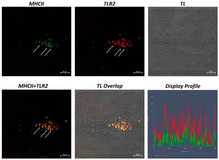
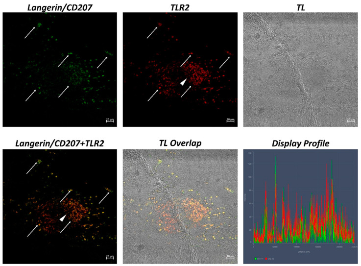
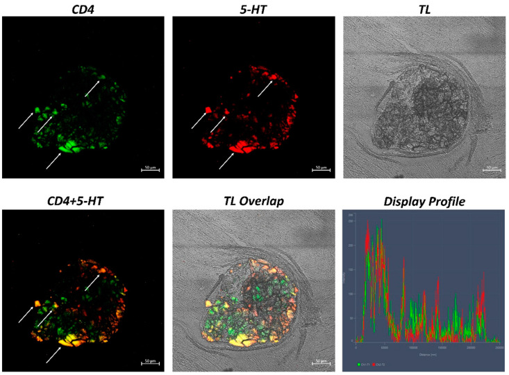
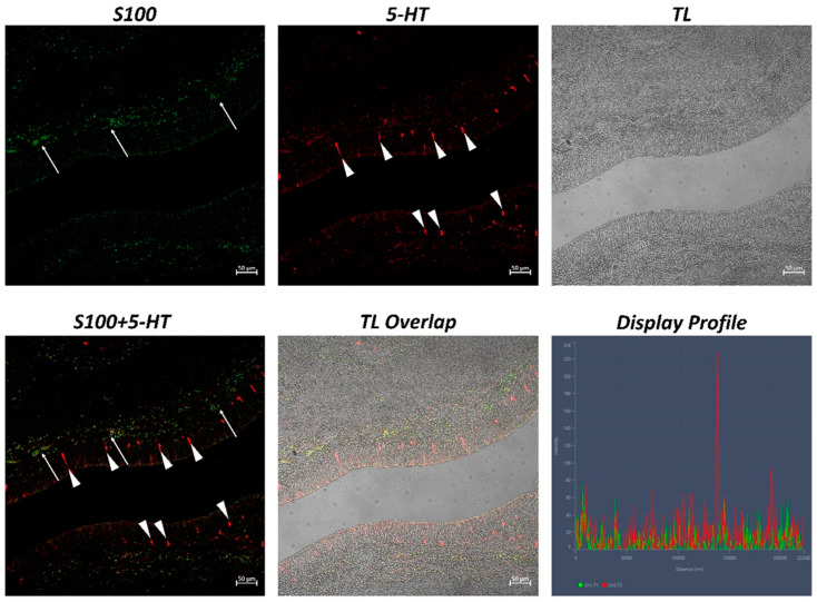
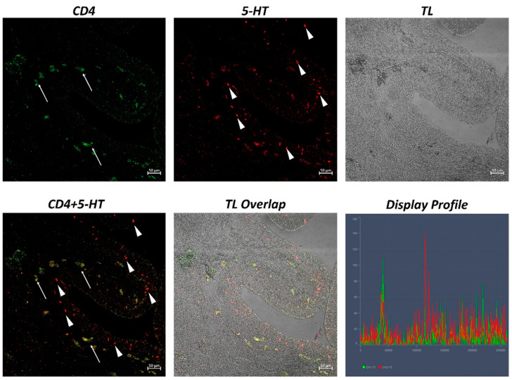
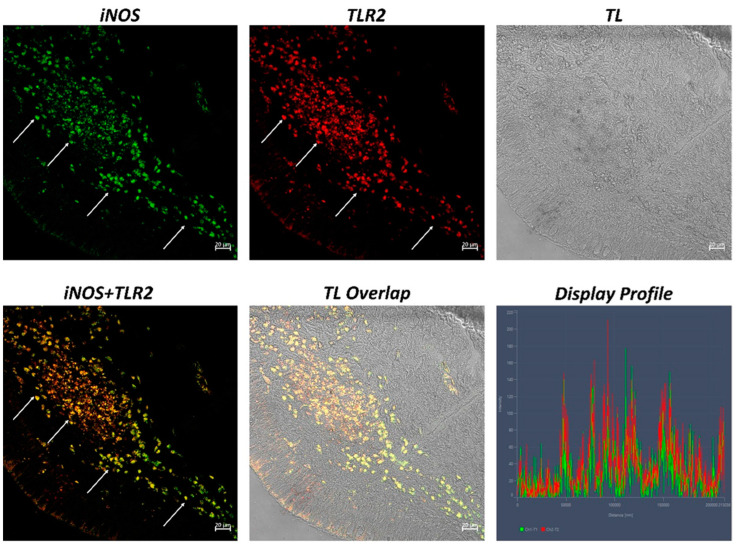
The examination of both transverse and longitudinal sections of the pyloric ceca and intestine terminal region (rectum) reveals massive aggregates of immune cells. GALT was seen in two different shapes: the immune cells present in the thickness of the submucosa and the lamina propria are arranged in circumscribed oval-shaped structures that are morphologically similar to the isolated lymphoid follicles (ILFs) present in birds and mammals. Furthermore, scattered or clustered immune cells are densely packed in the lamina propria and submucosa. The ILFs consist of non-encapsulated, dense clusters of lymphocytes of various sizes and macrophages. Double immunolabeling with antibodies against TLR2 and MHCII reveals positive DC-like cells in the aggregates of lymphoid tissue (Figure 1); these cells are strongly positive with TLR2 and Langerin/CD 207 (Figure 2). A large accumulation of lymphocytic cells that are marked with CD4 and colocalized with serotonin can be seen, especially in the cells located at the periphery of the lymphoid structure (Figure 3). Numerous S100 and CD4-positive cells are scattered under the epithelium and there is no colocalization with serotonin, which marks the neuroendocrine cells in the epithelium (Figure 4 and Figure 5). Strong colocalization between TLR2 and iNOS is evident in the submucosa cells (Figure 6).
Figure 1.
Section of the H. niloticus gut (immunofluorescence 40×, scale bar 20 μm). Macrophages that are positive for TLR2 and MHC II can be noted in the lymphoid tissue aggregates (marked by arrows). The “Display profile” function highlights the colocalization. TL = transmitted light.
Figure 2.
Section of the H. niloticus gut (immunofluorescence 20×, scale bar 20 μm). DC-like cells that are immunoreactive for Langerin and TLR2, organized in clusters, can be seen (arrows). Some immune cells are positive exclusively for TLR2 (see the arrowhead). The “Display profile” function highlights the colocalization. TL = transmitted light.
Figure 3.
Section of the H. niloticus gut (immunofluorescence, 40×, scale bar 50 μm. The presence of an important accumulation of densely packed lymphocytes is highlighted by CD4 and 5-HT positivity (arrows). The “Display profile” function highlights the colocalization. TL = transmitted light.
Figure 4.
Section of the H. niloticus gut (immunofluorescence 20×, scale bar 50 μm). Immune cells that are positive for S100 are evident in the submucosa (arrows), while the neuroendocrine cells (5-HT positive) are evident in the epithelium (arrowheads). The “Display profile” function highlights the absence of colocalization. TL = transmitted light.
Figure 5.
Section of the H. niloticus gut (immunofluorescence 20×, scale bar 50 μm). Lymphocytes that are positive for CD4 and 5-HT are scattered in the submucosa (arrows). The presence of neuroendocrine cells that are immunoreactive to 5-HT can also be noted (arrowheads). The “Display profile” function highlights the colocalization. TL = transmitted light.
Figure 6.
Section of the H. niloticus gut * immunofluorescence (* 40×, scale bar 20 μm). Numerous immune cells, which are densely organized, colocalize for iNOS and TLR2 (arrows) in the submucosa. The “Display profile” function highlights the colocalization. TL = transmitted light.
Quantitative analysis revealed an equal number of cells that were immunopositive for each antibody tested (Table 1).
Table 1.
Statistical analysis data. (Mean values ± standard deviation) (n = 3).
| No. of Positive Immune Cells | |
|---|---|
| TLR-2 | 476.38 ± 45.29 * |
| MHC class II | 447.44 ± 38.20 ** |
| Langerin | 329.46 ± 28.01 * |
| CD4 | 365.25 ± 33.06 * |
| 5-HT | 313.94 ± 29.36 * |
| S100 | 406.36 ± 39.85 * |
| iNOS | 418.42 ± 47.31 ** |
| TLR2 + MHC II | 416.03 ± 37.78 * |
| TLR2 + Langerin | 301.53 ± 30.25 ** |
| CD4 + 5-HT | 307.48 ± 20.34 * |
| TLR2 + iNOS | 403.94 ± 32.75 ** |
* p ≤ 0.05; ** p ≤ 0.01.
3. Discussion
H. niloticus is particularly interesting in the study of the phylogenesis of vertebrates because, although it presents primitive characteristics, it has anatomical specializations that are similar to those of the higher vertebrates. Some of the primitive features belonging to H. niloticus are an elongated and robust body, dorsal and anal fins that are elongated and posteriorly positioned, a rounded caudal fin, and strong, large scales. Furthermore, H. niloticus presents reduced lamellar surfaces and a large gas bladder that helps them to acquire O2 from the environment [53]. On the other hand, it presents a stomach consisting of a proventriculus and a ventriculus (gizzard), found in birds and reptiles, as an organ of digestion, due to its omnivorous feeding [2]. Moreover, the wall of the alimentary tract of H. niloticus shows a strong similarity to that of the higher vertebrates. It is composed of four layers, which, proceeding from the inside outward, comprise the mucosa, submucosa, inner circular layer, outer longitudinal layer of muscularis, and serosa [4,54]. In agreement with Guerrera et al. [4], in this study, massive aggregates of lymphoid and innate immune cells were observed in the intestinal mucosa and submucosa, constituting structures that were arranged in circumscribed oval-shaped forms resembling mammalian ILFs. Lymphoid aggregates have been found in all vertebrates, including amphibians, reptiles, and birds [55,56,57,58,59,60]. GALT is the main MALT of teleosts, being continuously exposed to a pathogen-rich environment in either freshwater or seawater. In teleosts, the highly organized lymphoid organs, such as mesenteric lymph nodes and Peyer’s patches, which have been observed in birds and mammals, are missing. However, in some studies, the existence of ILT [10,14,16,61,62], and the bursa in Atlantic salmon (Salmo salar, Linnaeus 1758), which are an analog of the avian bursa of Fabricius [63], were reported as the organized lymphoid structure of teleosts. In the phylogenesis of vertebrates, the lymphoid system has increased its complexity and structural organization, to become increasingly efficient and specialized. Intestinal lymphoid aggregates lack germinal centers, as described in the lamina propria of birds; those found in cold-blooded vertebrates may represent primitive versions of the cryptopatches that are present in the mammalian intestine [26,64]. Teleosts can present B and T lymphocytes in separate areas of the MALT of the gut, gills, and nasopharynges, as with those amphibians and reptiles that separate the B and T cell areas in the spleen [7]; finally, birds and mammals present lymphoid organs that are highly organized, with separated B and T cell areas and well-demarcated germinal centers [7,65]. Several studies have described the fish GALT as consisting of immune cells (lymphocytes, macrophages, dendritic cells, and plasma cells), which are presented in clusters along the mucosa of the alimentary canal and the intraepithelial lymphocytes distributed among the enterocytes [8,24,66]. The gut-associated lymphoid tissue of H. niloticus seems to be present in two different shapes. Our results showed scattered clusters of immune cells in the mucosal lamina propria of the pyloric ceca and rectum (the terminal part of the hindgut), using confocal immunofluorescence. The gut mucosal epithelia play a significant role in fish immunology [29,67]. Using confocal microscopy, we have immunohistochemically characterized the DC-like cells with antibodies against TLR2 and MHCII; the presence of these cells was confirmed by colocalization between TLR2 and Langerin/CD 207; furthermore, the S100 protein and iNOS have been used to mark the macrophages and CD4 antibodies to characterize the T lymphocytes. CD4+ helper T cells can be found in the teleost’s gastrointestinal lamina propria [13,68,69]. These cells express CD4 on their surface for specific antigen recognition. Toll-like receptors are involved in the recognition of pathogens, through specialized antigen-presenting cells (APCs). These antigen-presenting cells include macrophages, granulocytes, and dendritic-like cells, as well as B cells. The presentation of the antigen takes place via MHCII, activating T cells that proliferate and produce inflammatory cytokines [70]. The distribution of these innate and adaptive immune cells, as delineated by the pattern of anti-TLR2, anti-MHCII, anti-Langerin/CD 207, anti-iNOS, and anti-CD4 recognition, was similar to that documented in other studies [8,23,51]. Recent findings have shown rainbow trout to be a model for intestinal immune responses. In the intestine lamina propria, pathogens can be taken up by the antigen-presenting cells (APCs) and then presented to CD4-T cells. Consequently, the B lymphocytes, activated by the cytokines produced by T cells, proliferate and differentiate into plasma cell-like cells, resulting in the production of antibodies [16,71].
This interaction between the immune cells in fish is very similar to that found in mammals; thus, improving this knowledge could be useful for formulating new vaccines and finding new models by which to improve mammalian mucosal immunology. One very interesting finding is the positivity to 5-HT; the endocrine cells of the intestinal epithelium are positive for this neurotransmitter. Moreover, the lymphocytes that are localized in the peripheral zone of the lymphoid structures present colocalization between CD4 and serotonin. The immune cells express serotonin receptors and serotonin transporter (SERT), which are known serotonergic components of the immune cells [72,73,74]. T cells take up serotonin via SERT and then express numerous 5-HT receptors that are involved in the proliferation of T lymphocytes [74,75].
4. Materials and Methods
The paraffin-embedded tissue of males and females of the adult African bonytongue, H. niloticus (Cuvier, 1829), from a previous study was used for this research. In that study, the fish’s digestive system (from tongue to anus) was sampled and processed for routine histological study (for details, see Guerrera et al. [4]).
4.1. Immunofluorescence
To identify the localization of anti-TLR2, serotonin (5-HT), MHCII, Langerin/H-4, the S100 protein, and CD4 antibodies, an immunohistochemistry investigation was carried out on the pyloric ceca and intestine’s terminal region (rectum). Deparaffinized and rehydrated serial slices (10 µm thick) were rinsed in Tris-HCl solution (0.05 M, pH 7.5) with 0.1% bovine serum albumin and 0.2% Triton-X 100. Slices were incubated in a 0.3% H2O2 (PBS) solution to prevent endogenous peroxidase activity; finally, fetal bovine serum (F7524, Sigma-Aldrich, St. Louis, Missouri, USA) was added to the washed sections for 30 min to prevent nonspecific binding, then the primary antibodies were incubated. Anti-TLR2 and anti-serotonin (5-HT) polyclonal antibodies were used in the double-label experiments, with a monoclonal antibody for MHCII, Langerin/H-4, S100 protein, and CD4 (for details, see Table 2). A humid chamber was used for overnight incubation at 4°C. Subsequently, the sections were rinsed in buffer and incubated for 40 min at room temperature with Alexa Fluor IgG (H + L) secondary antibodies (for details, see Table 2) in a dark, humid chamber. Finally, Fluoromount Aqueous Mounting Medium was applied to mount the dehydrated sections (Sigma-Aldrich, Burlington, MA, USA).
Table 2.
Antibodies data.
| Primary Antibodies | Supplier | Catalog Number | Source | Dilution | Antibody ID |
| TLR-2 (pAb) | Active Motif | 40981 | Rabbit | 1:125 | AB_2750977 |
| Anti-serotonin (5HT) | Sigma-Aldrich | S5545 | Rabbit | 1:300 | AB_477522 |
| Langerin/H-4 | Santa Cruz Biotechnology | sc-271272 | Mouse | 1:250 | AB_10611518 |
| MHC class II (Y-Ae) | Santa Cruz Biotechnology | Sc-32247 | Mouse | 1:250 | AB_627939 |
| S100 (s161) | Santa Cruz Biotechnology | Sc-53438 | Mouse | 1:100 | AB_630214 |
| CD4 MT310 | Santa Cruz Biotechnology | Sc-19641 | Mouse | 1:100 | AB_627055 |
| iNOS | Santa Cruz Biotechnology | Sc7271 | Mouse | 1:200 | |
| Secondary Antibodies | Supplier | Source | Dilution | Antibody ID | |
| Alexa Fluor 488 anti-mouse IgG FITC conjugated |
Invitrogen | A-21202 | Donkey | 1:300 | AB_141607 |
| Alexa Fluor 594 anti-rabbit IgG TRITC conjugated |
Invitrogen | A32754 | Donkey | 1:300 | AB_2762827 |
4.2. Laser confocal immunofluorescence
Sections were analyzed and images were acquired using a Zeiss LSMDUO confocal laser scanning microscope with a META module (Carl Zeiss MicroImaging GmbH, Germany) microscope, the LSM700 AxioObserver. The Zen 2011 (LSM 700, Zeiss software, Oberkochen, Germany) built-in “colocalization view” was used to highlight the expression of both antibodies’ signals [76,77,78], in order to produce a “colocalization” signal, along with the display profile, the scatter plot and fluorescent signal measurements. Each image was rapidly acquired to minimize photodegradation.
4.3. Statistical Analysis
Data were gathered for quantitative analysis through the examination of ten sections and twenty fields per sample. The cell positivity was assessed using ImageJ software 1.53e. The plugin, “Analyze particles”, was used to count the number of cells. We enumerated the number of cells in each field that were positive for TLR2, Langerin/CD207, 5-HT, MHC II, S100, and CD4, using a SigmaPlot, version 14.0 (Systat Software, San Jose, CA, USA). A one-way ANOVA and Student’s t-test were used to assess the normally distributed data. Data means and standard deviations (SD) are shown as ** p 0.01 and * p 0.05.
5. Conclusions
For the first time in this study, immune cells of the GALT of H. niloticus were characterized, confirming the presence of organized lymphoid structures similar to those seen in the higher vertebrates. Further studies could be useful to better understand the phylogeny of the vertebrate immune system and to consider the possibility of vaccination strategies for highly commercial species, such as H. niloticus.
Author Contributions
Conceptualization, E.R.L. and M.C.G.; methodology, M.A., A.A. and A.M.; software, M.A., A.A. and A.M.; validation, E.R.L., A.A., M.C.G., A.G., M.A., K.Z.; M.K., G.Z., S.P. and A.M.; formal analysis, M.A., A.A. and A.M.; investigation, M.A., A.A. and A.M.; resources, E.R.L., M.C.G., A.G., K.Z. and M.K.; data curation, M.A., A.A. and A.M.; writing—original draft preparation, E.R.L., A.A., M.C.G. and M.A.; writing—review and editing, E.R.L., A.A., A.M., M.C.G., M.A., S.P. and A.G.; visualization, E.R.L., M.C.G. and A.G.; supervision, E.R.L., M.C.G., S.P., A.G. and G.Z.; project administration, E.R.L., M.C.G. and A.G. All authors have read and agreed to the published version of the manuscript.
Institutional Review Board Statement
Animals were exempt from the EU legislation governing the use of animals for scientific purposes because they were used only for their organs and tissues.
Informed Consent Statement
Not applicable.
Data Availability Statement
All data presented in this study are available from the corresponding author, upon responsible request.
Conflicts of Interest
The authors declare no conflict of interest.
Funding Statement
This research received no external funding.
Footnotes
Disclaimer/Publisher’s Note: The statements, opinions and data contained in all publications are solely those of the individual author(s) and contributor(s) and not of MDPI and/or the editor(s). MDPI and/or the editor(s) disclaim responsibility for any injury to people or property resulting from any ideas, methods, instructions or products referred to in the content.
References
- 1.Wikondi J., Tonfack D.J., Meutchieye F., Tomedi T.M. Aquaculture of Heterotis niloticus in Sub-Saharan Africa: Potentials and Perspectives. Genet. Biodivers. J. 2022;6:37–44. doi: 10.46325/gabj.v6i1.195. [DOI] [Google Scholar]
- 2.Agbugui M.O., Egbo H.O., Abhulimen F.E. The Biology of the African Bonytongue Heterotis niloticus (Cuvier, 1829) from the Lower Niger River at Agenebode in Edo State, Nigeria. Int. J. Zool. 2021;2021:1748736. doi: 10.1155/2021/1748736. [DOI] [Google Scholar]
- 3.Obermiller L.E., Pfeiler E. Phylogenetic relationships of elopomorph fishes inferred from mitochondrial ribosomal DNA sequences. Mol. Phylogenetics Evol. 2003;26:202–214. doi: 10.1016/S1055-7903(02)00327-5. [DOI] [PubMed] [Google Scholar]
- 4.Guerrera M.C., Aragona M., Briglia M., Porcino C., Mhalhel K., Cometa M., Abbate F., Montalbano G., Laurà R., Levanti M. The Alimentary Tract of African Bony-Tongue, Heterotis niloticus (Cuvier, 1829): Morphology Study. Animals. 2022;12:1565. doi: 10.3390/ani12121565. [DOI] [PMC free article] [PubMed] [Google Scholar]
- 5.Wilson J., Castro L. Fish Physiology. Volume 30. Elsevier; Amsterdam, The Netherlands: 2010. Morphological diversity of the gastrointestinal tract in fishes; pp. 1–55. [Google Scholar]
- 6.Adite A., ImorouToko I., Gbankoto A. Fish assemblages in the degraded mangrove ecosystems of the coastal zone, Benin, West Africa: Implications for ecosystem restoration and resources conservation. J. Environ. Prot. 2013;4:1461–1475. doi: 10.4236/jep.2013.412168. [DOI] [Google Scholar]
- 7.Silva-Sanchez A., Randall T.D. Chapter 2—Anatomical Uniqueness of the Mucosal Immune System (GALT, NALT, iBALT) for the Induction and Regulation of Mucosal Immunity and Tolerance. In: Kiyono H., Pascual D.W., editors. Mucosal Vaccines. 2nd ed. Academic Press; Cambridge, MA, USA: 2020. pp. 21–54. [Google Scholar]
- 8.Salinas I. The mucosal immune system of teleost fish. Biology. 2015;4:525–539. doi: 10.3390/biology4030525. [DOI] [PMC free article] [PubMed] [Google Scholar]
- 9.Mitchell C.D., Criscitiello M.F. Comparative study of cartilaginous fish divulges insights into the early evolution of primary, secondary and mucosal lymphoid tissue architecture. Fish Shellfish. Immunol. 2020;107:435–443. doi: 10.1016/j.fsi.2020.11.006. [DOI] [PubMed] [Google Scholar]
- 10.Koppang E.O., Fischer U., Moore L., Tranulis M.A., Dijkstra J.M., Köllner B., Aune L., Jirillo E., Hordvik I. Salmonid T cells assemble in the thymus, spleen and in novel interbranchial lymphoid tissue. J. Anat. 2010;217:728–739. doi: 10.1111/j.1469-7580.2010.01305.x. [DOI] [PMC free article] [PubMed] [Google Scholar]
- 11.Sepahi A., Salinas I. The evolution of nasal immune systems in vertebrates. Mol. Immunol. 2016;69:131–138. doi: 10.1016/j.molimm.2015.09.008. [DOI] [PMC free article] [PubMed] [Google Scholar]
- 12.Yu Y.-Y., Kong W.-G., Xu H.-Y., Huang Z.-Y., Zhang X.-T., Ding L.-G., Dong S., Yin G.-M., Dong F., Yu W., et al. Convergent Evolution of Mucosal Immune Responses at the Buccal Cavity of Teleost Fish. iScience. 2019;19:821–835. doi: 10.1016/j.isci.2019.08.034. [DOI] [PMC free article] [PubMed] [Google Scholar]
- 13.Lee P.T., Yamamoto F.Y., Low C.F., Loh J.Y., Chong C.M. Gut Immune System and the Implications of Oral-Administered Immunoprophylaxis in Finfish Aquaculture. Front. Immunol. 2021;12:773193. doi: 10.3389/fimmu.2021.773193. [DOI] [PMC free article] [PubMed] [Google Scholar]
- 14.Gomez D., Sunyer J.O., Salinas I. The mucosal immune system of fish: The evolution of tolerating commensals while fighting pathogens. Fish Shellfish. Immunol. 2013;35:1729–1739. doi: 10.1016/j.fsi.2013.09.032. [DOI] [PMC free article] [PubMed] [Google Scholar]
- 15.Tacchi L., Musharrafieh R., Larragoite E.T., Crossey K., Erhardt E.B., Martin S.A.M., LaPatra S.E., Salinas I. Nasal immunity is an ancient arm of the mucosal immune system of vertebrates. Nat. Commun. 2014;5:5205. doi: 10.1038/ncomms6205. [DOI] [PMC free article] [PubMed] [Google Scholar]
- 16.Parra D., Korytář T., Takizawa F., Sunyer J.O. B cells and their role in the teleost gut. Dev. Comp. Immunol. 2016;64:150–166. doi: 10.1016/j.dci.2016.03.013. [DOI] [PMC free article] [PubMed] [Google Scholar]
- 17.Salinas I., Fernández-Montero Á., Ding Y., Sunyer J.O. Mucosal immunoglobulins of teleost fish: A decade of advances. Dev. Comp. Immunol. 2021;121:104079. doi: 10.1016/j.dci.2021.104079. [DOI] [PMC free article] [PubMed] [Google Scholar]
- 18.German D.P., Horn M.H. Gut length and mass in herbivorous and carnivorous prickleback fishes (Teleostei: Stichaeidae): Ontogenetic, dietary, and phylogenetic effects. Mar. Biol. 2006;148:1123–1134. doi: 10.1007/s00227-005-0149-4. [DOI] [PubMed] [Google Scholar]
- 19.Sunyer J.O. Fishing for mammalian paradigms in the teleost immune system. Nat. Immunol. 2013;14:320–326. doi: 10.1038/ni.2549. [DOI] [PMC free article] [PubMed] [Google Scholar]
- 20.Mowat A.M., Agace W.W. Regional specialization within the intestinal immune system. Nat. Rev. Immunol. 2014;14:667–685. doi: 10.1038/nri3738. [DOI] [PubMed] [Google Scholar]
- 21.Picchietti S., Buonocore F., Guerra L., Belardinelli M.C., De Wolf T., Couto A., Fausto A.M., Saraceni P.R., Miccoli A., Scapigliati G. Molecular and cellular characterization of European sea bass CD3ε+ T lymphocytes and their modulation by microalgal feed supplementation. Cell Tissue Res. 2021;384:149–165. doi: 10.1007/s00441-020-03347-x. [DOI] [PubMed] [Google Scholar]
- 22.Graves C.L., Chen A., Kwon V., Shiau C.E. Zebrafish harbor diverse intestinal macrophage populations including a subset intimately associated with enteric neural processes. iScience. 2021;24:102496. doi: 10.1016/j.isci.2021.102496. [DOI] [PMC free article] [PubMed] [Google Scholar]
- 23.Zaccone G., Alesci A., Mokhtar D.M., Aragona M., Guerrera M.C., Capillo G., Albano M., de Oliveira Fernandes J., Kiron V., Sayed R.K.A., et al. Localization of Acetylcholine, Alpha 7-NAChR and the Antimicrobial Peptide Piscidin 1 in the Macrophages of Fish Gut: Evidence for a Cholinergic System, Diverse Macrophage Populations and Polarization of Immune Responses. Fishes. 2023;8:43. doi: 10.3390/fishes8010043. [DOI] [Google Scholar]
- 24.Moradkhani A., Abdi R., Abadi M.S.-A., Nabavi S.M., Basir Z. Quantification and description of gut-associated lymphoid tissue in, shabbout, Arabibarbus grypus (actinopterygii: Cypriniformes: Cyprinidae), in warm and cold season. Acta Ichthyol. Et Piscat. 2020;50:423–432. doi: 10.3750/AIEP/02910. [DOI] [Google Scholar]
- 25.Dai X., Shu M., Fang W. Histological and ultrastructural study of the digestive tract of rice field eel, Monopterus albus. J. Appl. Ichthyol. 2007;23:177–183. doi: 10.1111/j.1439-0426.2006.00830.x. [DOI] [Google Scholar]
- 26.Aghaallaei N., Agarwal R., Benjaminsen J., Lust K., Bajoghli B., Wittbrodt J., Feijoo C.G. Antigen-Presenting Cells and T Cells Interact in a Specific Area of the Intestinal Mucosa Defined by the Ccl25-Ccr9 Axis in Medaka. Front. Immunol. 2022;13:812899. doi: 10.3389/fimmu.2022.812899. [DOI] [PMC free article] [PubMed] [Google Scholar]
- 27.Tacchi L., Larragoite E.T., Muñoz P., Amemiya C.T., Salinas I. African Lungfish Reveal the Evolutionary Origins of Organized Mucosal Lymphoid Tissue in Vertebrates. Curr. Biol. 2015;25:2417–2424. doi: 10.1016/j.cub.2015.07.066. [DOI] [PMC free article] [PubMed] [Google Scholar]
- 28.Lauriano E., Faggio C., Capillo G., Spanò N., Kuciel M., Aragona M., Pergolizzi S. Immunohistochemical characterization of epidermal dendritic-like cells in giant mudskipper, Periophthalmodon schlosseri. Fish Shellfish. Immunol. 2018;74:380–385. doi: 10.1016/j.fsi.2018.01.014. [DOI] [PubMed] [Google Scholar]
- 29.Lauriano E., Pergolizzi S., Aragona M., Montalbano G., Guerrera M., Crupi R., Faggio C., Capillo G. Intestinal immunity of dogfish Scyliorhinus canicula spiral valve: A histochemical, immunohistochemical and confocal study. Fish Shellfish. Immunol. 2019;87:490–498. doi: 10.1016/j.fsi.2019.01.049. [DOI] [PubMed] [Google Scholar]
- 30.Lauriano E., Pergolizzi S., Capillo G., Kuciel M., Alesci A., Faggio C. Immunohistochemical characterization of Toll-like receptor 2 in gut epithelial cells and macrophages of goldfish Carassius auratus fed with a high-cholesterol diet. Fish Shellfish. Immunol. 2016;59:250–255. doi: 10.1016/j.fsi.2016.11.003. [DOI] [PubMed] [Google Scholar]
- 31.Lauriano E., Pergolizzi S., Cascio P.L., Kuciel M., Zizzo N., Guerrera M., Aragona M., Capillo G. Expression of Langerin/CD207 in airways, lung and associated lymph nodes of a stranded striped dolphin (Stenella coeruleoalba) Acta Histochem. 2020;122:151471. doi: 10.1016/j.acthis.2019.151471. [DOI] [PubMed] [Google Scholar]
- 32.Lauriano E., Silvestri G., Kuciel M., Żuwała K., Zaccone D., Palombieri D., Alesci A., Pergolizzi S. Immunohistochemical localization of Toll-like receptor 2 in skin Langerhans’ cells of striped dolphin (Stenella coeruleoalba) Tissue Cell. 2014;46:113–121. doi: 10.1016/j.tice.2013.12.002. [DOI] [PubMed] [Google Scholar]
- 33.Alesci A., Albano M., Savoca S., Mokhtar D.M., Fumia A., Aragona M., Lo Cascio P., Hussein M.M., Capillo G., Pergolizzi S., et al. Confocal Identification of Immune Molecules in Skin Club Cells of Zebrafish (Danio rerio, Hamilton 1882) and Their Possible Role in Immunity. Biology. 2022;11:1653. doi: 10.3390/biology11111653. [DOI] [PMC free article] [PubMed] [Google Scholar]
- 34.Alesci A., Capillo G., Mokhtar D.M., Fumia A., D’Angelo R., Lo Cascio P., Albano M., Guerrera M.C., Sayed R.K.A., Spanò N., et al. Expression of Antimicrobic Peptide Piscidin1 in Gills Mast Cells of Giant Mudskipper Periophthalmodon schlosseri (Pallas, 1770) Int. J. Mol. Sci. 2022;23:13707. doi: 10.3390/ijms232213707. [DOI] [PMC free article] [PubMed] [Google Scholar]
- 35.Alesci A., Pergolizzi S., Capillo G., Lo Cascio P., Lauriano E.R. Rodlet cells in kidney of goldfish (Carassius auratus, Linnaeus 1758): A light and confocal microscopy study. Acta Histochem. 2022;124:151876. doi: 10.1016/j.acthis.2022.151876. [DOI] [PubMed] [Google Scholar]
- 36.Alesci A., Pergolizzi S., Lo Cascio P., Fumia A., Lauriano E.R. Neuronal regeneration: Vertebrates comparative overview and new perspectives for neurodegenerative diseases. Acta Zool. 2022;103:129–140. doi: 10.1111/azo.12397. [DOI] [Google Scholar]
- 37.Alesci A., Pergolizzi S., Savoca S., Fumia A., Mangano A., Albano M., Messina E., Aragona M., Lo Cascio P., Capillo G., et al. Detecting Intestinal Goblet Cells of the Broadgilled Hagfish Eptatretus cirrhatus (Forster, 1801): A Confocal Microscopy Evaluation. Biology. 2022;11:1366. doi: 10.3390/biology11091366. [DOI] [PMC free article] [PubMed] [Google Scholar]
- 38.Alesci A., Cicero N., Salvo A., Palombieri D., Zaccone D., Dugo G., Bruno M., Vadalà R., Lauriano E.R., Pergolizzi S. Extracts deriving from olive mill waste water and their effects on the liver of the goldfish Carassius auratus fed with hypercholesterolemic diet. Nat. Prod. Res. 2014;28:1343–1349. doi: 10.1080/14786419.2014.903479. [DOI] [PubMed] [Google Scholar]
- 39.Alesci A., Lauriano E.R., Aragona M., Capillo G., Pergolizzi S. Marking vertebrates langerhans cells, from fish to mammals. Acta Histochem. 2020;122:151622. doi: 10.1016/j.acthis.2020.151622. [DOI] [PMC free article] [PubMed] [Google Scholar]
- 40.Pergolizzi S., Marino A., Capillo G., Aragona M., Marconi P., Lauriano E. Expression of Langerin/CD 207 and α-smooth muscle actin in ex vivo rabbit corneal keratitis model. Tissue Cell. 2020;66:1–6. doi: 10.1016/j.tice.2020.101384. [DOI] [PubMed] [Google Scholar]
- 41.Mokhtar D.M. Characterization of the fish ovarian stroma during the spawning season: Cytochemical, immunohistochemical and ultrastructural studies. Fish Shellfish. Immunol. 2019;94:566–579. doi: 10.1016/j.fsi.2019.09.050. [DOI] [PubMed] [Google Scholar]
- 42.Oliveira M., Ribeiro A., Hylland K., Guilhermino L. Single and combined effects of microplastics and pyrene on juveniles (0+ group) of the common goby Pomatoschistus microps (Teleostei, Gobiidae) Ecol. Indic. 2013;34:641–647. doi: 10.1016/j.ecolind.2013.06.019. [DOI] [Google Scholar]
- 43.Yang Z., Yan W.X., Cai H., Tedla N., Armishaw C., Di Girolamo N., Wang H.W., Hampartzoumian T., Simpson J.L., Gibson P.G., et al. S100A12 provokes mast cell activation: A potential amplification pathway in asthma and innate immunity. J. Allergy Clin. Immunol. 2007;119:106–114. doi: 10.1016/j.jaci.2006.08.021. [DOI] [PubMed] [Google Scholar]
- 44.Zaccone G., Lauriano E.R., Capillo G., Kuciel M. Air-breathing in fish: Air-breathing organs and control of respiration: Nerves and neurotransmitters in the air-breathing organs and the skin. Acta Histochem. 2018;120:630–641. doi: 10.1016/j.acthis.2018.08.009. [DOI] [PubMed] [Google Scholar]
- 45.Quintero-Villegas A., Valdés-Ferrer S.I. Role of 5-HT7 receptors in the immune system in health and disease. Mol. Med. 2019;26:2. doi: 10.1186/s10020-019-0126-x. [DOI] [PMC free article] [PubMed] [Google Scholar]
- 46.Alesci A., Pergolizzi S., Fumia A., Calabrò C., Lo Cascio P., Lauriano E.R. Mast cells in goldfish (Carassius auratus) gut: Immunohistochemical characterization. Acta Zool. 2022;103 doi: 10.1111/azo.12417. [DOI] [Google Scholar]
- 47.Lovy J., Wright G.M., Speare D.J. Comparative cellular morphology suggesting the existence of resident dendritic cells within immune organs of salmonids. Anat. Rec. 2008;291:456–462. doi: 10.1002/ar.20674. [DOI] [PubMed] [Google Scholar]
- 48.Kordon A.O., Scott M.A., Ibrahim I., Abdelhamed H., Ahmed H., Baumgartner W., Karsi A., Pinchuk L.M. Identification of Langerhans-like cells in the immunocompetent tissues of channel catfish, Ictalurus punctatus. Fish Shellfish. Immunol. 2016;58:253–258. doi: 10.1016/j.fsi.2016.09.033. [DOI] [PubMed] [Google Scholar]
- 49.Zaghloul D.M., Derbalah A.E., Rutland C.S. Unique characterization of Langerhans cells in the spleen of the African catfish (Clarias gariepinus) Matters Sel. 2017;3:e201703000005. doi: 10.19185/matters.201703000005. [DOI] [Google Scholar]
- 50.Soleto I., Granja A.G., Simón R., Morel E., Díaz-Rosales P., Tafalla C. Identification of CD8α+ dendritic cells in rainbow trout (Oncorhynchus mykiss) intestine. Fish Shellfish. Immunol. 2019;89:309–318. doi: 10.1016/j.fsi.2019.04.001. [DOI] [PMC free article] [PubMed] [Google Scholar]
- 51.Alesci A., Capillo G., Fumia A., Messina E., Albano M., Aragona M., Lo Cascio P., Spanò N., Pergolizzi S., Lauriano E.R. Confocal Characterization of Intestinal Dendritic Cells from Myxines to Teleosts. Biology. 2022;11:1045. doi: 10.3390/biology11071045. [DOI] [PMC free article] [PubMed] [Google Scholar]
- 52.Moore L.J., Dijkstra J.M., Koppang E.O., Hordvik I. CD4 homologues in Atlantic salmon. Fish Shellfish. Immunol. 2009;26:10–18. doi: 10.1016/j.fsi.2008.09.019. [DOI] [PubMed] [Google Scholar]
- 53.Zaccone G., Capillo G., Aragona M., Alesci A., Cupello C., Lauriano E.R., Guerrera M.C., Kuciel M., Zuwala K., Germana A., et al. Gill structure and neurochemical markers in the African bonytongue (Heterotis niloticus): A preliminary study. Acta Histochem. 2022;124:151954. doi: 10.1016/j.acthis.2022.151954. [DOI] [PubMed] [Google Scholar]
- 54.Nelson J.S., Grande T.C., Wilson M.V. Fishes of the World. John Wiley & Sons; Hoboken, NJ, USA: 2016. [Google Scholar]
- 55.Ardavin C.F., Zapata A., Villena A., Solas M.T. Gut-Associated lymphoid tissue (GALT) in the amphibian urodele Pleurodeles waltl. J. Morphol. 1982;173:35–41. doi: 10.1002/jmor.1051730105. [DOI] [PubMed] [Google Scholar]
- 56.Marshall J.A., Dixon K.E. Cell specialization in the epithelium of the small intestine of feeding Xenopus laevis tadpoles. J. Anat. 1978;126:133–144. [PMC free article] [PubMed] [Google Scholar]
- 57.Befus A.D., Johnston N., Leslie G., Bienenstock J. Gut-associated lymphoid tissue in the chicken. I. Morphology, ontogeny, and some functional characteristics of Peyer’s patches. J. Immunol. 1980;125:2626–2632. doi: 10.4049/jimmunol.125.6.2626. [DOI] [PubMed] [Google Scholar]
- 58.Borysenko M., Cooper E.L. Lymphoid tissue in the snapping turtle, Chelydra serpentina. J. Morphol. 1972;138:487–497. doi: 10.1002/jmor.1051380408. [DOI] [PubMed] [Google Scholar]
- 59.Colombo B.M., Scalvenzi T., Benlamara S., Pollet N. Microbiota and mucosal immunity in amphibians. Front. Immunol. 2015;6:111. doi: 10.3389/fimmu.2015.00111. [DOI] [PMC free article] [PubMed] [Google Scholar]
- 60.Arroyo Portilla C., Tomas J., Gorvel J.-P., Lelouard H. From species to regional and local specialization of intestinal macrophages. Front. Cell Dev. Biol. 2021;8:624213. doi: 10.3389/fcell.2020.624213. [DOI] [PMC free article] [PubMed] [Google Scholar]
- 61.Aas I.B., Austbø L., König M., Syed M., Falk K., Hordvik I., Koppang E.O. Transcriptional characterization of the T cell population within the salmonid interbranchial lymphoid tissue. J. Immunol. 2014;193:3463–3469. doi: 10.4049/jimmunol.1400797. [DOI] [PubMed] [Google Scholar]
- 62.Salinas I., Zhang Y.-A., Sunyer J.O. Mucosal immunoglobulins and B cells of teleost fish. Dev. Comp. Immunol. 2011;35:1346–1365. doi: 10.1016/j.dci.2011.11.009. [DOI] [PMC free article] [PubMed] [Google Scholar]
- 63.Løken O.M., Bjørgen H., Hordvik I., Koppang E.O. A teleost structural analogue to the avian bursa of Fabricius. J. Anat. 2020;236:798–808. doi: 10.1111/joa.13147. [DOI] [PMC free article] [PubMed] [Google Scholar]
- 64.Matsunaga T., Rahman A. What brought the adaptive immune system to vertebrates?—The jaw hypothesis and the seahorse. Immunol. Rev. 1998;166:177–186. doi: 10.1111/j.1600-065X.1998.tb01262.x. [DOI] [PubMed] [Google Scholar]
- 65.Pellicioli A.C.A., Bingle L., Farthing P., Lopes M.A., Martins M.D., Vargas P.A. Immunosurveillance profile of oral squamous cell carcinoma and oral epithelial dysplasia through dendritic and T-cell analysis. J. Oral Pathol. Med. 2017;46:928–933. doi: 10.1111/jop.12597. [DOI] [PubMed] [Google Scholar]
- 66.Rombout J.H.W.M., Yang G., Kiron V. Adaptive immune responses at mucosal surfaces of teleost fish. Fish Shellfish. Immunol. 2014;40:634–643. doi: 10.1016/j.fsi.2014.08.020. [DOI] [PubMed] [Google Scholar]
- 67.Nutsch K.M., Hsieh C.-S. T cell tolerance and immunity to commensal bacteria. Curr. Opin. Immunol. 2012;24:385–391. doi: 10.1016/j.coi.2012.04.009. [DOI] [PMC free article] [PubMed] [Google Scholar]
- 68.Pedini V., Scocco P., Gargiulo A., Ceccarelli P., Lorvik S. Glycoconjugate characterization in the intestine of Umbrina cirrosa by means of lectin histochemistry. J. Fish Biol. 2002;61:1363–1372. doi: 10.1111/j.1095-8649.2002.tb02482.x. [DOI] [Google Scholar]
- 69.Mabbott N., Donaldson D., Ohno H., Williams I., Mahajan A.M. Cells: Important Immunosurveillance Posts in the Intestinal Epithelium. Mucosal Immunol. 2013;6:666–677. doi: 10.1038/mi.2013.30. [DOI] [PMC free article] [PubMed] [Google Scholar]
- 70.Firmino J.P., Galindo-Villegas J., Reyes-López F.E., Gisbert E. Phytogenic bioactive compounds shape fish mucosal immunity. Front. Immunol. 2021;12:695973. doi: 10.3389/fimmu.2021.695973. [DOI] [PMC free article] [PubMed] [Google Scholar]
- 71.Abelli L., Picchietti S., Romano N., Mastrolia L., Scapigliati G. Immunohistochemistry of gut-associated lymphoid tissue of the sea bass Dicentrarchus labrax (L.) Fish Shellfish. Immunol. 1997;7:235–245. doi: 10.1006/fsim.1996.0079. [DOI] [Google Scholar]
- 72.Faraj B.A., Olkowski Z.L., Jackson R.T. Expression of a high-affinity serotonin transporter in human lymphocytes. Int. J. Immunopharmacol. 1994;16:561–567. doi: 10.1016/0192-0561(94)90107-4. [DOI] [PubMed] [Google Scholar]
- 73.Gordon J., Barnes N.M. Box 1. Where, when and why might lymphocytes’ see’serotonin and dopamine? Trends Immunol. 2003;24:438. doi: 10.1016/S1471-4906(03)00176-5. [DOI] [PubMed] [Google Scholar]
- 74.Herr N., Bode C., Duerschmied D. The Effects of Serotonin in Immune Cells. Front. Cardiovasc. Med. 2017;4:48. doi: 10.3389/fcvm.2017.00048. [DOI] [PMC free article] [PubMed] [Google Scholar]
- 75.Nordlind K., Sundström E., Bondesson L. Inhibiting Effects of Serotonin Antagonists on the Proliferation of Mercuric Chloride Stimulated Human Peripheral Blood T Lymphocytes. Int. Arch. Allergy Immunol. 1992;97:105–108. doi: 10.1159/000236104. [DOI] [PubMed] [Google Scholar]
- 76.Lauriano E.R., Capillo G., Icardo J.M., Fernandes J.M.O., Kiron V., Kuciel M., Zuwala K., Guerrera M.C., Aragona M., Germana’ A., et al. Neuroepithelial cells (NECs) and mucous cells express a variety of neurotransmitters and neurotransmitter receptors in the gill and respiratory air-sac of the catfish Heteropneustes fossilis (Siluriformes, Heteropneustidae): A possible role in local immune defence. Zoology. 2021;148:125958. doi: 10.1016/j.zool.2021.125958. [DOI] [PubMed] [Google Scholar]
- 77.Capillo G., Zaccone G., Cupello C., Fernandes J.M.O., Viswanath K., Kuciel M., Zuwala K., Guerrera M.C., Aragona M., Icardo J.M., et al. Expression of acetylcholine, its contribution to regulation of immune function and O2 sensing and phylogenetic interpretations of the African butterfly fish Pantodon buchholzi (Osteoglossiformes, Pantodontidae) Fish Shellfish. Immunol. 2021;111:189–200. doi: 10.1016/j.fsi.2021.02.006. [DOI] [PubMed] [Google Scholar]
- 78.Lauriano E., Guerrera M., Laurà R., Capillo G., Pergolizzi S., Aragona M., Abbate F., Germanà A. Effect of light on the calretinin and calbindin expression in skin club cells of adult zebrafish. Histochem. Cell Biol. 2020;154:495–505. doi: 10.1007/s00418-020-01883-9. [DOI] [PubMed] [Google Scholar]
Associated Data
This section collects any data citations, data availability statements, or supplementary materials included in this article.
Data Availability Statement
All data presented in this study are available from the corresponding author, upon responsible request.


