Abstract
Occlusions in the blood vessels caused by blood clots, referred to as thrombosis, and the subsequent outcomes are leading causes of morbidity and mortality worldwide. In vitro and in vivo models of thrombosis have advanced our understanding of the complex pathways involved in its development and allowed the evaluation of different therapeutic approaches for its management. This review summarizes different commonly used approaches to induce thrombosis in vivo and in vitro, without detailing the protocols for each technique or the mechanism of thrombus development. For ease of flow, a schematic illustration of the models mentioned in the review is shown below. Considering the number of available approaches, we emphasize the importance of standardizing thrombosis models in research per study aim and application, as different pathophysiological mechanisms are involved in each model, and they exert varying responses to the same carried tests. For the time being, the selection of the appropriate model depends on several factors, including the available settings and research facilities, the aim of the research and its application, and the researchers’ experience and ability to perform surgical interventions if needed.
Keywords: animal thrombosis models, endothelial dysfunction, hypercoagulation, in vivo thrombosis models, stasis, stenosis, thrombosis
1. Introduction
Thrombosis refers to pathological clot formation within the blood vasculature that may limit or block the blood flow. This may lead to severe conditions such as stroke, pulmonary embolism (PE), myocardial infarction (MI), organ and tissue ischemia, or other conditions depending on the site where the clot has formed. This disorder is among the leading causes of morbidity and mortality worldwide, as it was estimated to account for one in four deaths in 2010 [1]. Stroke and MI are among the most threatening thrombotic incidents [2]. Decreased quality of life, poor prognosis, and shorter life expectancy are associated with several thrombotic-related disorders, like chronic thromboembolic pulmonary hypertension (CTEPH), which occurs due to obstruction in major arteries that supply the lungs and is considered a long-term complication of PE [3,4].
Several factors that affect normal coagulation and homeostasis contribute to thrombosis, which may be acquired, inherited, or a combination of both. Homeostasis imbalance due to endothelial defects, changes in the blood flow, or alternation in fibrinolytic and coagulation components leading to a hypercoagulable state all contribute to thrombosis development [5].
Animal models of thrombosis are critical in vascular research for understanding the complex mechanism of hemostasis and thrombus formation and for anti-thrombotic drug screening. Several in vitro and in vivo models are routinely used in research involving thrombosis, which will be summarized in this review focusing on the most commonly used models.
2. Thrombosis Overview
The single-sheet endothelial layer lining the blood vessels acts as a barrier between the blood’s components and the underlying layers with their highly reactive elements [6]. A complex mechanism that involves an interaction between the endothelial layer, blood components, inflammatory factors, cytokines, coagulation factors, and plasma proteins is responsible for maintaining a normal blood flow within the vasculature. An imbalance between any of these components promotes a hypercoagulable state and thrombosis development [7].
The two main types of thrombosis are venous thrombosis and arterial thrombosis, which develop in the veins and arteries, respectively. Both types occur through a distinctive pathological mechanism. On one side, venous thrombosis has been associated with dysfunction in the endothelium and activation of the clotting system and the thrombus is referred to as red thrombus, being rich in blood cells; whereas arterial thrombosis is linked with platelet activation and is known as white thrombus [8,9,10]. However, they might share some common pathophysiological pathways and risk factors [11].
The treatment and management of thrombosis are different depending on many factors, including age, history, and underlying comorbidities, among others, but most importantly, they depend on the type of thrombosis. For instance, anticoagulants are mainly used for the management of venous thrombosis, while antiplatelet agents are used for arterial thrombosis. Treatment guidelines for the management of the different types and subtypes of thrombosis are constantly updated for appropriate control of this complicated disorder [12,13,14].
3. Methods and Selection Criteria
The main search term that was used to find the studies included in this review is ‘thrombosis models’, and this was combined with other words, including in vitro, in vivo, murine, porcine, swine, pigs, rodents, rats, mice, zebrafish, stasis, stenosis, hypercoagulation, endothelial injury, shunt model, thrombosis on a chip, microfluidics, and flow chamber. Studies in this review were included from all years; no restriction was placed on publication dates. For in vivo models, studies on animals other than mice, rats, pigs, and zebrafish were excluded.
4. Models for Thrombosis
4.1. In Vitro Models
Reproducing a vascular disorder outside the biological system is a complicated procedure, which usually requires microfluidic devices to mimic the biological system. Traditional in vitro models mostly evaluate a single aspect or component of the vascular disorder, like the platelet aggregation assay, which mainly evaluates the influence of agonists or antagonists on the platelets’ functions [15].
Among the earliest in vitro thrombosis models was the capillary thrombometer, described by Morawitz and Jürgens in 1930, which consisted of a horizontal glass capillary connected to two glass columns and was used to assess in vitro thrombus formation rate through the movement of blood back and forth in the glass capillary until the thrombus was formed [16].
On the other hand, the more recent thrombosis in vitro models, like thrombus on a chip, include more components, with better-controlled conditions to accurately mimic the biological system, including the vascular structure, blood cellular components, and signaling molecules.
4.1.1. Macrofluidic- and Microfluidic-Based Models
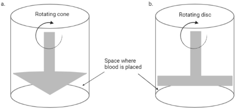
The emergence of microfluidics technology has allowed the development of in vitro models of the vascular system with precise manipulation of the blood flow and cellular components, also providing a major advantage of using smaller blood volume as compared to earlier methods. Before this technology, macrofluidic systems were used, including a cone-and-plate device and two-disc rheometer, as shown in Figure 1, which rely on the rotation of the cone or disc for shear stress induction, and have been used to evaluate the influence of varying shear stress on endothelial cells [17]. The cone-and-plate device was the first to report that endothelial cells’ function and morphology are modulated by altered shear stresses [18]; however, they are not as common nowadays as the microfluidic systems.
Figure 1.
A simplified diagram of a rotational rheometer. (a) Cone and plate rheometer; (b) two-disc rheometer (Created with BioRender.com).
A well-known model that was developed with the microfluidics technology is the ‘thrombosis on a chip’, which allowed the modification of the endothelial surface to mimic different prothrombic conditions, easily introducing foreign agents for study, and high-throughput drug screening, among many other applications [19,20].
Clearly, the design of microfluidic systems to study blood vascular disorders has gone through several developments throughout the years, from the use of glass capillaries to the three-dimensional endothelial cells lined with hydrogels to better mimic the biological system [21]. Mentioned below are the most used devices in research nowadays, which rely on macro- and microfluidics technologies.
Flow Chambers
This is among the earliest methods used to model thrombosis in vitro, which generally represents a channel which blood passes through. This device has gone through many modifications throughout time and is commonly used for quantitative assays to measure in vitro thrombus formation [22].
Different flow chamber-based devices have been developed, ranging from simple to more complicated systems. These devices vary in size and flow surface coatings, which include endothelial cells, collagen, or other synthetic peptides, and are applied mainly to assess platelets’ function and hemostasis [22,23]. Several devices are classified under this technology, including viscometers, parallel-plate, annular, and tubular flow chambers, some of which have a constant flow of fluid while others have a controlled flow rate [24]. Also, custom-made devices are developed by a number of laboratories relying on this method [25].
A common approach in designing a flow chamber-based thrombosis model is the perfusion of blood sample over a surface coated with a platelet-activating or thrombogenic agent, as shown in Figure 2, in which collagen is the most commonly used. However, other agents like fibrin, fibrinogen, von Willebrand factor (vWF), and others are also employed [26,27]. Generally, the method involves coating a glass coverslip with the thrombogenic agent, followed by blood perfusion over the coverslip under controlled shear rate, and in some cases the process is monitored in real time [28,29]. Some studies reported the use of cell-free homogenate of atherosclerotic plaque as a thrombogenic agent and were reported to generate in vitro thrombosis through direct activation of platelets; thus, this method can be used as a model for arterial thrombosis and to evaluate novel anti-thrombogenic agents [29,30].
Figure 2.
Schematic diagram of a flow chamber-based thrombosis model (Created with BioRender.com).
Considering the variations among the designed devices employing this technology, some initiatives have been undertaken to standardize these flow-based assays; recommendations have been given regarding standardizing the flow chambers and a cost-efficacy comparison has also been considered [22]. This is an important step in this area and, if adopted by the designing companies, will further enhance reproducibility in the carried tests.
Thrombosis on a Chip
These models also employ the same concept as flow chambers but can be considered unique in terms of the ‘chip’ feature. These chips can be designed using soft lithography or bioprinting technology where microchannels are created using different materials and can be functionalized with endothelial cells [31].
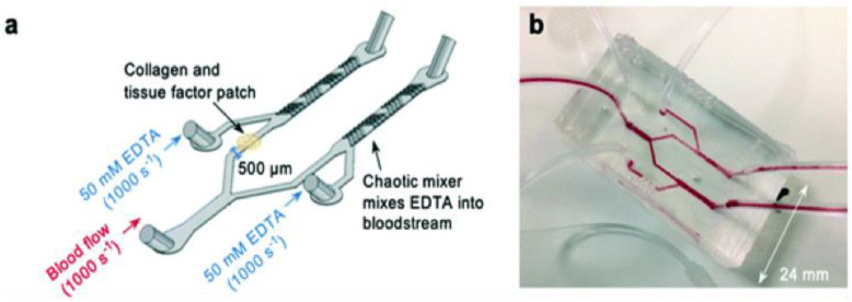
Figure 3 shows a thrombosis-on-a-chip model that employs a novel method to develop occlusive thrombosis and can be used to evaluate the inhibition of thrombus formation by different compounds. In this device, a bifurcation system is designed where blood enters through a single inlet, passes through two different branches, and leaves from two outlets, which is believed to induce the formation of occlusive thrombus [32]. In one channel, a patch of collagen and tissue factor (TF) is placed to mimic plaque rupture. Two additional inlets linked to each arm of the device were also designed to incorporate ethylenediaminetetraacetic acid (EDTA) downstream of the collagen and TF patch, which was used to assess the efficacy of an antiplatelet drug [33].
Figure 3.
Novel thrombosis-on-a-chip device to measure occlusion time. (a) A schematic illustration of the ‘EDTA-quenched’ device; (b) a photograph of the device. Reproduced from Ref. [33] with permission from the Royal Society of Chemistry.
The application of this system in thrombosis includes high-throughput screening of anti-thrombotic drugs, the assessment of changes in different cellular components under different conditions, like shear stress and/or testing chemical reagents, and replacement of standard clinical tests for clotting and hemostasis assessments [34,35], as human blood can be used in these devices in very small amounts.
The main disadvantage of this microfluidic-based system is that the small device size does not allow the exact recreation of the pathological conditions, especially regarding altering blood flow over a longer distance, which is related to many thrombotic pathological conditions [36], since in these devices the blood moves a very short distance that does not recapitulate the human condition.
Other Microfluidic-Based Models
Considering the pathophysiology of venous thrombosis and the time it usually takes to develop, in vitro venous thrombosis models are not as common as arterial thrombosis. However, some studies reported the development of in vitro models of venous thrombosis.
In one study, a microfluidic device with geometries similar to human venous valves was designed, employing primary and secondary vortex characterized by the point of vortical flow and the point of low shear rate, respectively, that are believed to mimic human conditions and support thrombus formation [37,38]. The study reported in vitro venous thrombosis development through a three-step process, including initial fibrin formation at the site of the secondary vortex, followed by platelet delivery at the site between the primary vortex and the formed fibrin with the aid of red blood cells, and then platelet adherence to the fibrin, resulting in thrombus growth and escape to the bulk flow [39]. This device is among the few in vitro thrombosis models that recapitulate venous thrombosis.
Some in vitro models were created to recapitulate shear stress induced by blood-contacting medical devices. This is an important issue that has been characterized and addressed by Chen et al., 2015, using a novel blood-shearing device designed to mimic the pathological flow conditions of high shear stress and short exposure time caused by medical devices like catheters, stents, heart valves, and many others, helping in understanding the tendency and mechanism of developing thrombosis in patients implanted with these devices [40,41,42].
A list of some in vitro thrombosis models is summarized in the Table 1 below.
Table 1.
A list of some in vitro thrombosis models.
| Type of In Vitro Model | Application | Reference |
|---|---|---|
| Parallel-plate flow chamber with endothelial cells matrix-covered surface | Compare various low-molecular-weight heparin and a pentasaccharide for suitability in the in vitro thrombosis model | [43] |
| Parallel-plate flow chamber-based model with fibrin- or fibrinogen-coated surface | Compare and characterize platelet adhesion to fibrin- and fibrinogen-coated surfaces under controlled flow | [26] |
| Parallel-plate flow chamber-based model with collagen- or plaque-coated surface | Compare the thrombogenic effect of different collagen fibers to atherosclerotic plaque | [30] |
| Flow chamber-based model with fibrinogen- or vWF-coated surface | Identify the mechanism of platelet adhesion to fibrinogen and vWF | [27] |
| Flow chamber-based model with collagen-coated surface | Identify the role of human collagen receptors GPVI and α2β1 in thrombus formation |
[29] |
| Fibrinogen-coated flow chambers | Assess platelet adhesion and aggregation following incubation with H2-rich saline | [44] |
| Microfluidic-based device with blood flow under pathophysiological shear rate |
Measurement of coagulation and platelet function | [34] |
| Microfluidic-based device with collagen-coated glass substrate | Measurement of platelet adhesion and blood viscosity | [35] |
| Microfluidic lung chip device lined with primary human alveolar epithelium | Monitor pulmonary thrombosis development and evaluate the effect of different pro-thrombotic and anti-thrombotic factors | [45] |
| Microfluidic device mimicking human venous valves | Develop a venous valvular stasis model and study the effect of platelets and red blood cells on thrombus development | [39] |
| Occlusive thrombosis-on-a-chip microfluidic device | Evaluation of anti-thrombotic drugs | [33] |
| Collagen-coated capillary with controlled rheological conditions | Examine the role of thrombin in platelet recruitment and thrombus stabilization | [46] |
| Collagen-coated glass stenosis model | Describe the structure of arterial thrombi | [47] |
| Endothelialized microfluidic device | Study the mechanism of FeCl3-induced thrombosis | [48] |
| Endothelialized microfluidic device | Study the effect of microplastics on thrombus properties | [49] |
| Endothelialized microfluidic device | A bioassay for hematological disorders and evaluating drug efficacy | [32] |
| In vitro human plasma clot formation assay |
Compare the effect of aprotinin and tranexamic acid on the coagulation pathway and thrombus formation | [50] |
| 3D-bioprinted thrombosis on a chip model coated with human endothelium embedded in a hydrogel | Develop a highly human biomimetic thrombosis model and study its pathophysiology and potential drug efficacy assessment | [51] |
| 3D-printed microfluidic chip coated with human umbilical vein endothelial cells | Recapitulate the three-dimensional structure of healthy and stenotic coronary arteries and assess platelet aggregation | [52] |
| Annular and rectangular perfusion chambers with steady flow | Study the effect of endothelial cells activation on thrombus formation | [53] |
| Multiplate aggregometer and platelet function analyzer (PFA-100) | Test platelet aggregation to investigate cilostazol’s anti-platelet effect | [54] |
| Blood-shearing device | Study the influence of non-physiological stress on platelets and vWF | [42] |
4.2. In Vivo Models
In vivo thrombosis models have been developed using various techniques that relied on the Virchow’s triad, which describes the three main elements involved in thrombus formation, which are damage to the vascular wall, disturbance in blood flow, and the presence of a hypercoagulable state [55]. Different methods can be applied to initiate any of these conditions, like promoting coagulation through the use of certain chemicals, directly injuring the vasculature to damage the endothelium, or artificially inducing stenosis to alter blood flow, among different other methods [56,57].
This review focuses on the animal models that are most used in research, including murine models, porcine models, and zebrafish.
4.2.1. Murine Models
Rats and mice are among the most common animal models that are used to recapitulate different human disorders; being mammals means they share some common physiological processes with humans. They also share similarities with human genetics, as almost every disease-associated gene in humans has a counterpart in rats and mice [36]. They also provide numerous opportunities for genetic manipulation, and their small size allows ease of handling for maintenance and performing of different tests.
As previously mentioned, in vivo thrombus development involves the creation of endothelial injury, promoting a local hypercoagulable state, or artificial induction of vascular stasis or stenosis, and all these parameters have been employed in murine thrombosis models either alone or in combination with each other using different techniques, as described below.
Induction of Endothelial Injury
This can be done with the use of laser- or photochemical-induced injuries, where a beam of laser is focused on a blood vessel that is mostly but not always exposed through surgery. In photochemical-induced injury, the model is first treated with a chemical, like the commonly used Rose Bengal, followed by subsequent light illumination to further promote endothelial damages [58]. In these cases, the developed thrombus can be monitored over time using intravital microscopy and fluorophores that specifically target the thrombus [59]. On the other hand, direct mechanical injuries can also be induced by scrapping the endothelial wall using forceps to pinch a surgically exposed blood vessel [60], or through excision and ligation [61] or any other method that physically damages the endothelium.
Chemicals like ferric chloride (FeCl3) are commonly used and have been proven to induce occlusive thrombus in a dose-dependent manner, where they can be applied to surgically expose vessels to promote endothelial injuries via free radical generation and induction of oxidative stress [62,63].
Promoting Hypercoagulation
Factors that promote hypercoagulation are normally used in combination with other methods, like stasis, stenosis, or endothelial injury, to accelerate thrombus development. Serum, tissue factor, and high-fat diet (HFD) are among the common agents that are used to promote coagulation.
Hyperlipidemia is known to induce a hypercoagulable state by promoting several coagulation factors and fibrin deposition on the vascular wall, and the state is shown to be reversed when the high fat is withdrawn from the diet [57]. When combined with arteriovenous (AV) shunt models or ferric chloride-treated models, diabetic fatty rats were shown to develop thrombosis at a faster rate [64].
Serum and tissue factor are also commonly used as hypercoagulable agents in animal models, where the administration of serum is usually accompanied with vascular ligation to facilitate thrombus formation. One study reported that heterologous serum obtained from human blood samples had a stronger thrombogenic potential in rats than homologous serum [65]. Alternating the serum components can help elucidate the mechanism of thrombus development and the factors involved in thrombogenicity [66]. On the other hand, tissue factor, which is normally a glycoprotein that is expressed at the site of vascular injury, is used to induce thrombosis by direct infusion into the blood vessel [65,67,68].
Also, any antifibrinolytic agents that inhibit fibrin degradation can be employed to promote hypercoagulation, like tranexamic acid, which acts by binding to the surface of plasminogen or plasmin, preventing it from binding to and degrading fibrin, and was used to induce acute hypercoagulation in rat models by a single intragastric administration [69].
Transgenic murine models with hypercoagulable states are also commonly employed in thrombosis studies. In this regard, one study reported that transgenic mice with altered thrombomodulin (TM) gene, that plays a role in anticoagulation, referred to as the TMPro/Pro model, has an increased vascular fibrin deposition and a higher tendency to develop thrombosis when endothelial injury or stasis is induced by FeCl3 treatment or arterial ligation, respectively [70]. A review on the available transgenic mouse models of venous thrombosis has been reported by Audrey et al., 2007 [71].
Induction of Stasis or Stenosis
This method is mostly employed in larger vessels and refers to complete or partial blockage of blood flow, which is most commonly accomplished by narrowing the vessels by tying them with a ligature or compressing with a forceps; the method was first introduced in 1976 [72], and is referred to as Folt’s model. One study applied vascular ligation in mice of different ages and found that older mice have a vascular environment that promotes thrombus development [73].
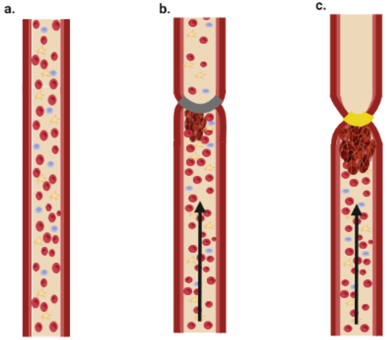
Figure 4 shows a simplified diagram of stasis and stenosis obtained through tightening of the blood vessel.
Figure 4.
Schematic illustration of stasis and stenosis models. (a) Normal blood flow; (b) stenosis model—thrombus is formed due to partial block of blood flow; (c) stasis model—thrombus is formed due to complete block of blood flow (Created with BioRender.com).
These models are mainly used to understand the mechanism of thrombus development due to complete or partial blockage of blood flow, to study the interaction between platelets and coagulation factors, or to evaluate antithrombotic drugs.
Even though murine models are among the most used animal models in research, their main disadvantage in thrombosis research is their small size, making it challenging and complicated to perform intravascular interventions. However, they do provide the advantage of ease of maintenance with low cost. Also, the feasibility of genetic alternations helps elucidate the molecular mechanism and factors involved in this pathogenesis.
4.2.2. Porcine Models
In addition to sharing more genetic similarities with humans than mice do [74], pigs also provide the advantage of large size, which means that thrombosis models resemble humans more closely than smaller species and vascular interventions can be employed more easily. Also, porcine hemodynamics, coagulation cascade, and the vascular system overall are closer to humans [75,76,77]. Considering their large size, porcine thrombosis models mostly involve surgical interventions and, thus, are considered more complicated and practically demanding in comparison to other smaller animal models.
Endothelial Dysfunction
In pigs, different methods can alter endothelial integrity and functions, thus promoting coagulopathies and thrombosis. One study reported that balloon angioplasty in carotid arteries promotes erosion of the endothelial cells at the inflation site, with some sites showing tears in the underlying layers and excessive platelet deposition, eventually leading to thrombus development with complete occlusion in some cases. Such models can be used to evaluate therapeutic options to prevent endothelial injury and vascular occlusion following angioplasty, which is considered a common consequence following this therapeutic procedure [78]. Other approaches involve the use of balloon angioplasty wrapped with metallic coil to facilitate endothelial injury upon balloon inflation [79]. Also, stents—whether bare metals or drug eluting stents—can be employed where they are implanted in the vessel and used as models to study restenosis and stent thrombosis that can occur following these surgical procedures, which limits their application [80].
Electrical stimulation can also induce endothelial injury and complete vascular occlusion without the need of vascular constriction. This phenomenon is believed to occur due to platelet deposition at the site of injury, thus making this model useful to compare between different antiplatelet drugs [81].
Other studies reported that pigs fed with a high-cholesterol diet for at least nine weeks display attenuated endothelial functions with non-responsiveness to serotonin or bradykinin in terms of vasorelaxation, which is a prominent sign of endothelial dysfunction [82,83].
Induction of Stasis or Stenosis
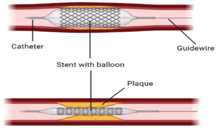
Balloon catheters or stent-based balloon catheters, which are normally used to dilate atherosclerotic blood vessels, as shown in Figure 5, are also used to limit blood flow or completely block it by controlling the degree of inflation. This method is employed in large animals like porcine models to study thrombosis development [84].
Figure 5.
A graphical demonstration of stent with balloon angioplasty to open narrowed arteries. (Created with BioRender.com).
Surgical ligature or surgical placement of ameroid constrictors, which are rings that swell as they absorb the body fluid and gradually limit the blood flow, are also used to generate reproducible stenosis [85,86,87]. Different procedures that can limit the blood flow can be employed to develop thrombosis models, like vascular plugs or stents made from a variety of materials with different shapes and characters; these can be placed via inflatable balloons in different vascular areas to develop novel thrombosis models [88,89].
Promoting Coagulation
Thrombin-induced deep-vein thrombosis (DVT) is a common model used in thrombosis studies, where thrombin, which is a multifunctional enzyme, acts primarily as a pro-coagulant factor aiding in clot formation by converting fibrinogen into fibrin [90]. In this method, a vein is usually surgically exposed followed by local thrombin injection, and in some cases, stasis is created by narrowing proximal veins using different methods [91,92]. Thrombus formation is monitored and can be released by mechanical bending of the muscle or by saline infusion to reach different areas in the vasculature [87]. Thrombosis can be created in different areas using this technique. These models can be used to study the complex pathophysiology of thrombosis, evaluate anti-thrombotic drugs, and test thrombolytic devices like catheters.
A novel model of pulmonary hypertension (PH) with vascular remodeling that manifests as thrombosis was developed employing stenosis, and devoid of the complex surgical procedures employed in different PH models. In this model, a combination of distal embolization using dextran microspheres that were infused weekly to the pulmonary artery through a catheter and coiling of pulmonary branches using silk sutures was employed over a four-week duration alternating between left and right pulmonary arteries. This model was used to develop a chronic PH phenotype that recapitulates the human pathophysiology, with obstructed vascular lumen and occlusions in the vascular arteries [93].
High-fat and high-cholesterol diets are also commonly used to induce arterial atherosclerosis, by activating platelets and clotting factors and reducing the anti-thrombotic properties of the endothelium [94,95,96]. One study reported that maintaining minipigs on high-fat/high-sucrose diets for 6 months resulted in the development of fatty aortic lesions, which can be used as a model for diabetes-accelerated atherosclerosis to study the mechanism underlying this condition [97]. Another study reported that feeding miniature swine with a high-fat/high-cholesterol diet for 10 to 12 months resulted in accelerated atherosclerosis in different arterial sites, with some developing occlusive thrombosis [98].
Ex Vivo Arteriovenous (AV) Shunt Model
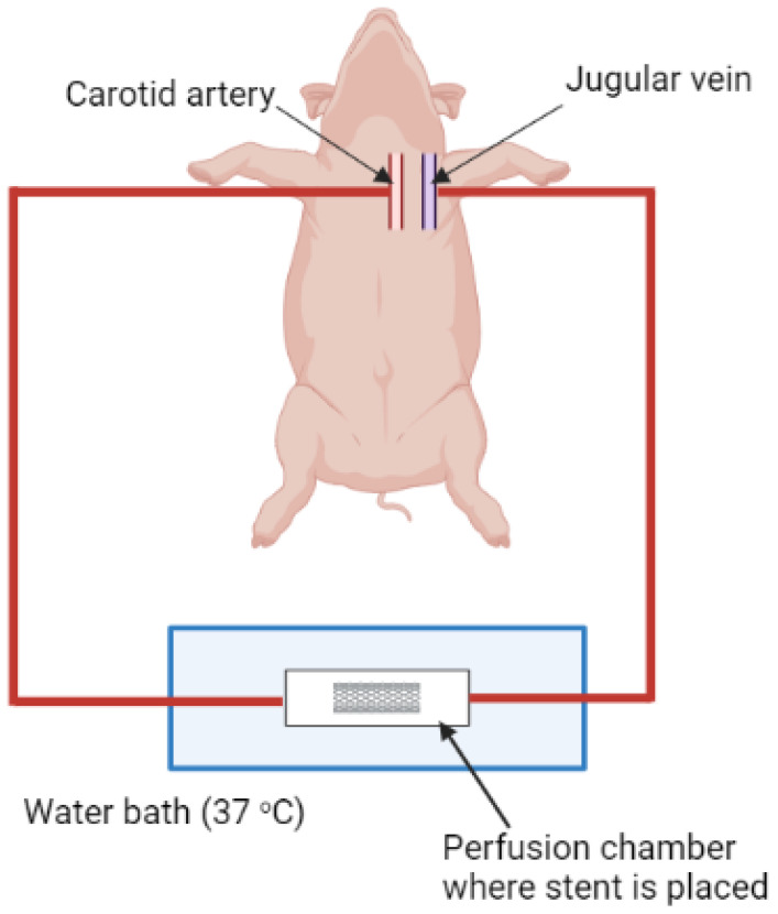
This is a more complicated but commonly used model that combines ex vivo and in vivo approaches. This model employs an extracorporeal perfusion system utilizing stents made from different materials and different morphologies that alter the shear rate of blood flow where the thrombus is developed. Figure 6 shows an illustration of the extracorporeal perfusion system, where stents can be placed in a perfusion chamber inside a water bath and an extracorporeal circuit is surgically utilized by connecting the carotid artery and jugular vein to external tubes, allowing the blood to pass through the perfusion chamber and return back to the jugular vein. Blood flow can be controlled and monitored using a pump and a flow meter connected to the extracorporeal circuit [99] (not shown in the figure).
Figure 6.
A diagram of porcine AV shunt model (Created with BioRender.com).
The main disadvantages of using porcine models in research are related to their large size, which requires larger facilities for maintenance and, thus, higher costs. Also, handling these animals requires well-trained researchers to perform tests and surgical interventions. Ethical concerns are another aspect to be considered, as there are more restrictions when dealing with larger animals compared to smaller animals, like rats, mice, and zebrafish.
4.2.3. Zebrafish
Zebrafish (Danio rerio) provide several advantages as model organisms, including their high reproduction rate, external fertilization, which allows ease of manipulation, and their transparency, which provides many advantages in labeling and visualization. Different human disorders have been modeled in zebrafish, including thrombosis.
Considering the small size of zebrafish, no methods involve surgical interventions or administration of an external device to induce stasis or stenosis. Most methods involve the use of chemicals, laser irradiation, or genetic alternations, as mentioned in detail below.
Induction of Endothelial Injury
As in murine models, this can be done by different techniques, where the most common is treatment with phenylhydrazine (PHZ), FeCl3, or laser irradiation. PHZ acts by generating superoxide radicals, causing damage to red blood cells through membrane lipid peroxidation, and enhancing thrombin generation [100]. The agent is also responsible for endothelial dysfunction leading to hypercoagulation, which all together act in thrombosis development [101]. Treatment of zebrafish 2 days post-fertilization with 1.5µM PHZ for 24 h developed a thrombosis model that was effective for screening of antithrombotic drugs [102].
FeCl3 and pulsed nitrogen laser irradiation were shown to promote vascular occlusion through endothelial injury, where FeCl3 causes damage throughout the vasculature, eventually forming a thrombus, while laser irradiation generates more of a local injury. Both models were effectively employed in genetic screening to identify genetic mutations involved in thrombosis [103].
Promoting a Hypercoagulable State
HFD can be used in zebrafish to induce hyperlipidemia and promote coagulation. A study showed that cholesterol appeared in zebrafish blood vessels on day five following a diet containing 8% cholesterol, and with time this developed into plaque, containing lipids and fibers [104]. Other parameters were also evaluated in this study and found to be consistent with human pathology of atherosclerosis (AS), where treatment with statin managed to effectively alleviate the symptoms. Thus, this approach can be used to model human AS and screen anti-AS drugs.
Genetic alternations, like inducing apoc2−/− mutation and liver X receptor deletion, are other methods used in zebrafish to develop dyslipidemia models and promote coagulation for various applications [105,106].
Zebrafish models provide several advantages over the other in vivo models, including the ease of thrombosis production with high reproducibility, as no method requires surgical interventions, and thrombus development can be monitored in real time. The combination of simplicity and efficiency makes them good choices for thrombosis research.
Table 2 shows a list of some in vivo models of thrombosis. Other in vivo models, like dogs, rabbits, and primates, will not be included in this review; however, developing thrombosis in these models usually employs the same methods used in porcine and murine models.
Table 2.
A list of some common animal thrombosis models.
| Method Employed in the Thrombosis Model | Mechanism of Thrombus Development | Application | Reference |
|---|---|---|---|
| Porcine | |||
| Balloon angioplasty-induced thrombosis | Endothelial injury | Evaluate angioplasty-induced thrombosis | [78] |
| Angioplasty balloon wrapped with a metallic wire coil | Endothelial injury | Determine the relationship between the degree of vascular injury and restenosis magnitude | [79] |
| Surgical ligation and thrombin administration followed by thrombus release to induce PE | Stasis and promoting a hypercoagulable state | Develop a new venous thromboembolism model for possible use in therapeutic testing | [87] |
| Surgical ligation and thrombin administration | Stasis and promoting a hypercoagulable state | Develop a new model of chronic venous thrombosis | [92] |
| Balloon catheter and thrombin administration | Stasis and promoting a hypercoagulable state | Monitor thrombolytic procedures with magnetic resonance imaging | [91] |
| Pulmonary artery embolization with dextran microspheres and surgical coiling of pulmonary branches | Stenosis | Develop a new model of chronic pulmonary hypertension with thrombosis | [93] |
| High-fat/high-sucrose diet-induced atherosclerosis | Promoting a hypercoagulable state | Develop a model of diabetic atherosclerosis | [97] |
| High-fat/high-cholesterol diet-induced atherosclerosis | Promoting a hypercoagulable state | Develop and characterize a diet-induced atherosclerosis model | [98] |
| Surgical ligation of femoral vein and thrombin administration | Stasis and promoting a hypercoagulable state | Develop a DVT model and assess changes in the femoral vein gene expression | [107] |
| Mechanical arterial injury in combination with stent placement followed by total occlusion | Endothelial injury and stasis | Characterize a stent thrombosis model | [108] |
| Balloon catheter and thrombin infusion | Stasis and promoting a hypercoagulable state | Evaluate a high-intensity ultrasound pulse (histotripsy) as a method of thrombolysis | [109] |
| Ischemia-reperfusion injured tissue model | Promoting a hypercoagulable state | Evaluate the role of fish oil in thrombosis development | [110] |
| Balloon catheter and thrombin infusion | Stasis and promoting a hypercoagulable state | Develop a survivable and reproducible iliocaval DVT model for possible use in therapeutic and imaging modalities’ evaluation | [84] |
| Electrical stimulation of the carotid artery endothelium | Endothelial injury | Compare the effect of cilostazol to ticlopidine in inhibiting occlusive thrombus formation | [81] |
| AV shunt model with nitinol stent exposed to arterial blood under high shear rate | Altering blood flow | Evaluate the effect of aspirin, clopidogrel, and combined therapy in inhibiting stent thrombosis development | [99] |
| AV shunt model | Altering blood flow | Compare the thrombogenicity of nitinol to stainless steel stents | [111] |
| Balloon catheter occlusion | Stasis | Evaluate oral administration of low-molecular-weight heparin with a carrier compound in DVT treatment | [112] |
| Self-expanding stent-graft device | Altering blood flow through stasis | Evaluate a thrombolytic therapy with urokinase | [88] |
| Balloon catheter injury | Endothelial injury | Study the effect of ionizing radiation on thrombosis development | [113] |
| Balloon catheter occlusion | Stasis | Use computed tomography to identify lung perfusion abnormalities | [114] |
| Balloon catheter occlusion and thrombin administration | Stasis and promoting a hypercoagulable state | Evaluate the safety and efficacy of microtripsy thrombolysis treatment | [115] |
| AV shunt model | Altering blood flow | Study the effect of rivaroxaban alone or in combination with dual antiplatelet therapy | [116] |
| Murine models | |||
| Laser-induced thrombosis in mice | Endothelial injury | Evaluation of anti-thrombotic drugs | [58] |
| Serum-induced thrombosis in rats | Promoting a hypercoagulable state | Compare the thrombogenicity of homologous and heterologous serum | [65] |
| Tissue factor-induced thrombosis in rats | Promoting a hypercoagulable state | Compare the anti-thrombotic effect of thrombin inhibitor and factor Xa inhibitor | [67] |
| Vascular ligation in mice | Stasis | Evaluate the influence of aging on thrombus resolution | [73] |
| FeCl3-induced thrombosis in mice | Endothelial injury | Develop a refined ferric chloride-induced thrombosis model and test it against anticoagulants | [63] |
| FeCl3 and laser-induced thrombosis in mice | Endothelial injury | Evaluate the potency and safety of anfibatide as an antithrombotic agent | [117] |
| Hypoxia-induced thrombosis in mice | Promoting a hypercoagulable state | Develop and study the mechanism of hypoxia-induced thrombosis | [118] |
| Thrombin-induced thrombosis in rats | Promoting a hypercoagulable state | Develop and characterize a thrombotic ischemia model that mimics human thromboembolic stroke | [119] |
| FeCl3-induced thrombosis in rats | Endothelial injury | Characterize the thrombus, evaluate a novel antithrombotic agent, and determine the relationship between vessel temperature and vascular occlusion | [62] |
| FeCl3-induced thrombosis in rats | Endothelial injury | Assessment of tiplaxtinin antithrombotic effect | [120] |
| Vascular ligation in rats | Stasis | Study the antithrombotic effect of grape seed proanthocyanidins extract | [121] |
| Zebrafish | |||
| PHZ-induced thrombosis | Endothelial injury and promoting a hypercoagulable state | Assessment of antithrombotic drugs | [102] |
| PHZ-induced thrombosis | Endothelial injury and promoting a hypercoagulable state | Evaluate the antithrombotic effect of Rubia cordifolia | [122] |
| FeCl3 or laser irradiation-induced thrombosis | Vascular injury | Genetic screening | [103] |
| Arachidonic acid-induced thrombosis | Promoting platelet aggregation | Evaluate the antithrombotic effect of danhong injection | [123] |
| Arachidonic acid-induced thrombosis | Promoting platelet aggregation | Evaluate the antithrombotic effect of Wuliangye Baijiu | [124] |
| Apoc2 mutant zebrafish | Promoting a hypercoagulable state | Characterization of apoc2 mutant zebrafish | [105] |
| Heg1 knockout zebrafish | Damaging the vascular endothelium integrity | Develop a zebrafish model of dilated cardiomyopathy and thrombosis and employ it in drug screening | [125] |
| High cholesterol and lipopolysaccharide diet | Promoting a hypercoagulable state | Drug screening | [104] |
4.3. Advantages and Disadvantages of In Vitro and In Vivo Models
4.3.1. In Vitro Models
Different cells grown on a dish do not represent the whole physiological system with all its complexity and interaction with different components, though they usually provide a general estimate of what might happen inside the body, which needs to be further confirmed through the use of a full organism. A major argument against the use of microfluidics is the microchannels themselves, which, despite providing the advantage of using smaller blood volume, do not represent thrombosis that usually develops in larger arteries and veins [126]. Considering these, it is important to consider in vivo models for confirmation of results obtained from in vitro studies.
4.3.2. In Vivo Models
Generally, no animal model can perfectly recapitulate any human disease, though their use has greatly advanced our understanding of human disorders and drug screening and development; thus, their use will continue to be central in research. Each animal model has its own limitations, where the choice will depend on several factors, including the aim of the study and availability of resources.
A brief comparison regarding the use of in vivo and in vitro models in thrombosis research are mentioned in the Table 3 below.
Table 3.
A comparison between the advantages and disadvantages of in vitro and in vivo models in research.
| Category | In Vitro Models | In Vivo Models |
|---|---|---|
| Reproducibility | Possible, especially when using the same device and conditions. | Variable, considering inter-species variations. |
| Ethical concerns | Minimal. | Strict, especially with larger animals. |
| Cost | Relatively cheap. | Relatively expensive. |
| Simplicity | Relatively simple, especially when using pre-designed devices. | Relatively complicated and time consuming. |
| Result translation | Results need to be further confirmed by in vivo studies. | Considering the settings, results can be more closely related to human conditions, with higher possibility of clinical translation. |
5. Conclusions
Approaches to generate thrombosis models in vivo and in vitro are wide and versatile, with many studies continuously developing novel techniques. Generally, methods that promote endothelial damage and activation of the coagulation pathway are used to induce venous thrombosis, while methods that promote platelet aggregation are used to induce arterial thrombosis. However, similar methods can sometimes be used to generate venous or arterial thrombosis, depending on the vessel type.
It should be noted that different methods generate thrombosis via different mechanisms and with different pathophysiology, as some studies have shown that thrombosis generated by different techniques responds differently to anti-thrombotic drugs. Thus, it is important to shift attention to standardize thrombosis models for each specific application, as there are large numbers of studies developing thrombosis models without specific and well-detailed characterization of the generated thrombus. Having a standard, reproducible model for each application can eliminate many of the ambiguities and non-consistent results in this field.
Acknowledgments
The authors acknowledge the staff from the department of pulmonary medicine, Josep Trueta University hospital de Girona, Santa Caterina hospital de Salt, and the Girona biomedical research institute (IDIBGI) for all the support provided. Figure 1, Figure 2, Figure 4, Figure 5 and Figure 6 were created with BioRender.com.
Abbreviations
| AV | Arteriovenous |
| AS | Atherosclerosis |
| CTEPH | Chronic thromboembolic pulmonary hypertension |
| EDTA | Ethylenediaminetetraacetic acid |
| DVT | Deep-vein thrombosis |
| HFD | High-fat diet |
| MI | Myocardial infarction |
| PHZ | Phenylhydrazine |
| PE | Pulmonary embolism |
| PH | Pulmonary hypertension |
| TM | Thrombomodulin |
| TF | Tissue factor |
| vWF | von Willebrand factor |
Author Contributions
O.T.C. contributed to drafting the concept and design of the paper; S.A. collected information and wrote the manuscript; and O.T.C., E.O. and R.O. were responsible for critical revision and final editing of the paper. All authors have read and agreed to the published version of the manuscript.
Conflicts of Interest
The authors declare no conflict of interest. The funders had no role in the writing of the manuscript.
Funding Statement
This research was supported by funding from a Miguel Servet grant from the Institute of Health Carlos III (CP17/00114). Sana Ayyoub was a recipient of a grant from Ajuts per a la contractació de personal investigador predoctoral en formació of Agència de Gestió d’Ajuts Universitaris i de Recerca (AGAUR; FI2022) from Generalitat de Cataluña (2022 FI_B 00941). Eduardo Oliver is a recipient of funds from a Ramón y Cajal grant (RYC2020-028884-I) funded by MCIN/AEI/10.13039/501100011033 and by “ESF Investing in your future”.
Footnotes
Disclaimer/Publisher’s Note: The statements, opinions and data contained in all publications are solely those of the individual author(s) and contributor(s) and not of MDPI and/or the editor(s). MDPI and/or the editor(s) disclaim responsibility for any injury to people or property resulting from any ideas, methods, instructions or products referred to in the content.
References
- 1.Wendelboe A.M., Raskob G.E. Global burden of thrombosis: Epidemiologic aspects. Circ. Res. 2016;118:1340–1347. doi: 10.1161/CIRCRESAHA.115.306841. [DOI] [PubMed] [Google Scholar]
- 2.Mackman N. Triggers, targets and treatments for thrombosis. Nature. 2008;451:914–918. doi: 10.1038/nature06797. [DOI] [PMC free article] [PubMed] [Google Scholar]
- 3.Zipes D.P. Braunwald’s heart disease: A textbook of cardiovascular medicine. BMH Med. J. 2018;5:63. [Google Scholar]
- 4.Klok F., Van der Hulle T., Exter P.D., Lankeit M., Huisman M., Konstantinides S. The post-PE syndrome: A new concept for chronic complications of pulmonary embolism. Blood Rev. 2014;28:221–226. doi: 10.1016/j.blre.2014.07.003. [DOI] [PubMed] [Google Scholar]
- 5.Rasche H. Haemostasis and thrombosis: An overview. Eur. Heart J. Suppl. 2001;3:Q3–Q7. doi: 10.1016/S1520-765X(01)90034-3. [DOI] [Google Scholar]
- 6.Wu K.K., Thiagarajan M.P. Role of endothelium in thrombosis and hemostasis. Annu. Rev. Med. 1996;47:315–331. doi: 10.1146/annurev.med.47.1.315. [DOI] [PubMed] [Google Scholar]
- 7.Ashorobi D., Ameer M.A., Fernandez R. StatPearls. StatPearls Publishing; Treasure Island, FL, USA: 2022. Thrombosis. [Google Scholar]
- 8.Migliacci R., Becattini C., Pesavento R., Davi G., Vedovati M.C., Guglielmini G., Falcinelli E., Ciabattoni G., Dalla Valle F., Prandoni P. Endothelial dysfunction in patients with spontaneous venous thromboembolism. Haematologica. 2007;92:812–818. doi: 10.3324/haematol.10872. [DOI] [PubMed] [Google Scholar]
- 9.Nieswandt B., Pleines I., Bender M. Platelet adhesion and activation mechanisms in arterial thrombosis and ischaemic stroke. J. Thromb. Haemost. 2011;9:92–104. doi: 10.1111/j.1538-7836.2011.04361.x. [DOI] [PubMed] [Google Scholar]
- 10.Vilahur G., Padro T., Badimon L. Atherosclerosis and thrombosis: Insights from large animal models. J. Biomed. Biotechnol. 2011;2011:907575. doi: 10.1155/2011/907575. [DOI] [PMC free article] [PubMed] [Google Scholar]
- 11.Prandoni P. Venous and arterial thrombosis: Is there a link? Thromb. Embolism Res. Clin. Pract. 2016;1:273–283. doi: 10.1007/5584_2016_121. [DOI] [PubMed] [Google Scholar]
- 12.Ortel T.L., Neumann I., Ageno W., Beyth R., Clark N.P., Cuker A., Hutten B.A., Jaff M.R., Manja V., Schulman S. American Society of Hematology 2020 guidelines for management of venous thromboembolism: Treatment of deep vein thrombosis and pulmonary embolism. Blood Adv. 2020;4:4693–4738. doi: 10.1182/bloodadvances.2020001830. [DOI] [PMC free article] [PubMed] [Google Scholar]
- 13.Mazzolai L., Ageno W., Alatri A., Bauersachs R., Becattini C., Brodmann M., Emmerich J., Konstantinides S., Meyer G., Middeldorp S. Second consensus document on diagnosis and management of acute deep vein thrombosis: Updated document elaborated by the ESC Working Group on aorta and peripheral vascular diseases and the ESC Working Group on pulmonary circulation and right ventricular function. Eur. J. Prev. Cardiol. 2022;29:1248–1263. doi: 10.1093/eurjpc/zwab088. [DOI] [PubMed] [Google Scholar]
- 14.Moster M., Bolliger D. Perioperative Guidelines on Antiplatelet and Anticoagulant Agents: 2022 Update. Curr. Anesthesiol. Rep. 2022:1–11. [Google Scholar]
- 15.Tsoupras A., Zabetakis I., Lordan R. Platelet aggregometry assay for evaluating the effects of platelet agonists and antiplatelet compounds on platelet function in vitro. MethodsX. 2019;6:63–70. doi: 10.1016/j.mex.2018.12.012. [DOI] [PMC free article] [PubMed] [Google Scholar]
- 16.Morawitz P., Jürgens R. Gibt es eine Thrombasthenie. Münch. Med. Wschr. 1930;77:2001. [Google Scholar]
- 17.Hosseini V., Mallone A., Nasrollahi F., Ostrovidov S., Nasiri R., Mahmoodi M., Haghniaz R., Baidya A., Salek M.M., Darabi M.A. Healthy and diseased in vitro models of vascular systems. Lab A Chip. 2021;21:641–659. doi: 10.1039/d0lc00464b. [DOI] [PubMed] [Google Scholar]
- 18.Dewey C., Jr., Bussolari S.R., Gimbrone M.A., Jr., Davies P.F. The dynamic response of vascular endothelial cells to fluid shear stress. J. Biomech. Eng. 1981;103:177–185. doi: 10.1115/1.3138276. [DOI] [PubMed] [Google Scholar]
- 19.Zhang B., Korolj A., Lai B.F.L., Radisic M. Advances in organ-on-a-chip engineering. Nat. Rev. Mater. 2018;3:257–278. [Google Scholar]
- 20.Lam W.A. Thrombosis-on-a-Chip: A new way to model a complex process. Blood. 2017;130:SCI-10. [Google Scholar]
- 21.Sebastian B., Dittrich P.S. Microfluidics to mimic blood flow in health and disease. Annu. Rev. Fluid Mech. 2018;50:483–504. [Google Scholar]
- 22.Roest M., Reininger A., Zwaginga J., King M., Heemskerk J. Flow Chamber-Based Assays to Measure Thrombus Formation In Vitro: Requirements for Standardization. Vol. 9. Wiley Online Library; New York, NY, USA: 2011. Biorheology Subcommittee of the SSC of the ISTH; pp. 2322–2324. [DOI] [PubMed] [Google Scholar]
- 23.Zwaginga J., Nash G., King M., Heemskerk J., Frojmovic M., Hoylaerts M., Sakariassen K. Biorheology Subcommittee of the SSC of the ISTH. Flow-based assays for global assessment of hemostasis. Part 1: Biorheologic considerations 1. J. Thromb. Haemost. 2006;4:2486–2487. doi: 10.1111/j.1538-7836.2006.02177.x. [DOI] [PubMed] [Google Scholar]
- 24.Slack S.M., Turitto V.T. Flow chambers and their standardization for use in studies of thrombosis. Thromb. Haemost. 1994;72:777–781. doi: 10.1055/s-0038-1648957. [DOI] [PubMed] [Google Scholar]
- 25.Van Kruchten R., Cosemans J.M., Heemskerk J.W. Measurement of whole blood thrombus formation using parallel-plate flow chambers–a practical guide. Platelets. 2012;23:229–242. doi: 10.3109/09537104.2011.630848. [DOI] [PubMed] [Google Scholar]
- 26.Jen C., Lin J. Direct observation of platelet adhesion to fibrinogen-and fibrin-coated surfaces. Am. J. Physiol. Heart Circ. Physiol. 1991;261:H1457–H1463. doi: 10.1152/ajpheart.1991.261.5.H1457. [DOI] [PubMed] [Google Scholar]
- 27.Savage B., Saldívar E., Ruggeri Z.M. Initiation of platelet adhesion by arrest onto fibrinogen or translocation on von Willebrand factor. Cell. 1996;84:289–297. doi: 10.1016/S0092-8674(00)80983-6. [DOI] [PubMed] [Google Scholar]
- 28.Kuijpers M.J., Schulte V., Bergmeier W., Lindhout T., Brakebusch C., Offermanns S., Fässler R., Heemskerk J.W., Nieswandt B. Complementary roles of platelet glycoprotein VI and integrin α2β1 in collagen-induced thrombus formation in flowing whole blood ex vivo. FASEB J. 2003;17:685–687. doi: 10.1096/fj.02-0381fje. [DOI] [PubMed] [Google Scholar]
- 29.Siljander P.R.-M., Munnix I.C., Smethurst P.A., Deckmyn H., Lindhout T., Ouwehand W.H., Farndale R.W., Heemskerk J.W. Platelet receptor interplay regulates collagen-induced thrombus formation in flowing human blood. Blood. 2004;103:1333–1341. doi: 10.1182/blood-2003-03-0889. [DOI] [PubMed] [Google Scholar]
- 30.Cosemans J.M., Kuijpers M.J., Lecut C., Loubele S.T., Heeneman S., Jandrot-Perrus M., Heemskerk J.W. Contribution of platelet glycoprotein VI to the thrombogenic effect of collagens in fibrous atherosclerotic lesions. Atherosclerosis. 2005;181:19–27. doi: 10.1016/j.atherosclerosis.2004.12.037. [DOI] [PubMed] [Google Scholar]
- 31.Zhang Y.S., Oklu R., Albadawi H. Bioengineered in vitro models of thrombosis: Methods and techniques. Cardiovasc. Diagn. Ther. 2017;7((Suppl. 3)):S329. doi: 10.21037/cdt.2017.08.08. [DOI] [PMC free article] [PubMed] [Google Scholar]
- 32.Tsai M., Kita A., Leach J., Rounsevell R., Huang J.N., Moake J., Ware R.E., Fletcher D.A., Lam W.A. In vitro modeling of the microvascular occlusion and thrombosis that occur in hematologic diseases using microfluidic technology. J. Clin. Investig. 2011;122:408–418. doi: 10.1172/JCI58753. [DOI] [PMC free article] [PubMed] [Google Scholar]
- 33.Berry J., Peaudecerf F.J., Masters N.A., Neeves K.B., Goldstein R.E., Harper M.T. An “occlusive thrombosis-on-a-chip” microfluidic device for investigating the effect of anti-thrombotic drugs. Lab A Chip. 2021;21:4104–4117. doi: 10.1039/D1LC00347J. [DOI] [PMC free article] [PubMed] [Google Scholar]
- 34.Jain A., Graveline A., Waterhouse A., Vernet A., Flaumenhaft R., Ingber D.E. A shear gradient-activated microfluidic device for automated monitoring of whole blood haemostasis and platelet function. Nat. Commun. 2016;7:1–10. doi: 10.1038/ncomms10176. [DOI] [PMC free article] [PubMed] [Google Scholar]
- 35.Yeom E., Park J.H., Kang Y.J., Lee S.J. Microfluidics for simultaneous quantification of platelet adhesion and blood viscosity. Sci. Rep. 2016;6:1–11. doi: 10.1038/srep24994. [DOI] [PMC free article] [PubMed] [Google Scholar]
- 36.Herbig B.A., Yu X., Diamond S.L. Using microfluidic devices to study thrombosis in pathological blood flows. Biomicrofluidics. 2018;12:42201. doi: 10.1063/1.5021769. [DOI] [PMC free article] [PubMed] [Google Scholar]
- 37.Lurie F., Kistner R.L., Eklof B., Kessler D. Mechanism of venous valve closure and role of the valve in circulation: A new concept. J. Vasc. Surg. 2003;38:955–961. doi: 10.1016/S0741-5214(03)00711-0. [DOI] [PubMed] [Google Scholar]
- 38.Karino T., Goldsmith H.L., Motomiya M., Mabuchi S., Sohara Y. Flow Patterns in Vessels of Simple and Complex Geometries a. Ann. N. Y. Acad. Sci. 1987;516:422–441. doi: 10.1111/j.1749-6632.1987.tb33063.x. [DOI] [PubMed] [Google Scholar]
- 39.Lehmann M., Schoeman R.M., Krohl P.J., Wallbank A.M., Samaniuk J.R., Jandrot-Perrus M., Neeves K.B. Platelets drive thrombus propagation in a hematocrit and glycoprotein VI–dependent manner in an in vitro venous thrombosis model. Arterioscler. Thromb. Vasc. Biol. 2018;38:1052–1062. doi: 10.1161/ATVBAHA.118.310731. [DOI] [PMC free article] [PubMed] [Google Scholar]
- 40.Chen Z., Mondal N.K., Ding J., Gao J., Griffith B.P., Wu Z.J. Shear-induced platelet receptor shedding by non-physiological high shear stress with short exposure time: Glycoprotein Ibα and glycoprotein VI. Thromb. Res. 2015;135:692–698. doi: 10.1016/j.thromres.2015.01.030. [DOI] [PMC free article] [PubMed] [Google Scholar]
- 41.Chen Z., Mondal N.K., Ding J., Koenig S.C., Slaughter M.S., Griffith B.P., Wu Z.J. Activation and shedding of platelet glycoprotein IIb/IIIa under non-physiological shear stress. Mol. Cell. Biochem. 2015;409:93–101. doi: 10.1007/s11010-015-2515-y. [DOI] [PMC free article] [PubMed] [Google Scholar]
- 42.Chen Z., Mondal N.K., Ding J., Koenig S.C., Slaughter M.S., Wu Z.J. Paradoxical Effect of Nonphysiological Shear Stress on Platelets and v on W illebrand Factor. Artif. Organs. 2016;40:659–668. doi: 10.1111/aor.12606. [DOI] [PMC free article] [PubMed] [Google Scholar]
- 43.Lozano M., Bos A., de Groot P.G., Van Willigen G., Meuleman D., Ordinas A., Sixma J. Suitability of low-molecular-weight heparin (oid) s and a pentasaccharide for an in vitro human thrombosis model. Arterioscler. Thromb. A J. Vasc. Biol. 1994;14:1215–1222. doi: 10.1161/01.ATV.14.7.1215. [DOI] [PubMed] [Google Scholar]
- 44.Wang Y., Wu Y.-P., Han J.-J., Zhang M.-Q., Yang C.-X., Jiao P., Tian H., Zhu C., Qin S.-C., Sun X.-J. Inhibitory effects of hydrogen on in vitro platelet activation and in vivo prevention of thrombosis formation. Life Sci. 2019;233:116700. doi: 10.1016/j.lfs.2019.116700. [DOI] [PubMed] [Google Scholar]
- 45.Jain A., Barrile R., van der Meer A.D., Mammoto A., Mammoto T., De Ceunynck K., Aisiku O., Otieno M.A., Louden C.S., Hamilton G.A. Primary human lung alveolus-on-a-chip model of intravascular thrombosis for assessment of therapeutics. Clin. Pharmacol. Ther. 2018;103:332–340. doi: 10.1002/cpt.742. [DOI] [PMC free article] [PubMed] [Google Scholar]
- 46.Wagner W., Hubbell J. Local thrombin synthesis and fibrin formation in an in vitro thrombosis model result in platelet recruitment and thrombus stabilization on collagen in heparinized blood. J. Lab. Clin. Med. 1990;116:636–650. [PubMed] [Google Scholar]
- 47.Kim D.A., Ku D.N. Structure of shear-induced platelet aggregated clot formed in an in vitro arterial thrombosis model. Blood Adv. 2022;6:2872–2883. doi: 10.1182/bloodadvances.2021006248. [DOI] [PMC free article] [PubMed] [Google Scholar]
- 48.Ciciliano J.C., Sakurai Y., Myers D.R., Fay M.E., Hechler B., Meeks S., Li R., Dixon J.B., Lyon L.A., Gachet C. Resolving the multifaceted mechanisms of the ferric chloride thrombosis model using an interdisciplinary microfluidic approach. Blood J. Am. Soc. Hematol. 2015;126:817–824. doi: 10.1182/blood-2015-02-628594. [DOI] [PMC free article] [PubMed] [Google Scholar]
- 49.Chen L., Zheng Y., Liu Y., Tian P., Yu L., Bai L., Zhou F., Yang Y., Cheng Y., Wang F. Microfluidic-based in vitro thrombosis model for studying microplastics toxicity. Lab. Chip. 2022;22:1344–1353. doi: 10.1039/D1LC00989C. [DOI] [PubMed] [Google Scholar]
- 50.Sperzel M., Huetter J. Evaluation of aprotinin and tranexamic acid in different in vitro and in vivo models of fibrinolysis, coagulation and thrombus formation. J. Thromb. Haemost. 2007;5:2113–2118. doi: 10.1111/j.1538-7836.2007.02717.x. [DOI] [PubMed] [Google Scholar]
- 51.Zhang Y.S., Davoudi F., Walch P., Manbachi A., Luo X., Dell’Erba V., Miri A.K., Albadawi H., Arneri A., Li X. Bioprinted thrombosis-on-a-chip. Lab. Chip. 2016;16:4097–4105. doi: 10.1039/C6LC00380J. [DOI] [PMC free article] [PubMed] [Google Scholar]
- 52.Costa P.F., Albers H.J., Linssen J.E., Middelkamp H.H., Van Der Hout L., Passier R., Van Den Berg A., Malda J., Van Der Meer A.D. Mimicking arterial thrombosis in a 3D-printed microfluidic in vitro vascular model based on computed tomography angiography data. Lab. Chip. 2017;17:2785–2792. doi: 10.1039/C7LC00202E. [DOI] [PubMed] [Google Scholar]
- 53.Zwaginga J., Sixma J., de Groot P.G. Activation of endothelial cells induces platelet thrombus formation on their matrix. Studies of new in vitro thrombosis model with low molecular weight heparin as anticoagulant. Arterioscler. Off. J. Am. Heart Assoc. Inc. 1990;10:49–61. doi: 10.1161/01.ATV.10.1.49. [DOI] [PubMed] [Google Scholar]
- 54.Kim C.-W., Yun J.-W., Bae I.-H., Park Y.-H., Jeong Y.S., Park J.W., Chung J.-H., Park Y.-H., Lim K.-M. Evaluation of anti-platelet and anti-thrombotic effects of cilostazol with PFA-100® and Multiplate® whole blood aggregometer in vitro, ex vivo and FeCl3-induced thrombosis models in vivo. Thromb. Res. 2011;127:565–570. doi: 10.1016/j.thromres.2011.02.004. [DOI] [PubMed] [Google Scholar]
- 55.Kushner A., West D., Pillarisetty L.S. StatPearls. StatPearls Publishing; Treasure Island, FL, USA: 2022. Virchow triad. [PubMed] [Google Scholar]
- 56.Albadawi H., Witting A.A., Pershad Y., Wallace A., Fleck A.R., Hoang P., Khademhosseini A., Oklu R. Animal models of venous thrombosis. Cardiovasc. Diagn. Ther. 2017;7((Suppl. 3)):S197. doi: 10.21037/cdt.2017.08.10. [DOI] [PMC free article] [PubMed] [Google Scholar]
- 57.De Curtis A., D’Adamo M.C., Amore C., Polishchuck R., Di Castelnuovo A., Donati M.B., Iacoviello L. Experimental Arterial Thrombosis in Genetically or Diet Induced Hyperlipidemia in Rats. Thromb. Haemost. 2001;86:1440–1448. doi: 10.1055/s-0037-1616747. [DOI] [PubMed] [Google Scholar]
- 58.Rosen E.D., Raymond S., Zollman A., Noria F., Sandoval-Cooper M., Shulman A., Merz J.L., Castellino F.J. Laser-induced noninvasive vascular injury models in mice generate platelet-and coagulation-dependent thrombi. Am. J. Pathol. 2001;158:1613–1622. doi: 10.1016/S0002-9440(10)64117-X. [DOI] [PMC free article] [PubMed] [Google Scholar]
- 59.Kamocka M., Mu J., Liu X., Chen N., Zollman A., Sturonas-Brown B., Dunn K., Xu Z., Chen D.Z., Alber M.S. Two-photon intravital imaging of thrombus development. J. Biomed. Opt. 2010;15:16020. doi: 10.1117/1.3322676. [DOI] [PMC free article] [PubMed] [Google Scholar]
- 60.Pierangeli S.S., Barker J.H., Stikovac D., Ackerman D., Anderson G., Barquinero J., Acland R., Harris E.N. Effect of human IgG antiphospholipid antibodies on an in vivo thrombosis model in mice. Thromb. Haemost. 1994;71:670–674. doi: 10.1055/s-0038-1642501. [DOI] [PubMed] [Google Scholar]
- 61.Sawyer P.N., Pate J.W., Weldon C.S. Relations of abnormal and injury electric potential differences to intravascular thrombosis. Am. J. Physiol. Leg. Content. 1953;175:108–112. doi: 10.1152/ajplegacy.1953.175.1.108. [DOI] [PubMed] [Google Scholar]
- 62.Kurz K., Main B., Sandusky G. Rat model of arterial thrombosis induced by ferric chloride. Thromb. Res. 1990;60:269–280. doi: 10.1016/0049-3848(90)90106-M. [DOI] [PubMed] [Google Scholar]
- 63.Wang X., Xu L. An optimized murine model of ferric chloride-induced arterial thrombosis for thrombosis research. Thromb. Res. 2005;115:95–100. doi: 10.1016/j.thromres.2004.07.009. [DOI] [PubMed] [Google Scholar]
- 64.Shang J., Chen Z., Wang M., Li Q., Feng W., Wu Y., Wu W., Graziano M.P., Chintala M. Zucker Diabetic Fatty rats exhibit hypercoagulability and accelerated thrombus formation in the Arterio-Venous shunt model of thrombosis. Thromb. Res. 2014;134:433–439. doi: 10.1016/j.thromres.2014.04.008. [DOI] [PubMed] [Google Scholar]
- 65.Millet J., Vaillot M., Theveniaux J., Brown N.L. Experimental venous thrombosis induced by homologous serum in the rat. Thromb. Res. 1996;81:497–502. doi: 10.1016/0049-3848(96)00023-0. [DOI] [PubMed] [Google Scholar]
- 66.Deykin D., Wessler S. Activation product, factor IX, serum thrombotic accelerator activity, and serum-induced thrombosis. J. Clin. Investig. 1964;43:160–166. doi: 10.1172/JCI104900. [DOI] [PMC free article] [PubMed] [Google Scholar]
- 67.Furugohri T., Fukuda T., Tsuji N., Kita A., Morishima Y., Shibano T. Melagatran, a direct thrombin inhibitor, but not edoxaban, a direct factor Xa inhibitor, nor heparin aggravates tissue factor-induced hypercoagulation in rats. Eur. J. Pharmacol. 2012;686:74–80. doi: 10.1016/j.ejphar.2012.04.031. [DOI] [PubMed] [Google Scholar]
- 68.Wessler S., Ward K., Ho C. Studies in intravascular coagulation. III. The pathogenesis of serum-induced venous thrombosis. J. Clin. Investig. 1955;34:647–651. doi: 10.1172/JCI103114. [DOI] [PMC free article] [PubMed] [Google Scholar]
- 69.Jing J., Du Z., Wen Z., Jiang B., He B. Dynamic changes of urinary proteins in a rat model of acute hypercoagulable state induced by tranexamic acid. J. Cell. Physiol. 2019;234:10809–10818. doi: 10.1002/jcp.27904. [DOI] [PubMed] [Google Scholar]
- 70.Weiler H., Lindner V., Kerlin B., Isermann B.H., Hendrickson S.B., Cooley B.C., Meh D.A., Mosesson M.W., Shworak N.W., Post M.J. Characterization of a mouse model for thrombomodulin deficiency. Arterioscler. Thromb. Vasc. Biol. 2001;21:1531–1537. doi: 10.1161/hq0901.094496. [DOI] [PubMed] [Google Scholar]
- 71.Cleuren A.C., van Vlijmen B.J., Reitsma P.H. Seminars in Thrombosis and Hemostasis. Thieme Medical Publishers, Inc.; New York, NY, USA: 2007. Transgenic mouse models of venous thrombosis: Fulfilling the expectations? pp. 610–616. [DOI] [PubMed] [Google Scholar]
- 72.Folts J.D., Crowell E.B., Jr., Rowe G.G. Platelet aggregation in partially obstructed vessels and its elimination with aspirin. Circulation. 1976;54:365–370. doi: 10.1161/01.CIR.54.3.365. [DOI] [PubMed] [Google Scholar]
- 73.McDonald A.P., Meier T.R., Hawley A.E., Thibert J.N., Farris D.M., Wrobleski S.K., Henke P.K., Wakefield T.W., Myers D.D., Jr. Aging is associated with impaired thrombus resolution in a mouse model of stasis induced thrombosis. Thromb. Res. 2010;125:72–78. doi: 10.1016/j.thromres.2009.06.005. [DOI] [PubMed] [Google Scholar]
- 74.Wernersson R., Schierup M.H., Jørgensen F.G., Gorodkin J., Panitz F., Stærfeldt H.-H., Christensen O.F., Mailund T., Hornshøj H., Klein A. Pigs in sequence space: A 0.66 X coverage pig genome survey based on shotgun sequencing. BMC Genom. 2005;6:1–7. doi: 10.1186/1471-2164-6-70. [DOI] [PMC free article] [PubMed] [Google Scholar]
- 75.Folts J., Rowe G. Circulation. American Heart Association; Dallas, TX, USA: 1983. Acute thrombus formation in stenosed pig coronary-arteries, causing sudden-death by ventricular-fibrillation; p. 264. [Google Scholar]
- 76.Leach C.M., Thorburn G.D. A comparative study of collagen induced thromboxane release from platelets of different species: Implications for human athero sclerosis models. Prostaglandins. 1982;24:47–59. doi: 10.1016/0090-6980(82)90176-9. [DOI] [PubMed] [Google Scholar]
- 77.Topaz O. Cardiovascular Thrombus: From Pathology and Clinical Presentations to imaging, Pharmacotherapy and Interventions. Academic Press; Cambridge, MA, USA: 2018. [Google Scholar]
- 78.Steele P.M., Chesebro J.H., Stanson A.W., Holmes D.R., Jr., Dewanjee M.K., Badimon L., Fuster V. Balloon angioplasty. Natural history of the pathophysiological response to injury in a pig model. Circ. Res. 1985;57:105–112. doi: 10.1161/01.RES.57.1.105. [DOI] [PubMed] [Google Scholar]
- 79.Schwartz R.S., Huber K.C., Murphy J.G., Edwards W.D., Camrud A.R., Vlietstra R.E., Holmes D.R. Restenosis and the proportional neointimal response to coronary artery injury: Results in a porcine model. J. Am. Coll. Cardiol. 1992;19:267–274. doi: 10.1016/0735-1097(92)90476-4. [DOI] [PubMed] [Google Scholar]
- 80.Miyauchi K., Kasai T., Yokayama T., Aihara K., Kurata T., Kajimoto K., Okazaki S., Ishiyama H., Daida H. Effectiveness of statin-eluting stent on early inflammatory response and neointimal thickness in a porcine coronary model. Circ. J. 2008;72:832–838. doi: 10.1253/circj.72.832. [DOI] [PubMed] [Google Scholar]
- 81.Kohda N., Tani T., Nakayama S., Adachi T., Marukawa K., Ito R., Ishida K., Matsumoto Y., Kimura Y. Effect of cilostazol, a phosphodiesterase III inhibitor, on experimental thrombosis in the porcine carotid artery. Thromb. Res. 1999;96:261–268. doi: 10.1016/S0049-3848(99)00109-7. [DOI] [PubMed] [Google Scholar]
- 82.Cohen R.A., Zitnay K.M., Haudenschild C.C., Cunningham L.D. Loss of selective endothelial cell vasoactive functions caused by hypercholesterolemia in pig coronary arteries. Circ. Res. 1988;63:903–910. doi: 10.1161/01.RES.63.5.903. [DOI] [PubMed] [Google Scholar]
- 83.Hasdai D., Mathew V., Schwartz R.S., Holmes D.R., Jr., Lerman A. The effect of basic fibroblast growth factor on coronary vascular tone in experimental hypercholesterolemia in vivo and in vitro. Coron. Artery Dis. 1997;8:299–304. doi: 10.1097/00019501-199705000-00007. [DOI] [PubMed] [Google Scholar]
- 84.Schwein A., Magnus L., Markovits J., Chinnadurai P., Autry K., Jenkins L., Barnes R., Vekilov D.P., Shah D., Chakfé N. Endovascular porcine model of iliocaval venous thrombosis. Eur. J. Vasc. Endovasc. Surg. 2022;63:623–630. doi: 10.1016/j.ejvs.2021.12.022. [DOI] [PubMed] [Google Scholar]
- 85.Omary R.A., Frayne R., Unal O., Warner T., Korosec F.R., Mistretta C.A., Strother C.M., Grist T.M. MR-guided angioplasty of renal artery stenosis in a pig model: A feasibility study. J. Vasc. Interv. Radiol. 2000;11:373–381. doi: 10.1016/S1051-0443(07)61433-X. [DOI] [PubMed] [Google Scholar]
- 86.Prasad P.V., Kim D., Kaiser A.M., Chavez D., Gladstone S., Li W., Buxton R.B., Edelman R.R. Noninvasive comprehensive characterization of renal artery stenosis by combination of STAR angiography and EPISTAR perfusion imaging. Magn. Reson. Med. 1997;38:776–787. doi: 10.1002/mrm.1910380514. [DOI] [PubMed] [Google Scholar]
- 87.Gromadziński L., Skowrońska A., Holak P., Smoliński M., Lepiarczyk E., Żurada A., Majewski M.K., Skowroński M.T., Majewska M. A New Experimental Porcine Model of Venous Thromboembolism. J. Clin. Med. 2021;10:1862. doi: 10.3390/jcm10091862. [DOI] [PMC free article] [PubMed] [Google Scholar]
- 88.Lin P.H., Chen C., Surowiec S.M., Conklin B., Bush R.L., Lumsden A.B. Evaluation of thrombolysis in a porcine model of chronic deep venous thrombosis: An endovascular model. J. Vasc. Surg. 2001;33:621–627. doi: 10.1067/mva.2001.109773. [DOI] [PubMed] [Google Scholar]
- 89.Kim W., Choi D., Jang Y., Nam C.M., Hur S.-H., Hong M.-K. Effect of intentional restriction of venous return on tissue oxygenation in a porcine model of acute limb ischemia. PloS ONE. 2020;15:e0243033. doi: 10.1371/journal.pone.0243033. [DOI] [PMC free article] [PubMed] [Google Scholar]
- 90.Al-Amer O.M. The role of thrombin in haemostasis. Blood Coagul. Fibrinolysis. 2022;33:145–148. doi: 10.1097/MBC.0000000000001130. [DOI] [PubMed] [Google Scholar]
- 91.Katoh M., Haage P., Spuentrup E., Günther R.W., Tacke J. A porcine deep vein thrombosis model for magnetic resonance-guided monitoring of different thrombectomy procedures. Investig. Radiol. 2007;42:727–731. doi: 10.1097/RLI.0b013e3180959a76. [DOI] [PubMed] [Google Scholar]
- 92.Geier B., Muth-Werthmann D., Barbera L., Bolle I., Militzer K., Philippou S., Mumme A. Laparoscopic ligation of the infrarenal vena cava in combination with transfemoral thrombin infusion: A new animal model of chronic deep venous thrombosis. Eur. J. Vasc. Endovasc. Surg. 2005;29:542–548. doi: 10.1016/j.ejvs.2005.02.006. [DOI] [PubMed] [Google Scholar]
- 93.Aguero J., Ishikawa K., Fish K.M., Hammoudi N., Hadri L., Garcia-Alvarez A., Ibanez B., Fuster V., Hajjar R.J., Leopold J.A. Combination proximal pulmonary artery coiling and distal embolization induces chronic elevations in pulmonary artery pressure in Swine. PLoS ONE. 2015;10:e0124526. doi: 10.1371/journal.pone.0124526. [DOI] [PMC free article] [PubMed] [Google Scholar]
- 94.Niewiarowski S., Rao A.K. Contribution of thrombogenic factors to the pathogenesis of atherosclerosis. Prog. Cardiovasc. Dis. 1983;26:197–222. doi: 10.1016/0033-0620(83)90006-3. [DOI] [PubMed] [Google Scholar]
- 95.Tandon N.N., Hoeg J.M., Jamieson G. Perfusion studies on the formation of mural thrombi with cholesterol-modified and hypercholesterolemic platelets. J. Lab. Clin. Med. 1985;105:146–156. [PubMed] [Google Scholar]
- 96.Shattil S., Anaya-Galindo R., Bennett J., Colman R.W., Cooper R. Platelet hypersensitivity induced by cholesterol incorporation. J. Clin. Investig. 1975;55:636–643. doi: 10.1172/JCI107971. [DOI] [PMC free article] [PubMed] [Google Scholar]
- 97.Xi S., Yin W., Wang Z., Kusunoki M., Lian X., Koike T., Fan J., Zhang Q. A minipig model of high-fat/high-sucrose diet-induced diabetes and atherosclerosis. Int. J. Exp. Pathol. 2004;85:223–231. doi: 10.1111/j.0959-9673.2004.00394.x. [DOI] [PMC free article] [PubMed] [Google Scholar]
- 98.Reitman J., Mahley R., Fry D. Yucatan miniature swine as a model for diet-induced atherosclerosis. Atherosclerosis. 1982;43:119–132. doi: 10.1016/0021-9150(82)90104-6. [DOI] [PubMed] [Google Scholar]
- 99.Makkar R., Eigler N., Kaul S., Frimerman A., Nakamura M., Shah P., Forrester J., Herbert J.-M., Litvack F. Effects of clopidogrel, aspirin and combined therapy in a porcine ex vivo model of high-shear induced stent thrombosis. Eur. Heart J. 1998;19:1538–1546. doi: 10.1053/euhj.1998.1042. [DOI] [PubMed] [Google Scholar]
- 100.Jain S.K. In vivo externalization of phosphatidylserine and phosphatidylethanolamine in the membrane bilayer and hypercoagulability by the lipid peroxidation of erythrocytes in rats. J. Clin. Investig. 1985;76:281–286. doi: 10.1172/JCI111958. [DOI] [PMC free article] [PubMed] [Google Scholar]
- 101.Sato H., Sakairi T., Fujimura H., Sugimoto J., Kume E., Kitamura K., Takahashi K. Hematological and morphological investigation of thrombogenic mechanisms in the lungs of phenylhydrazine-treated rats. Exp. Toxicol. Pathol. 2013;65:457–462. doi: 10.1016/j.etp.2012.01.004. [DOI] [PubMed] [Google Scholar]
- 102.Zhu X.-Y., Liu H.-C., Guo S.-Y., Xia B., Song R.-S., Lao Q.-C., Xuan Y.-X., Li C.-Q. A zebrafish thrombosis model for assessing antithrombotic drugs. Zebrafish. 2016;13:335–344. doi: 10.1089/zeb.2016.1263. [DOI] [PubMed] [Google Scholar]
- 103.Gregory M., Hanumanthaiah R., Jagadeeswaran P. Genetic analysis of hemostasis and thrombosis using vascular occlusion. Blood Cells Mol. Dis. 2002;29:286–295. doi: 10.1006/bcmd.2002.0568. [DOI] [PubMed] [Google Scholar]
- 104.Han J., Zhang R., Zhang X., Dong J., Chen M., Pan Y., Liao Z., Zhong M., He J., Wang F. Zebrafish Model for Screening Antiatherosclerosis Drugs. Oxidative Med. Cell. Longev. 2021;2021:9995401. doi: 10.1155/2021/9995401. [DOI] [PMC free article] [PubMed] [Google Scholar]
- 105.Liu C., Gates K.P., Fang L., Amar M.J., Schneider D.A., Geng H., Huang W., Kim J., Pattison J., Zhang J. Apoc2 loss-of-function zebrafish mutant as a genetic model of hyperlipidemia. Dis. Model. Mech. 2015;8:989–998. doi: 10.1242/dmm.019836. [DOI] [PMC free article] [PubMed] [Google Scholar]
- 106.Cruz-Garcia L., Schlegel A. Lxr-driven enterocyte lipid droplet formation delays transport of ingested lipids. J. Lipid Res. 2014;55:1944–1958. doi: 10.1194/jlr.M052845. [DOI] [PMC free article] [PubMed] [Google Scholar]
- 107.Gromadziński L., Paukszto Ł., Skowrońska A., Holak P., Smoliński M., Łopieńska-Biernat E., Lepiarczyk E., Lipka A., Jastrzębski J.P., Majewska M. Transcriptomic profiling of femoral veins in deep vein thrombosis in a porcine model. Cells. 2021;10:1576. doi: 10.3390/cells10071576. [DOI] [PMC free article] [PubMed] [Google Scholar]
- 108.Jeong M.H., Owen W.G., Staab M.E., Srivatsa S.S., Sangiorgi G., Stewart M., Holmes D.R., Jr., Schwartz R.S. Porcine model of stent thrombosis: Platelets are the primary component of acute stent closure. Catheter. Cardiovasc. Diagn. 1996;38:38–43. doi: 10.1002/(SICI)1097-0304(199605)38:1<38::AID-CCD9>3.0.CO;2-4. [DOI] [PubMed] [Google Scholar]
- 109.Maxwell A.D., Owens G., Gurm H.S., Ives K., Myers D.D., Jr., Xu Z. Noninvasive treatment of deep venous thrombosis using pulsed ultrasound cavitation therapy (histotripsy) in a porcine model. J. Vasc. Interv. Radiol. 2011;22:369–377. doi: 10.1016/j.jvir.2010.10.007. [DOI] [PMC free article] [PubMed] [Google Scholar]
- 110.Thorwest M., Balling E., Kristensen S.D., Aagaard S., Hakami A., Husted S.E., Marqversen J., Hjortdal V.E. Dietary fish oil reduces microvascular thrombosis in a porcine experimental model. Thromb. Res. 2000;99:203–208. doi: 10.1016/S0049-3848(00)00233-4. [DOI] [PubMed] [Google Scholar]
- 111.Thierry B., Merhi Y., Bilodeau L., Trepanier C., Tabrizian M. Nitinol versus stainless steel stents: Acute thrombogenicity study in an ex vivo porcine model. Biomaterials. 2002;23:2997–3005. doi: 10.1016/S0142-9612(02)00030-3. [DOI] [PubMed] [Google Scholar]
- 112.Salartash K., Lepore M., Gonze M.D., Leone-Bay A., Baughman R., Sternbergh III W.C., Bowen J.C., Money S.R. Treatment of experimentally induced caval thrombosis with oral low molecular weight heparin and delivery agent in a porcine model of deep venous thrombosis. Ann. Surg. 2000;231:789. doi: 10.1097/00000658-200006000-00002. [DOI] [PMC free article] [PubMed] [Google Scholar]
- 113.Vodovotz Y., Waksman R., Kim W.-H., Bhargava B., Chan R.C., Leon M. Effects of intracoronary radiation on thrombosis after balloon overstretch injury in the porcine model. Circulation. 1999;100:2527–2533. doi: 10.1161/01.CIR.100.25.2527. [DOI] [PubMed] [Google Scholar]
- 114.Screaton N.J., Coxson H.O., Kalloger S.E., Baile E.M., Nakano Y., Hiorns M., Mayo J.R. Detection of lung perfusion abnormalities using computed tomography in a porcine model of pulmonary embolism. J. Thorac. Imaging. 2003;18:14–20. doi: 10.1097/00005382-200301000-00002. [DOI] [PubMed] [Google Scholar]
- 115.Zhang X., Macoskey J.J., Ives K., Owens G.E., Gurm H.S., Shi J., Pizzuto M., Cain C.A., Xu Z. Non-invasive thrombolysis using microtripsy in a porcine deep vein thrombosis model. Ultrasound Med. Biol. 2017;43:1378–1390. doi: 10.1016/j.ultrasmedbio.2017.01.028. [DOI] [PMC free article] [PubMed] [Google Scholar]
- 116.Becker E., Perzborn E., Klipp A., Lücker C., Bütehorn U., Kast R., Badimon J., Laux V. Effects of rivaroxaban, acetylsalicylic acid and clopidogrel as monotherapy and in combination in a porcine model of stent thrombosis. J. Thromb. Haemost. 2012;10:2470–2480. doi: 10.1111/jth.12033. [DOI] [PubMed] [Google Scholar]
- 117.Lei X., Reheman A., Hou Y., Zhou H., Wang Y., Marshall A.H., Liang C., Dai X., Li B.X., Vanhoorelbeke K. Anfibatide, a novel GPIb complex antagonist, inhibits platelet adhesion and thrombus formation in vitro and in vivo in murine models of thrombosis. Thromb. Haemost. 2014;112:279–289. doi: 10.1160/TH13-06-0490. [DOI] [PubMed] [Google Scholar]
- 118.Lawson C.A., Yan S., Yan S.F., Liao H., Zhou Y.S., Sobel J., Kisiel W., Stern D.M., Pinsky D.J. Monocytes and tissue factor promote thrombosis in a murine model of oxygen deprivation. J. Clin. Investig. 1997;99:1729–1738. doi: 10.1172/JCI119337. [DOI] [PMC free article] [PubMed] [Google Scholar]
- 119.Zhang Z., Zhang R.L., Jiang Q., Raman S.B., Cantwell L., Chopp M. A new rat model of thrombotic focal cerebral ischemia. J. Cereb. Blood Flow Metab. 1997;17:123–135. doi: 10.1097/00004647-199702000-00001. [DOI] [PubMed] [Google Scholar]
- 120.Hennan J., Morgan G., Swillo R., Antrilli T., Mugford C., Vlasuk G., Gardell S., Crandall D. Effect of tiplaxtinin (PAI-039), an orally bioavailable PAI-1 antagonist, in a rat model of thrombosis. J. Thromb. Haemost. 2008;6:1558–1564. doi: 10.1111/j.1538-7836.2008.03063.x. [DOI] [PubMed] [Google Scholar]
- 121.Zhang Y., Shi H., Wang W., Ke Z., Xu P., Zhong Z., Li X., Wang S. Antithrombotic effect of grape seed proanthocyanidins extract in a rat model of deep vein thrombosis. J. Vasc. Surg. 2011;53:743–753. doi: 10.1016/j.jvs.2010.09.017. [DOI] [PubMed] [Google Scholar]
- 122.Chen Y., Chen P.-D., Bao B.-H., Shan M.-Q., Zhang K.-C., Cheng F.-F., Cao Y.-D., Zhang L., Ding A.-W. Anti-thrombotic and pro-angiogenic effects of Rubia cordifolia extract in zebrafish. J. Ethnopharmacol. 2018;219:152–160. doi: 10.1016/j.jep.2017.11.005. [DOI] [PubMed] [Google Scholar]
- 123.Qi Y., Zhao X., Liu H., Wang Y., Zhao C., Zhao T., Zhao B., Wang Y. Identification of a quality marker (Q-Marker) of danhong injection by the zebrafish thrombosis model. Molecules. 2017;22:1443. doi: 10.3390/molecules22091443. [DOI] [PMC free article] [PubMed] [Google Scholar]
- 124.Zhu H., Lan C., Zhao D., Wang N., Du D., Luo H., Lu H., Peng Z., Wang Y., Qiao Z. Wuliangye Baijiu but not ethanol reduces cardiovascular disease risks in a zebrafish thrombosis model. NPJ Sci. Food. 2022;6:1–8. doi: 10.1038/s41538-022-00170-2. [DOI] [PMC free article] [PubMed] [Google Scholar]
- 125.Lu S., Hu M., Wang Z., Liu H., Kou Y., Lyu Z., Tian J. Generation and application of the zebrafish heg1 mutant as a cardiovascular disease model. Biomolecules. 2020;10:1542. doi: 10.3390/biom10111542. [DOI] [PMC free article] [PubMed] [Google Scholar]
- 126.Mangin P.H., Neeves K.B., Lam W.A., Cosemans J.M., Korin N., Kerrigan S.W., Panteleev M.A., Biorheology S.O. In vitro flow-based assay: From simple toward more sophisticated models for mimicking hemostasis and thrombosis. J. Thromb. Haemost. 2021;19:582–587. doi: 10.1111/jth.15143. [DOI] [PubMed] [Google Scholar]


