
Figure 2. Modeling TME on tumor-on-a-chip with multiple components.
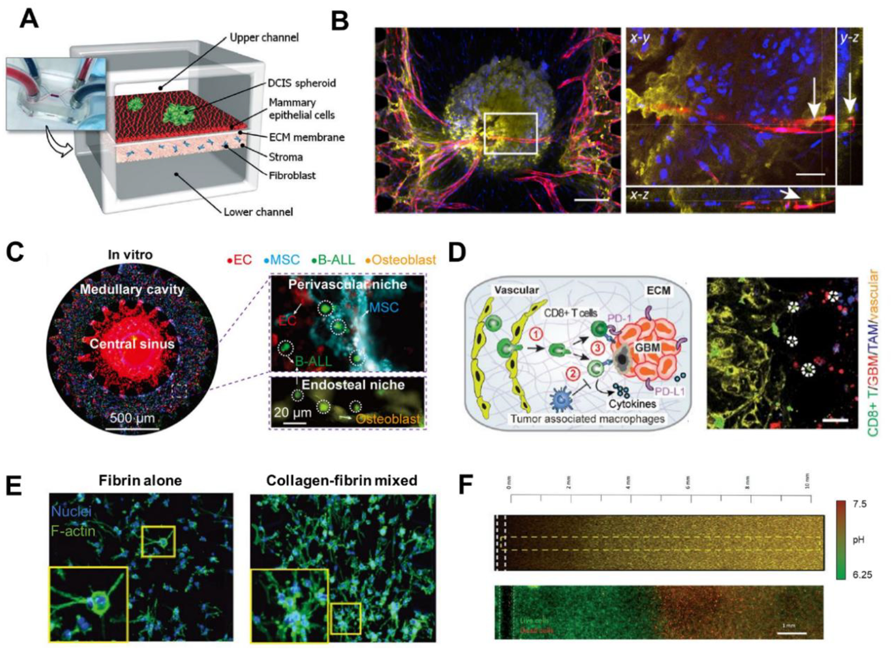
(A) A human breast-cancer-on-a-chip replicating the microarchitecture of breast ductal carcinoma with integration of tumor spheroids, mammary ductal epithelial cells, and mammary fibroblasts. Adapted with permission. Copyright 2015, the Royal Society of Chemistry. (Choi et al. 2015). (B) Immunofluorescence images of the tumor spheroid integrated with the blood vessels on a vascularized TOC. Lumen structure of vasculatures was observed, indicating the perfusability of vascular network. Adapted with permission. Copyright 2020, Elsevier. (Nashimoto et al. 2020). (C) Whole scan of the leukemic BM niche system. Compartments on chip resembled the in vivo counterparts like central sinus and medullary cavity with niche cells. Adapted under the terms of the CC-BY-NC license. Copyright 2020, the Authors. (Ma et al. 2020) (D) Immunosuppressive TME on GBM-on-a-chip. The reconstituted GBM TME recapitulated the infiltration of T cells and interactions among T cells, TAMs, and GBM tumor cells. Adapted under the terms of the CC-BY license. Copyright 2020, the Authors. (Cui et al. 2020) (E) Various components of ECM loaded on TOC revealed the distinct phenotypic changes of cancer cells in response to different ECM conditions. Adapted with permission. Copyright 2017, Wiley. (Chung et al. 2017). (F) pH gradient and necrotic region were formed on TOC under metabolic starvation gradients. Adapted with permission. Copyright 2019, the Royal Society of Chemistry. (Ayuso et al. 2019).