Abstract
Salt bridge (SB, double-charge-assisted hydrogen bonds) formation is one of the strongest molecular non-covalent interactions in biological systems, including ligand–receptor complexes. In the case of G-protein-coupled receptors, such an interaction is formed by the conserved aspartic acid (D3.32) residue and the basic moiety of the aminergic ligand. This study aims to determine the influence of the substitution pattern at the basic nitrogen atom and the geometry of the amine moiety at position 4 of 1H-pyrrolo[3,2-c]quinoline on the quality of the salt bridge formed in the 5-HT6 receptor and D3 receptor. To reach this goal, we synthetized and biologically evaluated a new series of 1H-pyrrolo[3,2-c]quinoline derivatives modified with various amines. The selected compounds displayed a significantly higher 5-HT6R affinity and more potent 5-HT6R antagonist properties when compared with the previously identified compound PZ-1643, a dual-acting 5-HT6R/D3R antagonist; nevertheless, the proposed modifications did not improve the activity at D3R. As demonstrated by the in silico experiments, including molecular dynamics simulations, the applied structural modifications were highly beneficial for the formation and quality of the SB formation at the 5-HT6R binding site; however, they are unfavorable for such interactions at D3R.
Keywords: 5-HT6R antagonists, D3R ligands, dual-acting compounds, molecular dynamics, salt bridge formation
1. Introduction
Serotonin type 6 receptor (5-HT6R) belongs to the family of G-protein-coupled receptors (GPCRs), which has emerged as a promising target for the treatment of cognitive decline associated with neurodegenerative (e.g., Alzheimer’s disease and Parkinson’s disease) and psychiatric disorders (e.g., depression and schizophrenia).
Apart from coupling to the Gs protein, 5-HT6R participates in other signaling pathways [1], including the mechanistic target of rapamycin (mTOR) [2], which accounts for the impact of the receptor in some cognition paradigms in rodents and cyclin-dependent kinase 5 (Cdk5) [3], which is involved in the neurogenesis process. One of the characteristic features of this receptor is its high level of constitutive activity, defined as the spontaneous activity of the receptor in the absence of an agonist [4]. In the hippocampus and the frontal cortex, 5-HT6Rs are localized on the neuronal dendrites and primary neuronal cilia of glutamatergic, GABA-ergic, and cholinergic neurons [5,6]. The ciliary location is of particular interest, as these sensory organelles are implicated in the neurodevelopmental process. The pharmacological blockade of 5-HT6R enhances cholinergic and glutamatergic neurotransmission, indicating that this mechanism is engaged in the improvement of cognitive functions displayed by 5-HT6R antagonists in animal models [7,8].
Recently, the development of dual-acting agents, which not only could relieve cognitive decline but may also produce antidepressant and anxiolytic effects [9] and antipsychotic [10] and neuroprotective properties [11], has gained considerable attention. A number of compounds combine antagonism at the 5-HT6R with serotonin type 2A receptor (5-HT2AR) antagonism [12,13], serotonin type 3 receptor (5-HT3R) antagonism [10], serotonin type 4 receptor (5-HT4R) agonism [14,15], GABA-A agonism [16] acetylcholinesterase inhibition [17], or monoaminoxidase type B (MAO-B) inhibition [18]
Simultaneous blockade of the dopamine D3 receptor (D3R) is one of the promising strategies in the elaboration of 5-HT6R antagonism-based dual-acting compounds for improved treatment of Alzheimer’s disease and other neurodegenerative disorders [19,20]. D3R is a GPCR localized in the limbic areas of the brain [21]. In addition to coupling to the Gi/0 protein, it additionally engages the Cdk5 [22] and mTOR [23] pathways, leading to the enhancement of acetylcholine and glutamate signaling [19,24].
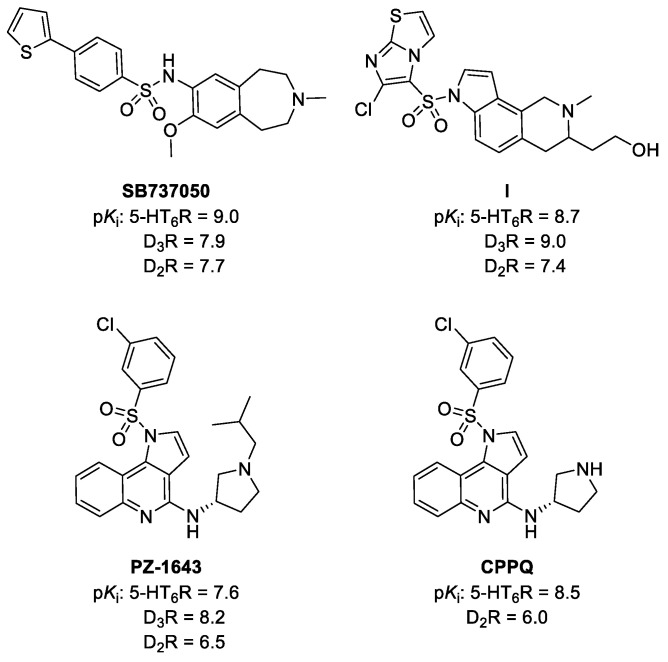
A quest for dual-acting 5-HT6/D3Rs antagonists has been initiated by the identification of compound SB737050 (Figure 1); however, it was burdened with a relatively high affinity for D2Rs [25]. Subsequently, a 2,3,4,7-tetrahydro-1H-pyrrolo[2,3-h]isoquinoline derivative I displaying a more balanced profile for 5-HT6 and D3Rs was described [19].
Figure 1.
Structures of dual-acting 5-HT6/D3Rs antagonists and 5-HT6R antagonist CPPQ.
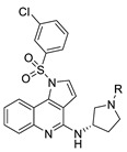
Recently disclosed compound PZ-1643, assigned as derivative 19 in [26], a dual-acting 5-HT6/D3R antagonist, was designed as a merged ligand in which the selective 5-HT6R neutral antagonist CPPQ ((S)-1-((3-chlorophenyl)sulfonyl)-N-(pyrrolidin-3-yl)-1H-pyrrolo[3,2-c]quinolin-4-amine, disclosed as compound 14 in [27], was combined with an alkyl chain, representing a characteristic structural feature of D3R antagonists.
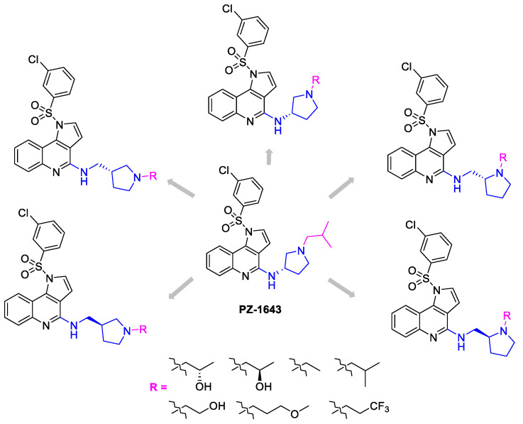
To further investigate the impact of the kind of the substituent at the basic nitrogen atom and the geometry of the amine fragment on 5-HT6R and D3R affinity, we designed a small series of compound PZ-1643 analogs. Structural modifications comprised the introduction of various alkyl-derived chains on the basic nitrogen atom and replacement of (S)-3-amino-1-Boc-pyrrolidine with enantiomers of 2-(aminomethyl)pyrrolidines and 3-(aminomethyl)pyrrolidines (Figure 2). The affinity for both targets was assessed in the binding experiments at 5-HT6R and D3R and was confirmed by molecular dynamics (MD) evaluation, which determined the quality of the salt bridge (SB) formed with D3.32 of 5-HT6R and D3R.
Figure 2.
Structural modifications in the amine fragment of novel 1H-pyrrolo[3,2-c]quinoline derivatives.
2. Results and Discussion
2.1. Chemistry
The key 1H-pyrrolo[3,2-c]quinoline synthon 5 was obtained in a multistep synthesis route, following the previously reported protocol (Scheme 1) [28].
Scheme 1.
Synthetic pathway leading to 1H-pyrrolo[3,2-c]quinoline 5: (i) Formamide, HCOOH, 120 °C, 12 h; (ii) TEA, POCl3, 0 °C, 30 min; (iii) Methyl propiolate, Ag2CO3, dioxane, 80 °C, 30 min; (iv) H2, Pd/C, MeOH, rt, 2 h; (v) AcOH, sec-BuOH, 60 °C, 3 h; (vi) POCl3, 105 °C, 4 h.
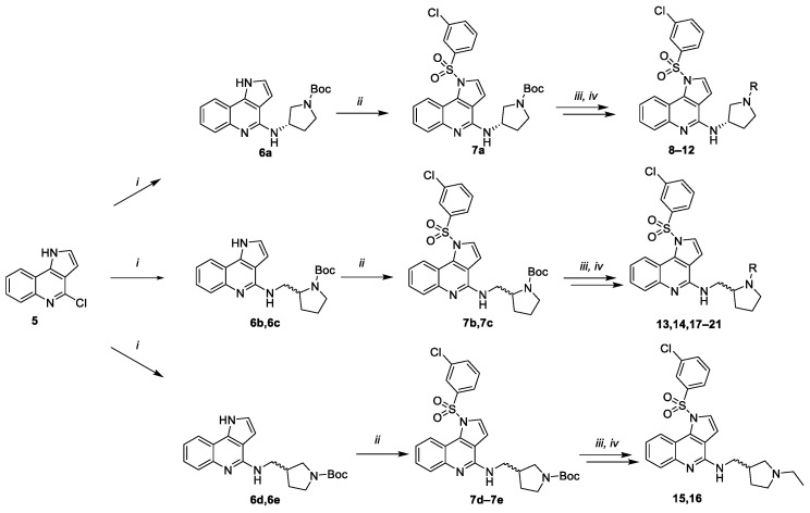
Heating of compound 5 with the excess of respective amine under microwave-assisted conditions yielded Boc-protected amine derivates, 6a–6e, which were further coupled with 3-chlorobenzenesulfonyl chloride in the presence of a phosphazene base yielding sulfonyl derivatives 7a–7e (Scheme 2). Treatment with 1M HCl in methanol afforded secondary amines, for which reductive amination was carried out with respective aldehydes.
Scheme 2.
Synthetic pathway leading to final compounds 8–21: (i) (S)-3-amino-1-Boc-pyrrolidine or (S)-2-(aminomethyl)pyrrolidine or (R)-2-(aminomethyl)pyrrolidine or (S)-3-(aminomethyl)pyrrolidine or (R)-3-(aminomethyl)pyrrolidine, acetonitrile, 140 °C, 7 h MW; (ii) 3-chlorobenzyl sulfonyl chloride, BTPP, DCM, 0 °C-rt; (iii) 1M HCl/MeOH; (iv) aldehyde: (S)-2-hydroxypropanal or (R)-2-hydroxypropanal or glycoaldehyde or 3-methoxypropanal or 3,3,3-trifluoropropanal or acetaldehyde or isobutyraldehyde, NaBH3CN, EtOH, rt, 12 h.
2.2. Determination of Affinity of Compounds for 5-HT6R and D3R and Assessment of the Impact of the Selected Compounds on 5-HT6R-Dependent Gs Signaling
The biological evaluation was initiated by assessing the affinity of the compounds for 5-HT6R in the [3H]-LSD radioligand binding assay. Experiments were performed in a stable HEK293 cell line expressing the human 5-HT6R [10]. The selected compounds, which showed the highest affinity for the serotoninergic target, were further tested for their affinity for D3R in the screening procedure using [3H]-methylspiperone as the radioligand. Experiments were performed in Chinese hamster ovary (CHO) cells with the stable expression of human D3R (Eurofins, Celle-L’Evescault, France) [29].
Antagonist properties of the most active compounds at 5-HT6R were evaluated in cAMP cellular assays, and their impact on cAMP production induced by 5-CT was studied [30]. The experiments were performed in 1321N1 cells expressing the human serotonin 5-HT6R. Finally, the impact of the selected derivatives on agonist-independent 5-HT6R-operated Gs signaling was tested in NG108-15 cells transiently expressing 5-HT6Rs, a cellular model in which 5-HT6R exhibits a high level of constitutive activity [5].
2.3. Structure–Activity Relationship Analysis
Designing dual-acting 5-HT6/D3R ligands in a group of 1H-pyrrolo[3,2-c]quinolines revealed that the introduction of an isobutyl chain on the nitrogen atom of pyrrolidine present in compound PZ-1643 maintained the high affinity for 5-HT6R and was beneficial for the affinity for D3R [26]. The analysis of molecular dynamics (MD) of the pairs of R and S enantiomers indicated that the S counterpart showed beneficial parameters for the distance and angle of the SB in 5-HT6R.
In the present study, initial efforts comprised the replacement of the isobutyl chain of the lead compound PZ-1643 (Figure 2) [26], with more polar substituents (Table 1). Encouragingly, the introduction of 2-hydroxyprop-1-yl enantiomers significantly increased the affinity for 5-HT6R (8, 9 vs. PZ-1643). Additionally, a preference for the R enantiomer was observed. The introduction of the 2-hydroxyethyl moiety was unfavorable for the interaction with 5-HT6R and decreased the affinity by threefold (10 vs. PZ-1643). Replacement of the hydroxyl group of 10 with a more hydrophobic trifluoromethyl substituent further decreased the 5-HT6R affinity (12 vs. 10). However, the introduction of 3-methoxyprop-1-yl was well tolerated (11). Being the most active compound from the evaluated series, 9 displayed a moderate affinity for D3R in the screening procedure.
Table 1.
Binding data of compounds 8−12 and reference compound PZ-1643 for 5-HT6 and D3 receptors.
| |||
| Compd | R |
Ki 5-HT6R
[nM] a |
%inh Binding
to D3R @1μM b |
| PZ-1643 |

|
27 c | 98% (7 nM) c |
| 8 |

|
11 | – |
| 9 |

|
6 | 54% |
| 10 |

|
84 | – |
| 11 |

|
17 | 25% |
| 12 |

|
556 | – |
a Mean Ki values based on at least three independent binding experiments (SEM ≤ 18%) performed in HEK293 cells with stable expression of human 5-HT6R. b Percentage displacement values at 10−6 M; performed at Eurofins in CHO cells with stable expression of human D3R. c Data taken from [26].
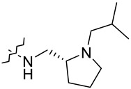
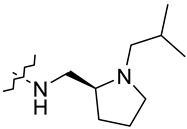
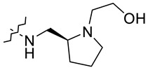
Further studies focusing on the modifications of the geometry of the amine fragment (Table 2) revealed that R enantiomers were preferred for binding to 5-HT6R (13 vs. 14, 15 vs. 16, 17 vs. 18, 19 vs. 20).
Table 2.
Binding data of compounds 13−21 for 5-HT6 and D3 receptors.

| |||
| Compd | R1 |
K
i
5-HT6R
[nM] a |
%inh Binding
to D3R @1μM b |
| 13 |

|
27 | 58% |
| 14 |

|
72 | – |
| 15 |

|
38 | – |
| 16 |

|
64 | – |
| 17 |
|
27 | 57% |
| 18 |
|
65 | – |
| 19 |

|
58 | – |
| 20 |
|
77 | – |
| 21 |

|
87 | – |
a Mean Ki values based on at least three independent binding experiments (SEM ≤ 18%) performed in HEK293 cells with stable expression of human 5-HT6R. b Percentage displacement values at 10−6 M; performed at Eurofins in CHO cells with stable expression of human D3R.
Our recent studies revealed that the introduction of an alkyl substituent on the basic nitrogen atom could be beneficial for dual 5-HT6R/D3R activity. In the evaluated series the ethyl chain on the basic center of (R)-2-(aminomethyl)pyrrolidinyl maintained the affinity for 5-HT6R when compared with PZ-1643 (13 vs. PZ-1643). The same modification applied in the 3-(aminomethyl)pyrrolidinyl derivatives decreased the affinity for 5-HT6R (15, 16 vs. PZ-1643) and indicated the preference of the (R)-2-(aminomethyl)-congener in the interaction with a serotoninergic target. Therefore, 2-(aminomethyl)pyrrolidinyl was selected as the amine fragment for further diversification and evaluation for 5-HT6R and D3R.
Further structural diversification at the basic nitrogen atom, which involved the replacement of ethyl with a more bulky isobutyl substituent in the R series of 2-(aminomethyl)pyrrolidine (17), maintained the affinity for 5-HT6R compared with compound PZ-1643. Subsequent modification, which comprised the introduction of 2-hydroxyethyl and 3-methoxyprop-1-yl, was unfavorable for the interaction with 5-HT6R (19, 20, 21 vs. PZ-1643).
Most active compounds of the evaluated series were functionalized with (R)-2-(aminomethyl)pyrrolidine at position 4 of the 1H-pyrrolo[3,2-c]quinoline core. Among them, derivatives containing an amine fragment substituted with ethyl (13) or isobutyl chains (17) displayed moderate affinity for D3R. Therefore, the applied structural modifications were favorable for the 5-HT6R affinity compared with PZ-1643; however, they did not improve the affinity for D3R.
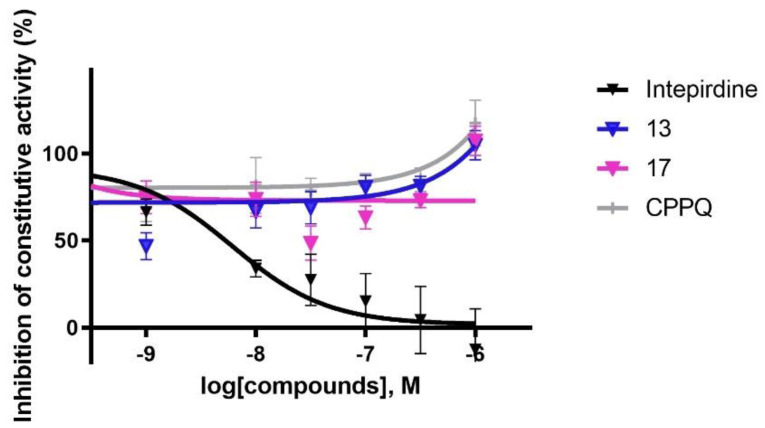
Taking into account the high affinity of 13 and 17 for 5-HT6R and the most potent affinity for D3Rs among the evaluated compounds, these derivatives were further tested for their antagonist properties at 5-HT6R in cellular assays performed in recombinant 1321N1 cells expressing the human serotonin 5-HT6R. The results of these assays were in line with those obtained from the binding experiments, since the evaluated compounds displayed nanomolar antagonist properties at 5-HT6R (13, Kb = 1.2 nM; 17, Kb = 3.8 nM). Further evaluation of the impact of the selected derivatives on 5-HT6R-operated Gs signaling revealed their neutral antagonist properties in this pathway (Figure 3) [10].
Figure 3.
Influence of 13, 17, CPPQ, and intepirdine on 5-HT6R constitutive activity at Gs signaling in NG108-15 cells.
2.4. In Silico Studies
To further investigate the influence of the kind of the substituent at the basic nitrogen atom and geometry of the amine fragment on the SB parameters (i.e., distance and angle) and receptor affinity, the molecular mechanism of action of selected structurally diverse compounds 9, 12, 13, and PZ-1643 was evaluated by flexible molecular docking (IFD procedure) to 5-HT6R (PDB ID: 7XTB) and D3R (PDB ID: 3PBL). The binding modes were coherent with our previously reported results [26] but did not show clear explanation of the structure–activity relationships (Figure S1). Therefore, a series of MD simulations was performed to determine the features responsible for changes in the potency.
A closer inspection of the MD trajectories showed that various alkyl chains on the nitrogen atom penetrated the narrow hydrophobic subpocket formed by transmembrane (TM) helixes 2, 3, and 7 in both receptors. As we proposed in our previous study, the higher binding activity of different derivatives as well as enantiomers originated from the quality of the SB formed with D3.32 [26,31,32]. Thus, the geometric parameters for the interaction with D3.32 observed during molecular dynamics simulations were analyzed (Table 3).
Table 3.
The mean geometric parameters of the salt bridge (SB) between basic nitrogen and carboxylic group of D3.32, observed during the molecular dynamics simulations for compounds 9, 12, 13, and PZ-1643.
| Compound | D3R | Freq. (%) | 5-HT6R | Freq. (%) | ||||||
|---|---|---|---|---|---|---|---|---|---|---|
| SB angle (o) | SB distance (Å) | SB angle (o) | SB distance (Å) | |||||||
| N-H···O= | N-H···O¯ | N···O= | N···O¯ | N-H···O= | N-H···O¯ | N···O= | N···O¯ | |||
| PZ-1643 | 145.7 | 146.9 | 3.8 | 3.8 | 84 | 149.8 | 144.1 | 3.5 | 3.9 | 98 |
| 9 | 135.8 | 129.6 | 5.2 | 4.1 | 30 | 131.2 | 145.7 | 3.8 | 3.4 | 89 |
| 12 | 142.2 | 143.0 | 4.8 | 5.8 | 0 | 139.6 | 129.9 | 5.3 | 3.6 | 77 |
| 13 | 145.0 | 145.5 | 4.2 | 4.3 | 44 | 155.9 | 161.5 | 3.8 | 3.2 | 98 |
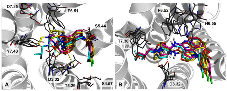
Regarding the interaction with 5-HT6R, compounds PZ-1643, 9, and 13 displayed the most favorable mean geometric parameters of the SB with D3.32 (i.e., both the distance and the angle of the SB lie in the favorable area of the interaction energy) [33]. In addition, 9 showed a hydrogen bond of the -OH group with T3.29. Molecular dynamics results further showed that the SB is a highly stable interaction, with a frequency of occurrence of more than 89% in complexes (Table 3). However, compound 12, bearing a 3,3,3-trifluoropropyl chain, showed an acceptable geometry of an SB, but in this case, a part of the salt bridge geometry might be distorted by the unfavorable polar interactions formed by the –CF3 group and the side chain of Y7.43 and T3.29 (the most distorted side chain among all of Y7.43; Figure 4A). In addition, significant stabilization of all derivatives by the hydrogen bond between S5.44 and the oxygen of sulfonamide groups and the halogen bond formed between the chlorine substituent and the carbonyl oxygen of S4.57 were noted. These interactions are not depicted in Figure 4, since their contribution to the MD trajectory, depending on the derivative, was between 20–40%.
Figure 4.
Superposition of the binding modes of 9 (magenta), 12 (orange), 13 (yellow), and PZ-1643 (cyan) in the 5-HT6 (A) and D3R (B) binding sites. Complexes were selected as the most populated conformations obtained by the clustering of the MD trajectories.
In the case of D3R, only PZ-1643 displayed the favorable geometric parameters of the SB. As revealed by MD simulations, the introduction of a 2-hydroxypropyl or 3,3,3-trifluoropropyl chain on the basic nitrogen atom led to the higher distortion during molecular dynamics, and thus less stable complexes (frequencies of the SB occurrence were substantially lower than for PZ-1643; Table 3). The conformations of the side chains of the respective amino acids in the D3R binding side were more distorted than in 5-HT6R.
3. Experimental Methods
3.1. Chemistry
General Method
The synthesis was conducted at room temperature, unless indicated otherwise. Organic solvents (from Chempur, Piekary Śląskie, Poland) were of reagent grade and were used without purification. All reagents (Sigma-Aldrich (Saint Louis, MI, USA), Fluorochem (Glossop, UK) and TCI (Zwijndrecht, Belgium) were of the highest purity. Column chromatography was performed on silica gel Merck 60 (70–230 mesh ASTM, Darmstadt, Germany).
UPLC and MS were carried out on a system consisting of a Waters Acquity UPLC coupled((Waters Corporation, Milford, MA, USA) to a Waters TQD mass spectrometer. All the analyses were carried out using an Acquity UPLC BEH C18 100 × 2.1 mm2 column at 40 °C. A flow rate of 0.3 mL/min and a gradient of (0−100) % B over 10 min was used: eluent A, water/0.1% HCOOH and eluent B, acetonitrile/0.1% HCOOH. Retention times, tR, were given in minutes. The UPLC/MS purity of all the test compounds and key intermediates was determined to be >95%.
The 1H NMR and 13C NMR spectra were recorded using JEOL JNM-ECZR 500 RS1 (ECZR version) at 500 and 126 MHz, respectively, as well as Bruker Advance III HD at 400 and 100 MHz, respectively. Chemical shifts are reported in parts per million using deuterated solvent for calibration (CD3OD). The J values are given in Hertz (Hz).
Compounds 1–5, 6a, and 7 were obtained according to the previously reported procedure and the analytical data are in accordance with the literature [26,28]. Compounds 13–21 were converted to hydrochloride salts.
(S)-1-((S)-3-((1-((3-chlorophenyl)sulfonyl)-1H-pyrrolo[3,2-c]quinolin-4-yl)amino)pyrrolidin-1-yl)propan-2-ol 8
Pale oil, 53% yield, tR = 3.84, C24H25ClN4O3S, MW 485.00, 1H NMR (400 MHz, CD3OD) δ (ppm) 1.17 (d, J = 6.1, 3H), 1.81–1.96 (m, 1H), 2.35–2.50 (m, 2H), 2.52–2.58 (m, 2H), 2.75–2.88 (m, 1H), 2.90–3.09 (m, 2H), 3.26–3.37 (m, 2H), 3.86–3.99 (m, 1H), 4.77–4.85 (m, 1H), 7.12–7.21 (m, 2H), 7.36–7.47 (m, 2H), 7.51–7.59 (m, 1H), 7.60–7.66 (m, 1H), 7.67–7.73 (m, 1H), 7.75–7.79 (s, 1H), 7.86–7.97 (d, J = 3.4 Hz, 1H), 8.71 (d, J = 8.3 Hz, 1H); 13C NMR (100 MHz, CD3OD) δ (ppm) 20.50, 31.44, 49.63, 53.61, 60.66, 63.50, 65.15, 105.92, 114.34, 116.12, 121.49, 122.77, 125.00, 126.5, 127.5, 128.23, 131.01, 134.26, 135.18, 139.60, 146.47, 151.22. Monoisotopic mass: 484.13, [M + H]+ = 485.1.
(R)-1-((S)-3-((1-((3-chlorophenyl)sulfonyl)-1H-pyrrolo[3,2-c]quinolin-4-yl)amino)pyrrolidin-1-yl)propan-2-ol 9
Pale oil, 57% yield, tR = 3.85, C24H25ClN4O3S, MW 485.00, 1H NMR (400 MHz, CD3OD) δ (ppm) 1.18 (d, J = 6.26 Hz, 3H), 1.81–1.94 (m, 1H), 2.36–2.48 (m, 2H), 2.48–2.64 (m, 2H), 2.79 (dd, J = 9.98, 3.72 Hz, 1H), 2.92–3.09 (m, 2H), 3.33 (dt, J = 3.28, 1.59 Hz, 1H), 3.88–3.99 (m, 1 H), 4.80–4.85 (m, 1H), 7.14–7.20 (m, 2H), 7.35–7.44 (m, 2H), 7.54 (d, J = 8.02 Hz, 1H), 7.60–7.65 (m, 1H), 7.68–7.73 (m, 1H), 7.75–7.80 (m, 1H), 7.92 (d, J = 3.72 Hz, 1H), 8.68 –8.76 (m, 1H), 8.75 (d, J = 8.3 Hz, 1H); 13C NMR (100 MHz, CD3OD) δ (ppm) 20.56, 31.51, 49.56, 52.89, 61.25, 63.46, 65.13, 105.96, 114.34, 116.15, 121.47, 122.77, 125.00, 126.48, 127.54, 128.21, 131.00, 134.25, 135.17, 135.28, 139.58, 146.50, 151.25. Monoisotopic mass: 484.13, [M + H]+ = 485.1.
(S)-2-(3-((1-((3-chlorophenyl)sulfonyl)-1H-pyrrolo[3,2-c]quinolin-4-yl)amino)pyrrolidin-1-yl)ethan-1-ol 10
Pale oil, 45% yield, tR = 3.86 min, C23H23ClN4O3S, MW 470.97, 1H NMR (400 MHz, CD3OD) δ (ppm) 2.23–2.38 (m, 1H), 2.52–2.62 (m, 4H), 3.27–3.37 (m, 2H), 3.72–3.89 (m, 4H), 7.19 (t, J = 7.73 Hz, 1H), 7.22–7.28 (m, 1H), 7.37 (q, J = 8.15 Hz, 2H), 7.51 (dd, J = 8.22, 0.98 Hz, 1H), 7.60 (d, J = 8.02 Hz, 1H), 7.70 (s, 1H), 7.81–7.91 (m, 2H), 8.64 (d, J = 8.41 Hz, 1H); 13C NMR (100 MHz, CD3OD) 29.06, 39.05, 49.95, 53.38, 56.33, 58.53, 106.28, 114.01, 123.05, 125.25, 126.56, 128.37, 129.06, 131.20, 134.61, 134.61, 135.36, 139.29, 149.81. Monoisotopic mass: 470.12, [M + H]+ = 471.1.
(S)-1-((3-chlorophenyl)sulfonyl)-N-(1-(3-methoxypropyl)pyrrolidin-3-yl)-1H-pyrrolo[3,2-c]quinolin-4-amine 11
Pale oil, 30% yield, tR = 3.95, C25H27ClN4O3S, MW 499.03, 1H NMR (400 MHz, CD3OD) δ (ppm) 1.76–1.99 (m, 2H), 2.15–2.30 (m, 1H), 2.41–2.58 (m, 1H), 3.03 (s, 2H), 3.11–3.19 (m, 2H), 3.21–3.24 (m, 3H), 3.24–3.29 (m, 1H), 3.41–3.57 (m, 2H), 3.63–3.80 (m, 1H), 4.78–4.83 (m, 1H), 7.02–7.12 (m, 1 H), 7.13–7.21 (m, 1H), 7.36 (dt, J = 10.86, 8.07 Hz, 2H), 7.46–7.54 (m, 1H), 7.54–7.65 (m, 2 H), 7.66–7.70 (m, 1 H), 7.83–7.89 (m, 1H), 8.62–8.72 (m, 1H); 13C NMR (100 MHz, CD3OD) 26.13, 29.22, 49.88, 52.89, 53.64, 57.61, 59.11, 69.62, 105.86, 110.01, 114.48, 116.08, 122.25, 122.96, 125.13, 126.46, 127.86, 128.63, 131.08, 134.39, 135.24, 139.48, 150.52. Monoisotopic mass 498.15, [M + H]+ = 499.1.
(S)-1-((3-chlorophenyl)sulfonyl)-N-(1-(3,3,3-trifluoropropyl)pyrrolidin-3-yl)-1H-pyrrolo[3,2-c]quinolin-4-amine 12
Pale oil, 30% yield, tR = 4.05, C24H22ClF3N4O2S, MW 522.97, 1H NMR (400 MHz, CD3OD) δ (ppm) 2.10 (dd, J = 12.89, 5.15 Hz, 1 H), 2.45–2.55 (m, 1H), 2.54–2.54 (m, 1H), 2.56–2.65 (m, 2H), 2.90–2.99 (m, 1H), 3.06–3.13 (m, 1H), 3.15–3.22 (m, 2H), 3.37 (ddd, J = 10.02, 8.45, 5.30 Hz, 1H), 4.76–4.83 (m, 1H), 7.15–7.22 (m, 2H), 7.35–7.43 (m, 2H), 7.51 (ddd, J = 8.16, 2.00, 1.00 Hz, 1H), 7.58–7.62 (m, 1H), 7.64 (dd, J = 8.31, 0.86 Hz, 1H), 7.72 (t, J = 1.86 Hz, 1H), 7.89 (d, J = 3.72 Hz, 1H), 8.68 (dd, J = 8.59, 1.15 Hz, 1H); 13C NMR (100 MHz, CD3OD) 21.64, 30.28, 50.03, 53.00, 59.36, 106.17, 114.24, 116.03, 122.34, 122.98, 125.16, 126.47, 128.66, 131.22, 134.49, 135.27, 139.37, 144.72, 150.53, 175.74, 177.46. Monoisotopic mass 522.11, [M + H]+ = 523.1.
(R)-1-((3-chlorophenyl)sulfonyl)-N-((1-ethylpyrrolidin-2-yl)methyl)-1H-pyrrolo[3,2-c]quinolin-4-amine hydrochloride 13
White solid, 47% yield, tR = 4.07, C24H26Cl2N4O2S, MW 505.46, 1H NMR (400 MHz, CD3OD) δ (ppm) 1.09 (t, J = 7.24 Hz, 3H), 1.69 (s, 3H), 1.82–1.98 (m, 1H), 2.36 (d, J = 1.56 Hz, 1H), 2.48 (d, J = 1.57 Hz, 1 H), 2.94–3.09 (m, 2H), 3.18 (d, J = 2.54 Hz, 1H), 3.46 (dd, J = 13.69, 5.48 Hz, 1H), 3.80 (dd, J = 13.89, 4.89 Hz, 1H), 7.00–7.11 (m, 2H), 7.21–7.33 (m, 2H), 7.34–7.43 (m, 1H), 7.47–7.56 (m, 2H), 7.64 (s, 1H), 7.76–7.83 (m, 1H), 8.60 (dd, J = 8.41, 0.78 Hz, 1H); 13C NMR (100 MHz, CD3OD) 11.55, 22.29, 28.00, 43.04, 49.18, 53.35, 64.59, 105.95, 114.42, 115.99, 121.63, 122.89, 125.03, 126.27, 128.39, 131.01, 134.28, 135.18, 135.27, 139.54, 146.19, 151.92. Monoisotopic mass 468.14, [M + H]+ = 469.4.
(S)-1-((3-chlorophenyl)sulfonyl)-N-((1-ethylpyrrolidin-2-yl)methyl)-1H-pyrrolo[3,2-c]quinolin-4-amine hydrochloride 14
White solid, 50% yield, tR = 4.08, C24H26Cl2N4O2S, MW 505.46, 1H NMR (500 MHz, CD3OD) δ (ppm) 1.26 (dt, J = 11.0, 5.5 Hz, 3H), 1.25–1.30 (s, 1H), 1.82–1.95 (s, 3H), 2.00–2.15 (s, 1H), 2.60–2.72 (s, 1H), 3.12–3.32 (m, 2H), 3.33–3.44 (s, 1H), 3.55–3.70 (m, 1H), 3.90 (dd, J = 14.0, 5.1 Hz, 1H), 4.53–4.60 (s, 1H), 7.12–7.17 (s, 1H), 7.19 (dd, J = 16.8, 9.0 Hz, 1H), 7.40–7.51 (m, 2H), 7.55–7.70 (m, 3H), 7.77–7.80 (s, 1H), 7.95 (d, J = 2.6 Hz, 1H), 8.75 (d, J = 8.5 Hz, 1H); 13C NMR (126 MHz, CD3OD) 11.57, 22.31, 28.02, 43.07, 49.22, 53.34, 64.62, 105.96, 114.42, 116.00, 121.66, 122.89, 125.05, 126.29, 128.41, 131.02, 134.30, 135.21, 135.30, 139.56, 146.21, 151.94. Monoisotopic mass 468.14, [M + H]+ = 469.4.
(R)-1-((3-chlorophenyl)sulfonyl)-N-((1-ethylpyrrolidin-3-yl)methyl)-1H-pyrrolo[3,2-c]quinolin-4-amine hydrochloride 15
White solid, 52% yield, tR = 4.09, C24H26Cl2N4O2S, MW 505.46, 1H NMR (500 MHz, CD3OD) δ (ppm) 1.22–1.29 (m, 1H), 1.31–1.41 (m, 3H), 1.85–2.12 (m, 1H), 2.46 (dd, J = 6.73, 5.30 Hz, 1H), 2.90–3.05 (m, 1H), 3.06–3.19 (m, 1H), 3.63 (dd, J = 5.87, 3.87 Hz, 1H), 3.66–3.74 (m, 1H), 3.74–3.83 (m, 1H), 3.83–3.90 (m, 1H), 3.83–3.90 (m, 1H), 3.91–4.04 (m, 1H), 3.92–3.95 (m, 1H), 7.45–7.51 (m, 1H), 7.51–7.57 (m, 2H), 7.62–7.71 (m, 2H), 7.83 (dd, J = 8.02, 1.15 Hz, 1H), 7.94 (t, J = 2.00 Hz, 1H), 8.17 (d, J = 3.72 Hz, 2H), 8.79–8.87 (m, 1H); 13C NMR (126 MHz, CD3OD) δ (ppm) 40.57, 44.46, 50.21, 56.29, 106.54, 115.09, 123.99, 125.68, 126.92, 130.12, 130.60, 131.56, 135.29, 138.78. Monoisotopic mass 468.14, [M + H]+ = 469.4.
(S)-1-((3-chlorophenyl)sulfonyl)-N-((1-ethylpyrrolidin-3-yl)methyl)-1H-pyrrolo[3,2-c]quinolin-4-amine hydrochloride 16
White solid, 52% yield, tR = 4.10, C24H26Cl2N4O2S, MW 505.46, 1H NMR (500 MHz, CD3OD) δ (ppm) 1.22–1.30 (m, 1H), 1.30–1.31 (m, 1H), 1.32–1.41 (m, 3H), 1.34–1.35 (m, 1H), 1.85–2.10 (m, 1H), 2.28–2.52 (m, 1H), 2.52–2.54 (m, 1H), 2.53–2.53 (m, 1H), 2.90–3.05 (m, 1H), 3.05–3.18 (m, 1H), 3.57–3.66 (m, 1H), 3.71 (dd, J = 5.30, 0.72 Hz, 1H), 3.75–3.87 (m, 1H), 3.76–3.86 (m, 1H), 3.87–4.02 (m, 2H), 7.46–7.52 (m, 1H), 7.52–7.56 (m, 2H), 7.62–7.71 (m, 2H), 7.80–7.86 (m, 1H), 7.94 (t, J = 1.86 Hz, 1H), 8.17 (d, J = 3.72 Hz, 2H), 8.10–8.16 (m, 1H), 8.83 (d, J = 8.59 Hz, 1H); 13C NMR (126 MHz, CD3OD) δ (ppm) 40.42, 44.90, 50.19, 56.30, 106.48, 113.13, 115.09, 119.07, 124.00, 126.92, 130.11, 130.60, 131.55, 135.29, 135.76, 138.78. Monoisotopic mass 468.14, [M + H]+ = 469.4.
(R)-1-((3-chlorophenyl)sulfonyl)-N-((1-isobutylpyrrolidin-2-yl)methyl)-1H-pyrrolo[3,2-c]quinolin-4-amine hydrochloride 17
White solid, 34% yield, tR = 4.43, C26H30Cl2N4O2S, MW 533.51, 1H NMR (500 MHz, CD3OD) δ (ppm) 0.95–1.07 (m, 6H), 1.23–1.30 (m, 1H), 1.26–1.30 (m, 1H), 1.98–2.12 (m, 2H), 2.12–2.13 (m, 1H), 2.12–2.22 (m, 2H), 2.42 (dd, J = 12.74, 6.44 Hz, 1H), 3.03–3.15 (m, 1H), 3.79–3.89 (m, 1H), 4.00–4.11 (m, 1H), 4.12–4.23 (m, 1H), 4.34–4.47 (m, 1H), 7.45–7.57 (m, 2H), 7.61–7.70 (m, 3H), 7.83 (d, J = 7.73 Hz, 1H), 7.93 (s, 1H), 8.17 (d, J = 3.72 Hz, 2H), 8.86 (d, J = 8.02 Hz, 1H); 13C NMR (126 MHz, CD3OD) δ (ppm) 19.83, 22.03, 25.51, 27.09, 29.43, 42.61, 54.79, 63.49, 106.83, 115.20, 123.94, 125.68, 126.89, 130.62, 131.57, 135.27, 135.72, 138.77. Monoisotopic mass 496.17, [M + H]+ = 497.4.
(S)-1-((3-chlorophenyl)sulfonyl)-N-((1-isobutylpyrrolidin-2-yl)methyl)-1H-pyrrolo[3,2-c]quinolin-4-amine hydrochloride 18
White solid, 34% yield, tR = 4.41, C26H30Cl2N4O2S, MW 533.51, 1H NMR (500 MHz, CD3OD) δ (ppm) 0.95–1.06 (m, 6H), 1.24–1.31 (m, 1H), 1.96–2.09 (m, 2H), 1.96–2.10 (m, 1H), 2.08–2.22 (m, 2H), 2.09–2.23 (m, 1–H), 2.38 (dd, J = 12.74, 6.44 Hz, 1H), 3.09 (dd, J = 12.89, 6.01 Hz, 1H), 3.74–3.86 (m, 1H), 3.91–4.00 (m, 1 H), 4.03–4.14 (m, 1H), 4.21–4.36 (m, 1H), 7.45–7.56 (m, 3H), 7.67 (d, J = 7.73 Hz, 2H), 7.83 (d, J = 8.02 Hz, 1H), 7.92 (s, 1 H), 8.18 (d, J = 3.72 Hz, 2H), 8.88 (d, J = 8.31 Hz, 1 H); 13C NMR (126 MHz, CD3OD) δ (ppm) 19.81, 22.02, 25.54, 27.12, 29.46, 42.63, 54.82, 63.52, 106.81, 115.22, 123.97, 125.69, 126.90, 130.64, 131.59, 135.29, 135.75, 138.80. Monoisotopic mass 496.17, [M + H]+ = 497.4.
(R)-2-(2-(((1-((3-chlorophenyl)sulfonyl)-1H-pyrrolo[3,2-c]quinolin-4 yl)amino)methyl)-pyrrolidin-1-yl)ethan-1-ol hydrochloride 19
White solid, 60% yield, tR = 3.86, C24H26Cl2N4O3S, MW 521.46, 1H NMR (500 MHz, CD3OD) δ (ppm) 1.78–1.94 (m, 3H), 1.97–2.15 (s, 1H), 2.60–2.76 (s, 1H), 2.80–2.95 (s, 1H), 3.17–3.30 (m, 1H), 3.35–3.47 (m, 2H), 3.59–3.74 (m, 1H), 3.80–3.86 (m, 2H), 3.87–3.92 (m, 1H), 4.50–4.74 (s, 1H), 7.18 (q, J = 6.2 Hz, 2H), 7.41 (t, J = 7.9 Hz, 2H), 7.56 (d, J = 8.0 Hz, 1H), 7.64 (d, J = 7.9 Hz, 1H), 7.72–7.76 (m, 1H), 7.77–7.80 (s, 1H), 7.92 (d, J = 3.7 Hz, 1H), 8.70 (d, J = 8.4 Hz, 1H); 13C NMR (126 MHz, CD3OD) δ (ppm) 22.53, 29.01, 43.12, 49.99, 52.28, 54.67, 56.29, 106.88, 114.12, 124.75, 125.87, 127.02, 130.12, 130.76, 131.63, 135.31, 139.79. Monoisotopic mass 484.13, [M + H]+ = 485.5.
(S)-2-(2-(((1-((3-chlorophenyl)sulfonyl)-1H-pyrrolo[3,2-c]quinolin-4 yl)amino)methyl)pyrrolidin-1-yl)ethan-1-ol hydrochloride 20
White solid, 60% yield, tR = 3.83, C24H26Cl2N4O3S, MW 521.46, 1H NMR (500 MHz, CD3OD) δ (ppm) 1.77–1.95 (m, 3H), 1.99–2.16 (s, 1H), 2.61–2.75 (s, 1H), 2.81–2.94 (s, 1H), 3.15–3.31 (m, 1H), 3.32–3.45 (m, 2H), 3.59–3.75 (m, 1H), 3.81–3.88 (m, 2H), 3.89–3.91 (m, 1H), 4.51–4.75 (s, 1H), 7.19 (q, J = 6.2 Hz, 2H), 7.42 (t, J = 7.9 Hz, 2H), 7.55 (d, J = 8.0 Hz, 1H), 7.65 (d, J = 7.9 Hz, 1H), 7.73–7.77 (m, 1H), 7.79–7.81 (s, 1H), 7.95 (d, J = 3.7 Hz, 1H), 8.73 (d, J = 8.4 Hz, 1H); 13C NMR (126 MHz, CD3OD) δ (ppm) 22.51, 28.98, 42.13, 49.89, 52.31, 54.87, 56.53, 106.70, 114.15, 124.83, 125.92, 127.01, 130.31, 130.82, 131.61, 135.77, 139.82. Monoisotopic mass 484.13, [M + H]+ = 485.5.
(R)-1-((3-chlorophenyl)sulfonyl)-N-((1-(3-methoxypropyl)pyrrolidin-2-yl)methyl)-1H-pyrrolo[3,2-c]quinolin-4-amine 21
White solid, 38% yield, tR = 4.25, C26H30Cl2N4O3S, MW 549.51, 1H NMR (500 MHz, CD3OD) δ (ppm) 1.80–1.98 (m, 5H), 2.05–2.18 (m, 1H), 2.58–2.73 (s, 1H), 2.73–2.90 (s, 1H), 3.16–3.20 (s, 3H), 3.20–3.27 (m, 1H), 3.33–3.47 (m, 4H), 3.48–3.59 (m, 1H), 3.60–3.79 (m, 1H), 3.91 (dd, J = 14.2, 5.5 Hz, 1H), 7.16–7.26 (m, 2H), 7.46 (t, J = 8.0 Hz, 2H), 7.56–7.62 (m, 1H), 7.68 (d, J = 8.2 Hz, 2H), 7.81 (t, J = 1.9 Hz, 1H), 7.97 (d, J = 3.7 Hz, 1H), 8.76 (d, J = 8.4 Hz, 1H); 13C NMR (126 MHz, CD3OD) δ (ppm) 22.95, 27.22, 42.79, 53.88, 57.42, 69.30, 101.97, 106.09, 114.61, 115.92, 120.53, 122.26, 123.10, 124.03, 125.22, 126.55, 127.38, 128.72, 129.83, 131.18, 133.80, 134.48, 135.31, 139.54, 152.31. Monoisotopic mass: 512.16, [M + H]+ = 513.4.
3.2. In Silico Evaluation
3.2.1. Structures of the Receptors
The structure of D3R in the complex with antagonist eticlopride (PDB code: 3PBL) and 5-HT6R in the complex with agonist serotonin (PDB code: 7XTB) were retrieved from the Protein Data Bank [34].
3.2.2. Molecular Docking
The 3-dimensional structures of the ligands were prepared using LigPrep v3.6 [35], and the appropriate ionization states at pH=7.4 ± 1.0 were assigned using Epik v3.4 [36,37]. The Protein Preparation Wizard was used to assign the bond orders and appropriate amino acid ionization states and to check for steric clashes. The receptor grid was generated (OPLS4 force field) by centering the grid box with a size of 12 Å on the D3.32 side chain. Automated flexible docking was performed using Glide v6.9 [38,39] at the SP level, and ten poses per ligand were generated. All ligands were docked using the induced fit docking (IFD) [40] protocol with SP with an OPLS4 force field [41]. The L-R complexes selected in the IFD procedure were next used in molecular dynamics simulations.
3.2.3. Molecular Dynamics
A 100 ns long molecular dynamics (MD) simulation was performed using Schrödinger Desmond software [42]. Each ligand–receptor complex was immersed into a POPC (309.5 K) membrane bilayer, the position of which was calculated using the PPM web server (https://opm.phar.umich.edu/ppm_server, accessed 20 May 2022) [43]. The system was solvated by water molecules described by the TIP4P potential and the OPLS4 force field was used for all atoms. An amount of 0.15 M NaCl was added to mimic the ionic strength inside the cell. The output trajectories were hierarchically clustered into 10 groups according to the ligand using the trajectory analysis tool from Schrödinger Suite. Based on obtained trajectories, the mean geometrical parameters of the salt bridge (distance and angle) with D3.32 were calculated using the Simulation Event Analysis tool in Maestro Schrödinger Suite.
3.3. In Vitro Pharmacological Evaluation
3.3.1. The 5-HT6Rs Affinity Evaluation
Cell Culture and Preparation of Cell Membranes for Radioligand Binding Assays
HEK293 cells with stable expression of human 5-HT6 receptors (prepared with the use of Lipofectamine 2000) were maintained at 37 °C in a humidified atmosphere with 5% CO2 and grown in Dulbecco’s modified Eagle medium containing 10% dialyzed fetal bovine serum and 500 μg/mL G418 sulfate. For membrane preparation, cells were sub-cultured in 150 cm2 flasks, grown to 90% confluence, washed twice with phosphate buffered saline (PBS), prewarmed to 37 °C, and pelleted by centrifugation (200× g) in PBS containing 0.1 mM EDTA and 1 mM dithiothreitol. Prior to membrane preparation, pellets were stored at −80 °C.
Radioligand Binding Assays
The cell pellets were thawed and homogenized in 10 volumes of assay buffer using an Ultra Turrax tissue homogenizer(IKA, Warsaw, Poland), centrifuged twice at 35,000× g for 15 min at 4 °C, and incubated for 15 min at 37 °C between centrifugation rounds [10]. The composition of the assay buffers was 50 mM Tris HCl, 0.5 mM EDTA, and 4 mM MgCl2. The assays were incubated in a total volume of 200 μL in 96-well microtiter plates for 1 h at 37 °C. The process of equilibration was terminated by rapid filtration through Unifilter plates with a 96-well cell harvester, and radioactivity retained on the filters was quantified on a Microbeta plate reader (PerkinElmer, Waltham, MA, USA). For displacement studies, the assay samples contained as radioligands (PerkinElmer, USA) 2 nM [3H]-LSD (83.6 Ci/mmol). Nonspecific binding was defined with 10 μM methiothepine. Each compound was tested in triplicate at 7 concentrations (10−10 to 10−4 M). The inhibition constants (Ki) were calculated from the Cheng−Prusoff equation [30]. Results were expressed as means of at least two independent experiments.
3.3.2. Evaluation of Antagonism at Functional Activity on 5-HT6Rs
The functional properties of compounds on 5-HT6R were evaluated using its ability to inhibit cAMP production induced by 5-CT (1000 nM), a 5-HT6R agonist [10]. The compound was tested in triplicate at 8 concentrations (10−11 to 10−4 M). The level of adenylyl cyclase activity was measured using frozen recombinant 1321N1 cells expressing the human serotonin 5-HT6R (PerkinElmer). Total cAMP was measured using the LANCE cAMP detection kit (PerkinElmer), according to the manufacturer’s directions. For quantification of cAMP levels, cells (5 μL) were incubated with a mixture of compounds (5 μL) for 30 min at room temperature in 384-well white opaque microtiter plates. After incubation, the reaction was stopped, and cells were lysed by the addition of 10 μL of working solution (5 μL of Eu-cAMP and 5 μL of ULight-anti-cAMP). The assay plate was incubated for 1 h at room temperature. Time-resolved fluorescence resonance energy transfer (TR-FRET) was detected by an Infinite M1000 Pro (Tecan, Männedorf, Switzerland) using instrument settings from LANCE cAMP detection kit manual(PerkinElmer, Waltham, MA, USA).
3.3.3. Determination of 5-HT6R Constitutive Activity at Gs Signaling
Neuroblastoma cells (NG108-15) were grown in DMEM (Dulbecco’s modified Eagle’s medium) supplemented with 10% heat-inactivated fetal bovine serum, 2% HAT (hypoxanthine/aminopterin/thymidine, Life technologies), glutamine, and antibiotics at 37 °C under 5% of CO2. cAMP measurement was performed in cells transiently transfected with a construct expressing the CAMYEL bioluminescence resonance energy transfer (BRET) sensor for cAMP [44] (3 μg DNA/million cells) alone or in combination with a plasmid encoding the human 5-HT6R (0.5 μg DNA/million cells). Transfection of the NG108-15 cells was conducted in suspension using Lipofectamine 2000, according to the manufacturer’s protocol. Plasmids and lipofectamine were diluted in Opti-MEM Reduced Serum Media (Gibco) and incubated at room temperature for 20 min before being added to the cells. Transfected cells were subsequently plated in white 96-well plates (Greiner) at a density of 50,000 cells per well. Then, 48 h after transfection, cells were washed with PBS containing calcium and magnesium. A triplicate of well was treated with the tested compound diluted in PBS containing calcium and magnesium at concentrations ranging from 0.1 nM to 10 μM. Intepirdine was used as a control for inverse agonist activity. Coelanterazine H (Molecular Probes) was added in each well at a final concentration of 5 μM and incubated at room temperature for 5 min before measuring BRET in a Mithras LB 940 plate reader (Berthold Technologies, Bad Wildbad, Germany). The decrease in CAMYEL BRET induced by the coexpression of the probe with the 5-HT6R as compared to the BRET measured in cells expressing the probe alone was used as an index of the 5-HT6R constitutive activity.
4. Conclusions
To investigate the impact of structural diversification of the amine fragment of the previously reported compound PZ-1643, a dual 5-HT6R/D3R antagonist, the new series of 1H-pyrrolo[3,2-c]quinolines modified at position 4 with various pyrrolidine-derived moieties was evaluated for the affinity for 5-HT6 and D3Rs. The selected compounds displayed a higher affinity and more potent antagonist properties for 5-HT6R than the previously reported lead compound; however, their affinity for D3R was not improved. As observed in the subsequent molecular dynamics simulations, the structural modifications applied, which were favorable for the interaction with 5-HT6R, showed a negative impact on the interactions with D3R. These effects result from the differences in the distance and angles formed between the basic center of the molecule and the respective residues of aspartic acid in the receptor binding sites. These changes in the geometry parameters affected the quality of the formed SB. The outcomes of this study provide structural hints for designing of dual-acting 5-HT6R/D3R antagonists to evaluate a contribution of the combination of 5-HT6R antagonism and D3R antagonism to the neurodegenerative processes.
Acknowledgments
L.K. thanks the Erasmus program for the possibility of international exchange between Faculty of Pharmacy, Jagiellonian University Medical College and Université de Montpellier.
Supplementary Materials
The following supporting information can be downloaded at: https://www.mdpi.com/article/10.3390/molecules28031096/s1, Figure S1: Superposition of the binding modes of compounds 9, 12 and 13; Figures S2–S7: 1H NMR and 13C NMR spectra of representative compounds
Author Contributions
Conceptualization: K.G. and P.Z.; Methodology: W.P. and R.K.; Software: W.P. and R.K.; Investigation: K.G., W.P., L.K., O.B., G.S., X.B., J.G., A.N., S.C.-D. and R.K.; Writing—original draft: K.G.; Writing—review & editing: S.C.-D., R.K. and P.Z.; Supervision: F.L., A.J.B., P.M., S.C.-D., R.K. and P.Z.; Project administration: K.G. and P.Z. All authors have read and agreed to the published version of the manuscript.
Institutional Review Board Statement
Not applicable.
Informed Consent Statement
Not applicable.
Data Availability Statement
The data presented in this study are available in the Supplementary Materials.
Conflicts of Interest
The authors declare no conflict of interest.
Funding Statement
The study was financially supported by the National Science Center, Grant No. DEC-2019/03/X/NZ7/01894, the Priority Research Area qLife under the program “Excellence Initiative Research University” at the Jagiellonian University in Krakow, the Université de Montpellier, Centre National de la Recherche Scientifique (CNRS), the French National Research Agency (“Investissements d’avenir” programme with the reference ANR-16-IDEX-0006).
Footnotes
Disclaimer/Publisher’s Note: The statements, opinions and data contained in all publications are solely those of the individual author(s) and contributor(s) and not of MDPI and/or the editor(s). MDPI and/or the editor(s) disclaim responsibility for any injury to people or property resulting from any ideas, methods, instructions or products referred to in the content.
References
- 1.Chaumont-Dubel S., Dupuy V., Bockaert J., Bécamel C., Marin P. The 5-HT6 receptor interactome: New insight in receptor signaling and its impact on brain physiology and pathologies. Neuropharmacology. 2020;172:107839. doi: 10.1016/j.neuropharm.2019.107839. [DOI] [PubMed] [Google Scholar]
- 2.Meffre J., Chaumont-Dubel S., Mannoury la Cour C., Loiseau F., Watson D.J.G., Dekeyne A., Séveno M., Rivet J.M., Gaven F., Déléris P., et al. 5-HT6 Receptor recruitment of mTOR as a mechanism for perturbed cognition in schizophrenia. EMBO Mol. Med. 2012;4:1043. doi: 10.1002/emmm.201201410. [DOI] [PMC free article] [PubMed] [Google Scholar]
- 3.Duhr F., Déléris P., Raynaud F., Séveno M., Morisset-Lopez S., Mannoury La Cour C., Millan M.J., Bockaert J., Marin P., Chaumont-Dubel S. Cdk5 Induces constitutive activation of 5-HT6 receptors to promote neurite growth. Nat. Chem. Biol. 2014;10:590. doi: 10.1038/nchembio.1547. [DOI] [PubMed] [Google Scholar]
- 4.De Deurwaerdère P., Bharatiya R., Chagraoui A., Di Giovanni G. Constitutive activity of 5-HT receptors: Factual analysis. Neuropharmacology. 2020;168:107967. doi: 10.1016/j.neuropharm.2020.107967. [DOI] [PubMed] [Google Scholar]
- 5.Deraredj Nadim W., Chaumont-Dubel S., Madouri F., Cobret L., De Tauzia M.L., Zajdel P., Benedetti H., Marin P., Morisset-Lopez S. Physical interaction between neurofibromin and serotonin 5-HT6 receptor promotes receptor constitutive activity. Proc. Natl. Acad. Sci. USA. 2016;113:12310. doi: 10.1073/pnas.1600914113. [DOI] [PMC free article] [PubMed] [Google Scholar]
- 6.Lesiak A.J., Brodsky M., Cohenca N., Croicu A.G., Neumaier J.F. Restoration of physiological expression of 5-HT6 receptor into the primary cilia of null mutant neurons lengthens both primary cilia and dendrites. Mol. Pharmacol. 2018;94:731–742. doi: 10.1124/mol.117.111583. [DOI] [PMC free article] [PubMed] [Google Scholar]
- 7.Codony X., Vela J.M., Ramírez M.J. 5-HT6 Receptor and cognition. Curr. Opin. Pharmacol. 2011;11:94. doi: 10.1016/j.coph.2011.01.004. [DOI] [PubMed] [Google Scholar]
- 8.de Jong I.E.M., Mørk A. Antagonism of the 5-HT6 receptor—Preclinical rationale for the treatment of Alzheimer’s disease. Neuropharmacology. 2017;125:50. doi: 10.1016/j.neuropharm.2017.07.010. [DOI] [PubMed] [Google Scholar]
- 9.Partyka A., Jastrzębska-Więsek M., Antkiewicz-Michaluk L., Michaluk J., Wąsik A., Canale V., Zajdel P., Kołaczkowski M., Wesołowska A. Novel antagonists of 5-HT6 and/or 5-HT7 receptors affect the brain monoamines metabolism and enhance the anti-immobility activity of different antidepressants in rats. Behav. Brain Res. 2019;359:9. doi: 10.1016/j.bbr.2018.10.004. [DOI] [PubMed] [Google Scholar]
- 10.Zajdel P., Grychowska K., Mogilski S., Kurczab R., Satała G., Bugno R., Kos T., Gołębiowska J., Malikowska-Racia N., Nikiforuk A., et al. Structure-based design and optimization of FPPQ, a dual-acting 5-HT3 and 5-HT6 receptor antagonist with antipsychotic and procognitive properties. J. Med. Chem. 2021;64:18. doi: 10.1021/acs.jmedchem.1c00224. [DOI] [PMC free article] [PubMed] [Google Scholar]
- 11.Vanda D., Canale V., Chaumont-Dubel S., Kurczab R., Satała G., Koczurkiewicz-Adamczyk P., Krawczyk M., Pietruś W., Blicharz K., Pękala E., et al. Imidazopyridine-based 5-HT6 receptor neutral antagonists: Impact of N1-benzyl and N1-phenylsulfonyl fragments on different receptor conformational states. J. Med. Chem. 2021;64:1180. doi: 10.1021/acs.jmedchem.0c02009. [DOI] [PubMed] [Google Scholar]
- 12.Staroń J., Kurczab R., Warszycki D., Satała G., Krawczyk M., Bugno R., Lenda T., Popik P., Hogendorf A.S., Hogendorf A., et al. Virtual screening-driven discovery of dual 5-HT6/5-HT2A receptor ligands with pro-cognitive properties. Eur. J. Med. Chem. 2020;185:111857. doi: 10.1016/j.ejmech.2019.111857. [DOI] [PubMed] [Google Scholar]
- 13.Kucwaj-Brysz K., Ali W., Kurczab R., Sudoł-Tałaj S., Wilczyńska-Zawal N., Jastrzębska-Więsek M., Satała G., Mordyl B., Żesławska E., Olejarz-Maciej A., et al. An exit beyond the pharmacophore model for 5-HT6R agents—A new strategy to gain dual 5-HT6/5-HT2A action for triazine derivatives with procognitive potential. Bioorg. Chem. 2022;121:105695. doi: 10.1016/j.bioorg.2022.105695. [DOI] [PubMed] [Google Scholar]
- 14.Yahiaoui S., Hamidouche K., Ballandonne C., Davis A., Sopkova de Oliveira Santos J., Freret T., Boulouard M., Rochais C., Dallemagne P. Design, synthesis, and pharmacological evaluation of multitarget-directed ligands with both serotonergic subtype 4 receptor (5-HT4R) partial agonist and 5-HT6R antagonist activities, as potential treatment of Alzheimer’s disease. Eur. J. Med. Chem. 2016;121:283. doi: 10.1016/j.ejmech.2016.05.048. [DOI] [PubMed] [Google Scholar]
- 15.Claeysen S., Bockaert J., Giannoni P. Serotonin: A New Hope in Alzheimer’s Disease? ACS Chem. Neurosci. 2015;6:940. doi: 10.1021/acschemneuro.5b00135. [DOI] [PubMed] [Google Scholar]
- 16.Marcinkowska M., Mordyl B., Fajkis-Zajączkowska N., Siwek A., Karcz T., Gawalska A., Bucki A., Żmudzki P., Partyka A., Jastrzębska-Więsek M., et al. Hybrid molecules combining GABA-A and serotonin 5-HT6 receptors activity designed to tackle neuroinflammation associated with depression. Eur. J. Med. Chem. 2023;247:115071. doi: 10.1016/j.ejmech.2022.115071. [DOI] [PubMed] [Google Scholar]
- 17.Wichur T., Pasieka A., Godyń J., Panek D., Góral I., Latacz G., Honkisz-Orzechowska E., Buski A., Siwek A., Głuch-Litwin M., et al. Discovery of 1-(phenylsulfonyl)-1H-indole-based multifunctional ligands targeting cholinesterases and 5-HT6 receptor with anti-aggregation properties against amyloid-beta and tau. Eur. J. Med. Chem. 2021;225:113783. doi: 10.1016/j.ejmech.2021.113783. [DOI] [PubMed] [Google Scholar]
- 18.Canale V., Grychowska K., Kurczab R., Ryng M., Raheem Keeri A., Satała G., Olejarz-Maciej A., Koczurkiewicz P., Drop M., Blicharz K., et al. A dual-acting 5-HT6 receptor inverse agonist/MAO-B inhibitor displays glioprotective and pro-cognitive properties. Eur. J. Med. Chem. 2020;208:112765. doi: 10.1016/j.ejmech.2020.112765. [DOI] [PubMed] [Google Scholar]
- 19.Millan M.J., Dekeyne A., Gobert A., Brocco M., Mannoury la Cour C., Ortuno J.-C., Watson D., Fone K.C.F. Dual-acting agents for improving cognition and real-world function in Alzheimer’s disease: Focus on 5-HT6 and D3 receptors as hubs. Neuropharmacology. 2020;177:108099. doi: 10.1016/j.neuropharm.2020.108099. [DOI] [PubMed] [Google Scholar]
- 20.Saavedra O.M., Karila D., Brossard D., Rojas A., Dupuis D., Gohier A., Mannoury la Cour C., Millan M.J., Ortuno J.-C., Hanessian S. Design and synthesis of novel N-sulfonyl-2-indoles that behave as 5-HT6 receptor ligands with significant selectivity for D3 over D2 receptors. Bioorg. Med. Chem. 2017;25:38. doi: 10.1016/j.bmc.2016.10.010. [DOI] [PubMed] [Google Scholar]
- 21.Chagraoui A., Di Giovanni G., De Deurwaerdère P. Neurobiological and pharmacological perspectives of D3 receptors in Parkinson’s disease. Biomolecules. 2022;12:243. doi: 10.3390/biom12020243. [DOI] [PMC free article] [PubMed] [Google Scholar]
- 22.Chen P.-C., Lao C.-L., Chen J.-C. The D3 dopamine receptor inhibits dopamine release in PC-12/hD3 cells by autoreceptor signaling via PP-2B, CK1, and Cdk-5. J. Neurochem. 2009;110:1180. doi: 10.1111/j.1471-4159.2009.06209.x. [DOI] [PubMed] [Google Scholar]
- 23.Salles M.J., Hervé D., Rivet J.M., Longueville S., Millan M.J., Girault J., Mannoury la Cour C. Transient and rapid activation of Akt/GSK-3β and mTORC1 signaling by D3 dopamine receptor stimulation in dorsal striatum and nucleus accumbens. J. Neurochem. 2013;125:532. doi: 10.1111/jnc.12206. [DOI] [PubMed] [Google Scholar]
- 24.Sokoloff P., Leriche L., Diaz J., Louvel J., Pumain R. Direct and indirect interactions of the dopamine D3 receptor with glutamate pathways: Implications for the treatment of schizophrenia. Naunyn-Schmiedeberg’s Arch. Pharmacol. 2013;386:107. doi: 10.1007/s00210-012-0797-0. [DOI] [PMC free article] [PubMed] [Google Scholar]
- 25.Dupuis D.S., Mannoury la Cour C., Verrièle C.L., Lavielle G., Millan M.J. Actions of novel agonists, antagonists and antipsychotic agents at recombinant rat 5-HT6 receptors: A comparative study of coupling to Gαs. Eur. J. Pharmacol. 2008;588:170. doi: 10.1016/j.ejphar.2008.04.039. [DOI] [PubMed] [Google Scholar]
- 26.Grychowska K., Chaumont-Dubel S., Kurczab R., Koczurkiewicz P., Deville C., Krawczyk M., Pietruś W., Satała G., Buda S., Piska K., et al. Dual 5-HT6 and D3 receptor antagonists in a group of 1H-pyrrolo[3,2-c]quinolines with neuroprotective and procognitive activity. ACS Chem. Neurosci. 2019;10:3183. doi: 10.1021/acschemneuro.8b00618. [DOI] [PubMed] [Google Scholar]
- 27.Grychowska K., Satała G., Kos T., Partyka A., Colacino E., Chaumont-Dubel S., Bantreil X., Wesołowska A., Pawłowski M., Martinez J., et al. Novel 1H-pyrrolo[3,2-c]quinoline based 5-HT6 receptor antagonists with potential application for the treatment of cognitive disorders associated with Alzheimer’s disease. ACS Chem. Neurosci. 2016;7:972. doi: 10.1021/acschemneuro.6b00090. [DOI] [PubMed] [Google Scholar]
- 28.Grychowska K., Olejarz-Maciej A., Blicharz K., Pietruś W., Karcz T., Kurczab R., Koczurkiewicz P., Doroz-Płonka A., Latacz G., Raheem Keeri A., et al. Overcoming undesirable hERG affinity by incorporating fluorine atoms: A case of MAO-B inhibitors derived from 1H-pyrrolo-[3,2-c]quinolines. Eur. J. Med. Chem. 2022;236:114329. doi: 10.1016/j.ejmech.2022.114329. [DOI] [PubMed] [Google Scholar]
- 29.MacKenzie R.G., Van Leeuwen D., Pugsley T.A., Shih Y.H., Demattos S., Tang L., Todd R.D., O’Malley K.L. Characterization of the human dopamine D3 receptor expressed in transfected cell lines. Eur. J. Pharmacol. 1994;266:79. doi: 10.1016/0922-4106(94)90212-7. [DOI] [PubMed] [Google Scholar]
- 30.Cheng Y., Prusoff W.H. Relationship between the inhibition constant (Ki) and the concentration of inhibitor which causes 50% inhibition (I50) of an enzymatic reaction. Biochem. Pharmacol. 1973;22:3099. doi: 10.1016/0006-2952(73)90196-2. [DOI] [PubMed] [Google Scholar]
- 31.Grychowska K., Kurczab R., Śliwa P., Satała G., Dubiel K., Matłoka M., Moszczyński-Pętkowski R., Pieczykolan J., Bojarski A.J., Zajdel P. Pyrroloquinoline scaffold-based 5-HT6R ligands: Synthesis, quantum chemical and molecular dynamic studies, and influence of nitrogen atom position in the scaffold on affinity. Bioorg. Med. Chem. 2018;26:3588. doi: 10.1016/j.bmc.2018.05.033. [DOI] [PubMed] [Google Scholar]
- 32.Hogendorf A.S., Hogendorf A., Kurczab R., Kalinowska-Tluscik J., Popik P., Nikiforuk A., Krawczyk M., Satala G., Lenda T., Knutelska J., et al. 2-Aminoimidazole-based antagonists of the 5-HT6 Receptor—A new concept in aminergic GPCR ligand design. Eur. J. Med. Chem. 2019;179:1. doi: 10.1016/j.ejmech.2019.06.001. [DOI] [PubMed] [Google Scholar]
- 33.Kurczab R., Śliwa P., Rataj K., Kafel R., Bojarski A.J. The salt bridge in ligand-protein complexes—Systematic theoretical and statistical investigations. J. Chem. Inf. Model. 2018;58:2224. doi: 10.1021/acs.jcim.8b00266. [DOI] [PubMed] [Google Scholar]
- 34.Chien E.Y.T., Liu W., Zhao Q., Katrich V., Won Han G., Hanson M.H., Shi L., Newman A.H., Javitch J.A., Cherezov V., et al. Structure of the human dopamine D3 receptor in complex with a D2/D3 selective antagonist. Science. 2010;30:1091. doi: 10.1126/science.1197410. [DOI] [PMC free article] [PubMed] [Google Scholar]
- 35.Murphy R.B., Philipp D.M., Friesner R.A. A Mixed Quantum Mechanics/Molecular Mechanics (QM/MM) Method for Large-Scale Modeling of Chemistry in Protein Environments. J. Comput. Chem. 2000;21:1442–1457. doi: 10.1002/1096-987X(200012)21:16<1442::AID-JCC3>3.0.CO;2-O. [DOI] [Google Scholar]
- 36.Anighoro A., Bajorath J., Rastelli G. Polypharmacology: Challenges and opportunities in drug discovery. J. Med. Chem. 2014;57:7874. doi: 10.1021/jm5006463. [DOI] [PubMed] [Google Scholar]
- 37.Shelley J.C., Cholleti A., Frye L.L., Greenwood J.R., Timlin M.R., Uchimaya M. Epik: A software program for pK(a) prediction and protonation state generation for drug-like molecules. J. Comput. Aided. Mol. Des. 2007;21:681. doi: 10.1007/s10822-007-9133-z. [DOI] [PubMed] [Google Scholar]
- 38.Halgren T. New method for fast and accurate binding-site identification and analysis. Chem. Biol. Drug Des. 2007;69:146. doi: 10.1111/j.1747-0285.2007.00483.x. [DOI] [PubMed] [Google Scholar]
- 39.Friesner R.A., Banks J.L., Murphy R.B., Halgren T.A., Klicic J.J., Mainz D.T., Repasky M.P., Knoll E.H., Shelley M., Perry J.K., et al. Glide: A new approach for rapid, accurate docking and scoring. 1. Method and assessment of docking accuracy. J. Med. Chem. 2004;47:1749. doi: 10.1021/jm0306430. [DOI] [PubMed] [Google Scholar]
- 40.Sherman W., Day T., Jacobson M.P., Friesner R.A., Farid R. Novel procedure for modeling ligand/receptor induced fit effects. J. Med. Chem. 2006;49:534–553. doi: 10.1021/jm050540c. [DOI] [PubMed] [Google Scholar]
- 41.Harder E., Damm W., Maple J., Wu C., Reboul M., Xiang J.Y., Wang L., Lupyan D., Dahlgren M.K., Knight J.L., et al. OPLS3: A force field providing broad coverage of drug-like small molecules and proteins. J. Chem. Theor. Comput. 2016;12:281. doi: 10.1021/acs.jctc.5b00864. [DOI] [PubMed] [Google Scholar]
- 42.Bowers K.J., Chow D.E., Xu H., Dror R.O., Eastwood M.P., Gregersen B.A., Klepeis J.L., Kolossvary I., Moraes M.A., Sacerdoti F.D., et al. Scalable Algorithms for Molecular Dynamics Simulations on Commodity Clusters; Proceedings of the ACM/IEEE SC 2006 Conference (SC’06); Tampa, FL, USA. 11–17 November 2006 ; p. 43. [Google Scholar]
- 43.Lomize M.A., Pogozheva I.D., Joo H., Mosberg H.I., Lomize A.L. OPM database and PPM web server: Resources for positioning of proteins in membranes. Nucleic Acids Res. 2011;40:370. doi: 10.1093/nar/gkr703. [DOI] [PMC free article] [PubMed] [Google Scholar]
- 44.Jiang L.I., Collins J., Davis R., Lin K.-M., DeCamp D., Roach T., Hsueh R., Rebres R.A., Ross E.M., Taussig R., et al. Use of a cAMP BRET sensor to characterize a novel regulation of cAMP by the sphingosine 1-phosphate/G13 pathway. J. Biol. Chem. 2007;282:10576. doi: 10.1074/jbc.M609695200. [DOI] [PMC free article] [PubMed] [Google Scholar]
Associated Data
This section collects any data citations, data availability statements, or supplementary materials included in this article.
Supplementary Materials
Data Availability Statement
The data presented in this study are available in the Supplementary Materials.


