Abstract
Objectives
Interferon Regulatory Factors (IRFs) regulate transcription of type-I interferons (IFNs) and IFN-stimulated genes. We previously reported that IFN-regulatory factor 7 (IRF7) is significantly upregulated in the brain of HIV-1 transgenic (HIV-1Tg) rats compared to F344 control rats in a region dependent manner [Li MD, Cao J, Wang S, Wang J, Sarkar S, Vigorito M, et al. Transcriptome sequencing of gene expression in the brain of the HIV-1 transgenic rat. PLoS One 2013]. The RNA deep-sequencing data were deposited in the NCBI SRA database with Gene Expression Omnibus (GEO) number GSE47474. Our current study utilized QIAGEN CLC Genomics Workbench and Ingenuity Pathway Analysis (IPA) to identify molecular pathways underlying the involvement of IRF7 in the HIV antiviral response.
Methods
The differential RNA expression data between HIV-1Tg and F344 rats as well as HAND+ and HIV+ cognitively normal patients was collected from GSE47474 and GSE152416, respectively. The “Core Expression Data Analysis” function identified the significant canonical pathways in the datasets with or without IRF7 and its 455 associated molecules.
Results
It was found that IRF7 and its 455 associated molecules altered the expression of pathways involving neurotransmission, neuronal survival, and immune function.
Conclusions
This in-silico study reveals that IRF7 is involved in the promotion of macrophage activity, neuronal differentiation, the modulation of the Th-1/Th-2 ratio, and the suppression of HIV-1 translation. Furthermore, we demonstrate that bioinformatics tools such as IPA can be employed to simulate the complete knockout of a target molecule such as IRF7 to study its involvement in biological pathways.
Keywords: bioinformatics, gene knockout, HIV, ingenuity pathway analysis, IRF7, neuronal survival
Background
IRF7, also known as Interferon Regulatory Factor 7, is a well characterized transcription factor that is known to regulate type I interferons (IFNs) against pathogenic infections from parasites, fungi, bacteria, and viruses. This antimicrobial and antiviral response begins with the recognition of microbial products and DNA by pathogen recognition receptors (PRRs) and Toll-like receptors (TLRs) [1]. These signals transduce molecules of the IFN-regulatory factor family (IRF), such as IRF3 and IRF7, in order to activate genes encoding IFN alpha/beta [2]. These signals lead to IRF7 becoming phosphorylated and translocated into the nucleus from its latent form in the cytoplasm [3]. Additionally, increased evidence suggests the role of the phosphatidylinositol 3 kinase pathway in the activation of IRFs, including IRF7 [4]. The induction of high levels of IFN alpha/beta regulate the immune response and their levels are dramatically increased due to the positive feedback loop created by IRF7 [5].
One of the most prevalent pathogenic viral infections worldwide is caused by Human Immunodeficiency Virus (HIV). According to current data, more than 75 million people around the world have been infected with HIV and 38 million people are currently living with the infection [6]. HIV is a retrovirus with the ability to integrate its DNA into the host genome after viral entry [7]. The virus is characterized by causing the cellular death of CD4+ Helper T-cell and modulating the ratio of CD4+/CD8+ cells in patients. It primarily gains entry through CD4 receptors on T lymphocytes, although it is also able to co-infect dendritic cells [8]. Additionally, research has shown that HIV can establish latent infection within memory CD4+ T cells that enter long periods of proliferation and renewal [9].
The immune response to HIV infection begins with PRR’s recognition of pathogen associated molecular patterns (PAMPs) in CD4+ cells [9]. Viral transcriptase products are sensed by intracellular PRRs including interferon inducible protein 16 (IFI16) and cyclic GMP–AMP synthase (cGAS) [10]. As previously stated, the activation of PRRs leads to signaling of IRFs, including IRF7, that induce the expression of type 1 IFN genes that effect many antiviral actions. Host intrinsic restriction factors are expressed that limit HIV replication and spread. These include proteins such as APOBEC3, TRIM5a, SAMHD1, tetherin, SLFN11, IFITM and MX2 [11]. These proteins express their viral restriction activity in various ways. SLFN11 binds to tRNAs and modulates their availability for HIV protein synthesis, while IFITM block HIV entry by colocalizing with HIV viral proteins Env and Gag and MX2 blocks the viral uncoating process to prevent viral integration into the host genome [12, 13].
HIV-1 transgenic (HIV-1Tg) rats appear to have significant behavioral deficits, as indicated in their decreased performance in the Morris Water Maze [14, 15]. RNA sequencing analysis of the HIV-1Tg rat brain found that IRF7 is significantly upregulated in the prefrontal cortex, striatum, and hippocampus compared to the control Fischer 344 (F344) rat [16]. The resulting abnormal gene expression suggested that IRF7 may be involved in deficits relating to learning, memory, and vulnerability to drug addiction observed in the HIV-1Tg rat model [17, 18] and HIV-positive individuals with diagnoses of HIV-Associated Neurocognitive Disorders (HANDs) [19]. The deep RNA-sequencing data were deposited in the NCBI SRA database with Gene Expression Omnibus (GEO) number GSE47474. Another study simulating the partial knockout of IRF7 via CRISPR/Cas9 gene editing in the human embryonic 293FT cell line found that IRF7 is involved in nicotine’s attenuation of the innate antiviral immune response following poly I:C stimulation, a synthetic double stranded RNA analogue that induces an innate immune response [20].
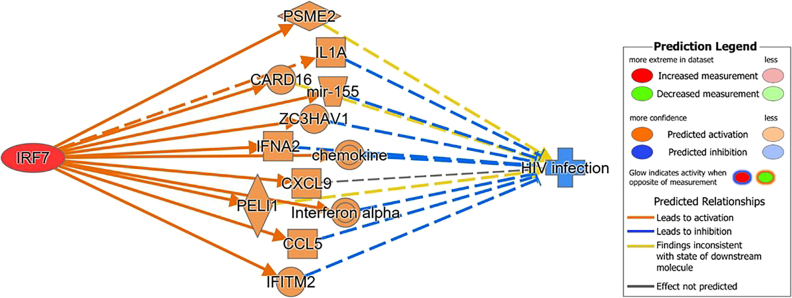
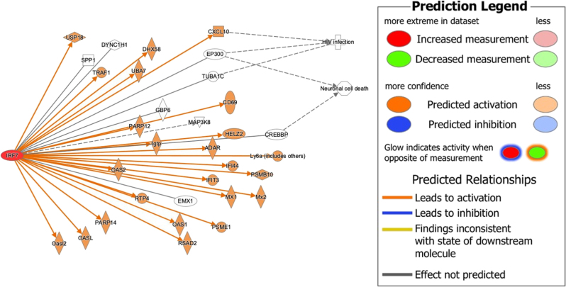
Given the previous literature on IRF7, this study seeks to elucidate what role IRF7 plays in antiviral response to HIV. This study utilized QIAGEN’s CLC Genomics workbench to collect the differential expression between F344 rats and HIV-1Tg rats from the genomics data deposited in the GSE47474 dataset. Likewise, the differential expression between control patients and individuals positive for HIV was collected using the genomics data deposit in the GSE152461 dataset [19]. QIAGEN’s Ingenuity Pathway Analysis (IPA) was used for data mining and the generation of connectivity mapping between IRF7 and HIV-infection and Neuronal Cell Death based on the manually curated publications stored in the QIAGEN Knowledge Base (QKB). In reference to the QKB, the molecules associated with IRF7 and with HIV-1 infection pathology were identified. Figure 1 displays the overlapping molecules associated with IRF7 and HIV-infection pathology. Using Molecular Activity Predictor (MAP) tool to simulate upregulation of IRF7 leading to inhibition of HIV-infection pathology through their overlapping associated molecules, This demonstrated IRF7's possible antiviral role toward HIV infection. With this premise, the molecules associated with IRF7 were removed from the molecular datasets (GSE47474 and GSE152416) to simulate the complete knockout of IRF7. Canonical analyses with and without IRF7 and its associated molecules were conducted and compared to determine the changes brought upon by IRF7 and its associated molecules in biological systems.
Figure 1:
The molecules associated with IRF7 and HIV-infection pathology were identified in reference to the QKB. The overlapping molecules between these two sets of associated molecules were found. The simulation of the upregulation of IRF7 was shown to inhibit HIV-infection pathology through their overlapping associated molecules, demonstrating that IRF7 could play an antiviral role against HIV.
Methods and materials
CLC Genomics Workbench
The CLC Genomics Workbench CL license was purchased from QIAGEN for the use of all features and tools in the CLC Genomics Workbench (version 22). CLC Genomics Workbench is a bioinformatics tool used to analyze, compare, and visualize genomics data. In this study, the “SRA Search” tool was used to download the GSE 47474 dataset (accession ID: SRR869044), comprising of RNA sequencing data from the prefrontal cortex (PFC), striatum (ST), and hippocampus (HIPP) of F344 Saline and HIV1-Tg transgenic rats, and the GSE152461 dataset (accession ID: SRR854758), comprising of RNA sequencing data from the prefrontal cortex of patients with HIV. Using the “Differential Expression” tool, a statistical differential expression test was run between the experimental (HIV-1Tg rat) and control (F344 rat) samples in the GSE47474 dataset, separated by region (PFC, ST, and HIPP) to identify the gene expression changes in the respective regions. Likewise, a second statistical differential expression test was run between the experimental (HIV + HAND) and control (HIV + cognitively normal) samples from the GSE152461 dataset. The significant gene expression changes for p<0.05 and p<0.005) were identified and uploaded to IPA for further analysis.
Ingenuity Pathway Analysis Software
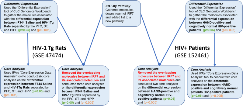
The IPA Analysis Match CL license was purchased from QIAGEN for the use of all features and tools in the IPA software (QIAGEN Inc., https://www.qiagenbioinformatics.com/products). IPA is a bioinformatics tool used to analyze data and biological processes using data from the QKB. The flow of steps used to identify and simulate the manipulation of IRF7-related genes and molecules is outlined in Figure 2. The genes involved in the GSE47474 dataset and GSE152461 were uploaded to IPA to be used in conjunction with data from the QKB on IRF7. The “Core Expression Data Analysis” feature of IPA was used to analyze the molecular datasets. The “Core Analysis” provides a “Canonical Pathway Analysis”, an “Upstream Analysis”, and a list of significant “Disease and Functions”. The “Canonical Pathway Analysis” reveals the canonical pathways within the molecular dataset that are statistically predicted to be involved using a −log(p value) calculated by the Benjamini–Hochberg Corrected Fisher’s Exact Test. Core analyses were run on the differential expression datasets of each brain region (prefrontal cortex, striatum, and hippocampus) with a p-value of 0.05 and 0.005. The “My Pathway” tool was used to identify the molecules associated with IRF7 from the QKB. After removing IRF7 and its associated molecules from each of the datasets, an additional six core analyses were run. Using the “My Pathway” tool of IPA, IRF7, CREBBP, HIV-Infection, and Neuronal Cell Death were added to a pathway. Next, the “Path Explorer” tool of IPA was used to connect IRF7 to CREBBP and CREBBP to HIV-Infection and Neuronal Cell Death. Finally, the Molecular Activity Predictor (MAP) tool was used to simulate the increased expression of IRF7 and determine its effects on the expression of CREBBP, HIV-Infection, and Neuronal Cell Death nodes. All data used for this study were retrieved from the QKB between December 23, 2021 until February 23, 2022.
Figure 2:
The flow of steps involved in using the bioinformatics and data collection tools offered by QIAGEN to simulate the complete knockout of IRF7 and its associated molecules in F344 rats and HIV-positive individuals. First, the “Differential Expression” tool of CLC genomics workbench was used to determine the differential expression between F344 Saline and HIV-1Tg rats and between HAND-positive and cognitively normal HIV-positive individuals. Then, the a “Canonical Pathway Analysis” was performed on the molecular datasets. Next, the “My Pathway” tool of IPA was used to identify the 455 molecules associated with IRF7. The overlapping molecules between the molecules associated with IRF7 and the molecules in the differential expression datasets were removed. Finally, the “Canonical Pathway Analysis” was performed on the datasets without the 455 molecules associated with IRF7.
Results
Overlap between molecules associated with IRF7 and the molecules associated with the differential expression between F344 Saline Rats and the HIV-1 TG rats in the GSE47474 dataset and the GSE152461 dataset.
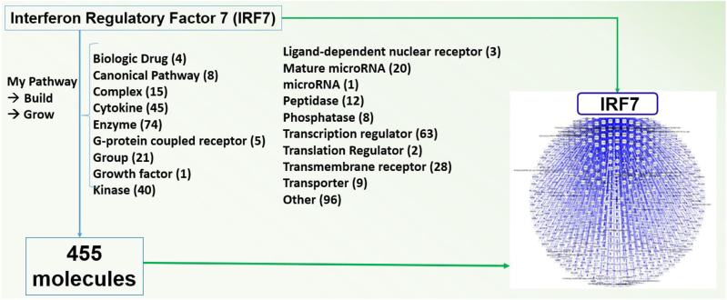
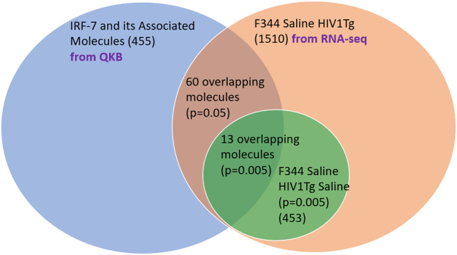
Using IPA’s “Build” tool to create a custom pathway, 511 molecules were associated with IRF7 (Figure 3). Of these 511 molecules, 56 molecules were removed for not naturally occurring in biological systems (i.e., chemical drugs and toxicants). Of the remaining 455 molecules associated with IRF7, 60 molecules were found to be overlapping between the GSE47474 dataset (Figure 4 and Table 1). However, within each of the three regions of the brain samples collected in the GSE47474 dataset, there were differences in the number of overlapping molecules. The number of overlapping molecules between the molecules associated with IRF7 and the molecules in the three different brain regions (PFC, ST, HP) in the GSE47474 dataset was 42, 22, and 18, respectively. All of the 455 molecules associated with IRF7 were found to be overlapping with the GSE152461 dataset (Figure 5).
Figure 3:
Using the QKB and the build tool of IPA, 511 molecules were found to be associated with IRF7. The above figure lists the 455 molecules of the 511 molecules that naturally occur in biological systems.
Figure 4:
Overlap of the biologically relevant molecules associated with IRF7 with the molecules in the differential expression between F344 and HIV-1Tg rats.
Table 1:
Overlapping Molecules between the GSE47474 dataset and Molecules Associated with IRF7.
| Molecule | Prefrontal cortex | Striatum | Hippocampus | Entrez gene name | Molecular family |
|---|---|---|---|---|---|
| ACADS | ✓ | ✓ | Acyl-CoA dehydrogenase short chain | Enzyme | |
| ACKR2 | ✓ | Atypical chemokine receptor 2 | G-protein coupled receptor | ||
| ADAR | ✓ | Adenosine deaminase RNA specific | Enzyme | ||
| Ccl2 | ✓ | Chemokine (C-C motif) ligand 2 | Cytokine | ||
| CD69a | ✓ | CD69 molecule | Transmembrane receptor | ||
| CREBBPa | ✓ | CREB binding protein | Transcription regulator | ||
| CSF1 | ✓ | Colony stimulating factor 1 | Cytokine | ||
| CXCL10a | ✓ | C-X-C motif chemokine ligand 10 | Cytokine | ||
| DHX58 | ✓ | ✓ | DExH-box helicase 58 | Enzyme | |
| DYNC1H1 | ✓ | Dynein cytoplasmic 1 heavy chain 1 | Peptidase | ||
| EFNA5a | ✓ | Ephrin A5 | Kinase | ||
| EMX1 | ✓ | Empty spiracles homeobox 1 | Transcription regulator | ||
| EP300a | ✓ | ✓ | E1A binding protein p300 | Transcription regulator | |
| FLT3 | ✓ | fms related receptor tyrosine kinase 3 | Kinase | ||
| GBP6 | ✓ | Guanylate binding protein family member 6 | Enzyme | ||
| HELZ2 | ✓ | Helicase with zinc finger 2 | Nucleus | ||
| IFI44 | ✓ | Interferon induced protein 44 | Other | ||
| IFIT3 | ✓ | Interferon induced protein with tetratricopeptide repeats 3 | Other | ||
| IFNGa | ✓ | Interferon gamma | Cytokine | ||
| Igtp | ✓ | Interferon gamma induced GTPase | Enzyme | ||
| IL2RG | ✓ | Interleukin 2 receptor subunit gamma | Transmembrane receptor | ||
| IQSEC1 | ✓ | IQ motif and Sec7 domain ArfGEF 1 | Other | ||
| IRF7a | ✓ | ✓ | ✓ | Interferon regulatory factor 7 | Transcription regulator |
| INSR | ✓ | Insulin receptor | Kinase | ||
| KL | ✓ | Klotho | Enzyme | ||
| Ly6a | ✓ | Lymphocyte antigen 6 complex, locus A | Other | ||
| MAP3K7 | ✓ | Mitogen-activated protein kinase kinase kinase 7 | Kinase | ||
| MAP3K8a | ✓ | Mitogen-activated protein kinase kinase kinase 8 | Kinase | ||
| MMP2 | ✓ | Matrix metallopeptidase 2 | Peptidase | ||
| MX1 | ✓ | ✓ | ✓ | MX dynamin-like GTPase 1 | Enzyme |
| Mx2 | ✓ | MX dynamin like GTPase 1 | Enzyme | ||
| NCOA2a | ✓ | Nuclear receptor coactivator 2 | Transcription regulator | ||
| NSD2 | ✓ | Nuclear receptor binding SET domain protein 2 | Enzyme | ||
| OAS1 | ✓ | ✓ | ✓ | 2′-5′-oligoadenylate synthetase 1 | Enzyme |
| OAS2 | ✓ | ✓ | 2′-5′-oligoadenylate synthetase 2 | Enzyme | |
| OASL | ✓ | 2′-5′-oligoadenylate synthetase like | Enzyme | ||
| OASI2 | ✓ | ✓ | ✓ | 2′-5′ oligoadenylate synthetase-like 2 | Enzyme |
| PARP12 | ✓ | poly(ADP-ribose) polymerase family member 12 | Enzyme | ||
| PARP14 | ✓ | ✓ | ✓ | poly(ADP-ribose) polymerase family member 14 | Enzyme |
| PDLIM2 | ✓ | PDZ and LIM domain 2 | Other | ||
| PGRa | ✓ | Progesterone receptor | Ligand-dependent nuclear receptor | ||
| PMAIP1 | ✓ | Phorbol-12-myristate-13-acetate-induced protein 1 | Other | ||
| PSEN1a | ✓ | Presenilin 1 | Peptidase | ||
| PSMB10 | ✓ | Proteasome 20S subunit beta 10 | Peptidase | ||
| PSME1 | ✓ | Proteasome activator subunit 1 | Peptidase | ||
| PTGER4 | ✓ | Prostaglandin E receptor 4 | G-protein coupled receptor | ||
| PRL | ✓ | Prolactin | Cytokine | ||
| RSAD2 | ✓ | ✓ | ✓ | Radical S-adenosyl methionine domain containing 2 | Enzyme |
| RTP4 | ✓ | ✓ | ✓ | Receptor transporter protein 4 | Other |
| SAMSN1 | ✓ | SAM domain, SH3 domain and nuclear localization signals 1 | Other | ||
| SATB1 | ✓ | SATB homeobox 1 | Transcription regulator | ||
| SPP1 | ✓ | Secreted phosphoprotein 1 | Cytokine | ||
| STAT2a | ✓ | Signal transducer and activator of transcription 2 | Transcription regulator | ||
| STAT6a | ✓ | ✓ | Signal transducer and activator of transcription 6 | Transcription regulator | |
| TLR3 | ✓ | Toll like receptor 3 | Transmembrane receptor | ||
| TLR5 | ✓ | Toll like receptor 5 | Transmembrane receptor | ||
| TRAF1 | ✓ | TNF receptor associated factor 1 | Other | ||
| TUBA1C | ✓ | ✓ | Tubulin alpha 1c | Other | |
| UBA7 | ✓ | Ubiquitin like modifier activating enzyme 7 | Enzyme | ||
| USP18 | ✓ | ✓ | ✓ | Ubiquitin specific peptidase 18 | Peptidase |
| ✓ Indicates the IRF7-associated molecules identified for each brain region | |||||
aIndicates molecules associated with IRF7 with a significance of p=0.005 for every brain region identified.
Figure 5:
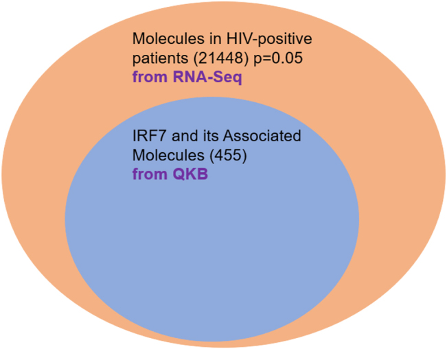
Overlap of the biologically relevant molecules associated with IRF7 with the molecules in the differential expression between HAND-positive and cognitively normal HIV-positive patients.
Canonical pathways involved in the differential expression with and without the molecules associated IRF7.
The core analyses performed with and without the molecules associated with IRF7 revealed significant changes in several canonical pathways (Table 2). In the GSE47474 dataset, 12, 4, and 4 pathways were found to be significantly impacted (Z-score ≥ +/−2) by the presence of IRF7 and its associated molecules in the PFC, ST, and HIPP, respectively. In the GSE152461 dataset, 13 pathways were significantly impacted. Furthermore, some of the significantly impacted pathways in each region and even between the datasets were overlapping. For example, the Estrogen Receptor Signaling pathway increased in the presence of the molecules associated with IRF7 in the PFC and ST of the GSE47474 dataset as well as in the GSE152461 dataset (PFC). The Dopamine-DARPP32 Feedback in cAMP Signaling pathway was found to increase in the PFC of both the GSE47474 and GSE152461 datasets, while the Neuroinflammation signaling was found to decrease in the PFC of both datasets. The Senescence pathway was found to increase in the PFC of the GSE47474 and the GSE152461 dataset. The CREB signaling in neurons pathway was found to increase in the GSE152461 dataset but only in the HIPP of the HIV-1Tg rat dataset.
Table 2:
Change in Z Score of Significant Canonical pathways in the Presence of IRF7 and its associated molecules.
| Canonical pathway | Categorization of canonical pathway | −Log(p-Value) | Change in Z-Score |
|---|---|---|---|
| 2a: Prefrontal cortex | |||
| Opioid signaling pathway | Neurotransmission | 12.8 | 0.239 |
| Dopamine-DARPP32 feedback in cAMP signaling | Neurotransmission | 12.3 | 0.120 |
| Synaptogenesis signaling pathway | Neuronal survival | 10.10 | 0.202 |
| Calcium signaling | Neurotransmission | 9.07 | 0.256 |
| Estrogen receptor signaling | Neurotransmission | 9.93 | 0.469 |
| Synaptic long-term potentiation | Neuronal survival | 9.89 | 0.322 |
| Senescence pathway | Neuronal survival | 8.58 | 0.268 |
| Neuroinflammation signaling pathway | Neuronal survival | 7.45 | −0.181 |
| VDR/RXR activation | Neurotransmission | 6.31 | −0.700 |
| Th1 pathway | Immune | 6.45 | 0.114 |
| Th2 pathway | Immune | 6.28 | −0.387 |
| Xenobiotic metabolism AHR signaling pathway | Immune | 6.10 | 1.216 |
| 2b: Striatum | |||
| IL-17 signaling | Immune | 8.51 | 2.449 |
| Xenobiotic metabolism CAR signaling pathway | Immune | 8.30 | 1.342 |
| Pulmonary healing signaling pathway | Neurotransmission | 7.31 | 2.236 |
| Estrogen receptor signaling | Neurotransmission | 6.65 | 2.449 |
| 2c: Hippocampus | |||
| Role of hypercytokinemia/hyperchemokinemia in the pathogenesis of influenza | Immune | 6.646 | 2.646 |
| Neuroinflammation signaling pathway | Neuronal survival | 6.488 | 2.121 |
| Phagosome formation | Immune | 6.177 | 2.673 |
| CREB signaling in neurons | Neuronal survival | 5.147 | 3.464 |
| 2d: GSE152461 dataset (prefrontal cortex) | |||
| Dopamine-DARPP32 feedback in cAMP signaling | Neurotransmission | 10.89 | 0.720 |
| CREB signaling in neurons | Neuronal survival | 9.24 | 0.330 |
| Synaptic long term depression | Neuronal survival | 9.60 | −0.165 |
| Role of PKR in interferon induction and antiviral response | Immune | 9.42 | 0.905 |
| Role of hypercytokinemia/hyperchemokinemia in the pathogenesis of influenza | Immune | 8.64 | 2.646 |
| Adrenomedullin signaling pathway | Immune | 7.43 | −0.482 |
| Calcium signaling | Neurotransmission | 7.28 | 0.278 |
| Estrogen receptor signaling | Neurotransmission | 7.13 | 0.469 |
| Synaptic long term potentiation | Neuronal survival | 6.89 | 0.322 |
| Senescence pathway | Neuronal survival | 6.58 | 0.268 |
| Neuroinflammation signaling pathway | Neuronal survival | 6.34 | −0.181 |
| VDR/RXR activation | Neurotransmission | 6.31 | −0.700 |
| Th1 pathway | Immune | 6.45 | 0.114 |
IRF7 and its associated molecules
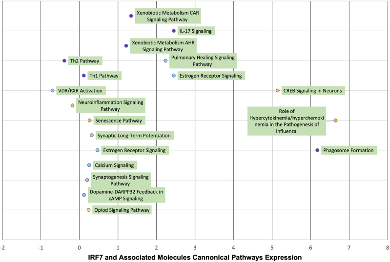
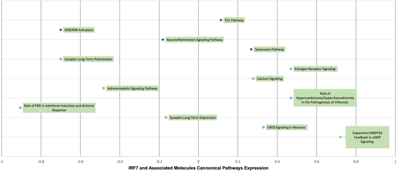
Using the “Core Analysis: Expression Analysis Tool”, IRF7 and its 60 associated molecules were compared to the 705 well-defined canonical pathways within QIAGEN’s Knowledge Base (QKB). The most significant pathways were chosen, which were those that displayed the largest –log(p-value) and demonstrated the strongest overlap between the IRF7 associated molecules. Then, the Z-scores associated with these pathways were found [21, 22]. Additionally, the same “Core Analysis” function was run but without IRF7 and its causally-associated molecules to simulate the knockout of IRF7. Again, the Z-scores associated with these pathways were found. Then these differences in Z-scores (Figures 6 and 7) were calculated to demonstrate the effect that IRF7 and its associated molecules had on pathways related to immune function (purple points), neuronal survival (orange) and neurotransmission (light blue).
Figure 6:
Visual illustration of the quantitative analysis on the relationship between the 60 overlapping molecules between the molecules associated with IRF7 and the differential expression between F344 Saline and HIV-1Tg rats in the GSE47474 dataset. The significant canonical pathways (Z-score >/=+/−2) in the core analyses with IRF7 and its associated molecules were identified, and their corresponding Z-scores were identified. Likewise, the Z-scores of the same canonical pathways in the datasets without IRF7 and its associated molecules were identified. The above figure lists the difference in the Z-scores with and without IRF7 and its associated molecules to determine the changes brought upon by IRF7 and its associated molecules.
Figure 7:
Visual illustration of the quantitative analysis on the relationship between the 455 overlapping molecules between the molecules associated with IRF7 and the differential expression between control patients and HIV-positive patients in the GSE152461 dataset. The significant canonical pathways (Z-score >/=+/−2) in the core analyses with IRF7 and its associated molecules were identified, and their corresponding Z-scores were identified. Likewise, the Z-scores of the same canonical pathways in the datasets without IRF7 and its associated molecules were identified. The above figure lists the difference in the Z-scores with and without IRF7 and its associated molecules to determine the changes brought upon by IRF7 and its associated molecules.
The pathways shown in Figure 6 convey Z-score expression changes for the canonical pathways associated with the GSE47474 dataset. The majority of the pathways experienced increased expression or upregulation while 3 pathways of note experienced decreased expression or downregulation which were the Th2 pathway (immune), VDR/RXR pathway (neurotransmission), and Neuroinflammation pathway (neuronal survival). Additionally, pathways of note experiencing increased expression were the Th1 pathway (immune), Synaptic Long-term Potentiation (neuronal survival), and CREB signaling in Neurons (neuronal survival).
Similarly, the pathways shown in Figure 7 convey Z-score expression changes for the canonical pathways associated with the GSE152461 dataset. For this dataset, 7 significant canonical pathways experienced increased expression when associated with IRF7 and its molecules while 6 canonical pathways experienced decreased expression. Similar to the GSE47474 dataset, the VDR/RXR pathway and Neuroinflammation pathway experienced decreased expression while the Th1 pathway experienced increased expression. However, while the Synaptic Long-term Potentiation pathway had an expression increase in the GSE47474 dataset, it expressed a decrease in the GSE152461 dataset.
Discussion
Previously, the deep sequencing of HIV-1Tg rats found that IRF7 was one of the only genes that was significantly upregulated in the prefrontal cortex, striatum, and hippocampus of the HIV-1Tg rats compared to the F344 rats. Given that IRF7 is the master regulator of type I interferon production and aberrant production of type I interferons is associated with autoimmune disorders, IRF7 appeared to be involved in the antiviral response to HIV. In another study, the partial knockout of IRF7 via CRISPR/Cas9 gene editing in the human embryonic kidney 293FT cell line found that in the absence of IRF7, the antiviral response in HIV-1Tg rats decreased [20]. To fully investigate what role IRF7 plays, we simulated the complete knockout of IRF7 and its associated molecules via an in-silico approach by using the data compiled in the QKB as well as data retrieved from rat in vivo and human in-vitro studies. Although taken from different species, the data shows how IRF7 and its associated molecules impact several pathways including neurotransmission-related pathways, immune-related pathways, and neuronal survival related pathways.
Neurotransmission and neuronal survival-related pathways
A common pattern in the core analyses of the rat PFC (GSE47474) and the human PFC (GSE152461) dataset in the presence of the molecules associated with IRF7 was the involvement of cAMP Response Element Binding Protein (CREB), which binds to CREB Binding Protein (CREBBP), a molecule downstream of IRF7. The Dopamine-DARPP32 Feedback in cAMP Signaling and the Calcium signaling pathway results in an increase in dopamine and intracellular calcium levels, respectively, both of which lead to increased CREB levels. Increased levels of CREB also appear due to the upregulation of the synaptic long term potentiation pathway by IRF7. In neurons, CREB functions as a transcription factor and binds to cAMP Response Element (CRE), leading to the transcription of several cell survival genes involved in neurogenesis and metabolism [23, 24]. As seen in Figure 8, IRF7 is connected to Neuronal Cell Death via CREBBP. Although IPA did not note a predicted activation or inhibition of CREBBP, it does present a connection between IRF7 and Neuronal Cell Death. Taken together, we find that IRF7 and its associated molecules could possibly reduce neuronal cell death by increasing the expression of CREB via the CREBBP gene.
Figure 8:
Connectivity map of the relationship between IRF7 and the 13 overlapping molecules between the molecules associated with IRF7 and the molecules in the differential expression between F344 Saline and HIV-1Tg rats in the GSE47474 dataset that influence CREB, neuronal cell death, and HIV infection pathology.
In addition to increasing CREB, IRF7 and its associated molecules also upregulated the expression of CCL2 in the PFC of F344 rats and in HIV-positive individuals (Table 2). CCL2 is known to induce neuronal differentiation, oligodendrocyte maturation, and myelin production. Low myelin production has been shown to result in neurocognitive changes, even in HIV-positive individuals receiving anti-retroviral therapy [16, 25, 26]. Therefore, the upregulation of CCL2 by IRF7 and its associated molecules may reduce neurodegeneration in the CNS, reducing the cognitive deficits brought upon by HIV-Associated Neurocognitive Disorders (HANDs).
Immune-related pathways
IRF7 and its associated molecules lead to the activation of canonical pathways involved in the cellular-mediated response to infection. The upregulation of the Th1 pathway in the PFC of HIV-1 Tg rats and HIV-positive individuals with mild neurocognitive deficits (Table 2a and d) leads to the development of T-Helper 1 cells, which aid in the production of IFN-γ, a cytokine that recruits macrophages for the destruction of pathogens and presentation to T-Lymphocytes. The increase in macrophage activity is also reflected in the increase of the phagosome formation pathway (Table 2c). At the same time, there was a decrease in the Th2 pathway in the PFC of F344 rats (Table 2a), leading to the decrease of Th-2 cells, which is involved in the humoral response. A widely accepted hypothesis suggests that the switch from Th1 activity to Th2 activity leads to the progression of Acquired Immunodeficiency Disorder (AIDS) in individuals affected with HIV [27, 28]. Therefore, the simultaneous decrease of Th2 activity and the simultaneous increase of Th1 activity in the PFC suggests that IRF7 and its associated molecules may slow or prevent the development of AIDS in HIV-positive individuals.
The upregulation of the Interleukin-17 signaling pathway was observed in the STR of F344 rats (Table 2b). Given that the low frequency of IL-17-producing cytotoxic T-cells is associated with the persistent immune activation in HIV-positive individuals despite receiving Highly Active Antiretroviral Therapy (HAART) [29], the upregulation of genes in the Interleukin-17 signaling pathway suggests that IRF7 and its associated molecules may prevent chronic immune activation in HIV-positive individuals.
It is hypothesized that HIV-1 translation is modulated by the inducible Protein Kinase RNA-activated (PKR), which phosphorylates the alpha subunit of eukaryotic translation initiation factor 2 (eIF2a). Upon phosphorylation, eIF2a hinders the ternary tRNAmet-GTP-eIF2 complex, resulting in the formation of stress granules (SGs) which encapsulate viral RNA and transcription and translation-related proteins, thus decreasing viral replication [30, 31]. In this study, we observe the upregulation of the Role of PKR in Interferon Induction and Antiviral Response signaling pathway in HIV-positive individuals (Table 2d), suggesting that IRF7 and its associated molecules may decrease HIV-1 translation by encapsulating the viral RNA and transcription and translation-related proteins needed for viral production.
Using the path explorer tool, a connectivity map between IRF7 and HIV-infection was generated using the data curated from the QKB (Figure 1). The simulated activation of IRF7 lead to the activation of several molecules downstream of IRF7, including several molecules found to be overlapping between the GSE47474 dataset and IRF7 and its associated molecules (Table 1) such as CXCL10 and molecules closely related to the overlapping molecules, such as CXCL9, CXCL8, CD80, CCL8, etc. The activation of said molecules leads to the inhibition of HIV-infection. Although this connectivity map was not generated from the data from the GSE47474 and GSE152461 datasets, it supports our findings that IRF7 can play an antiviral role against HIV-infection via the overlapping molecules between the GSE47474 dataset and IRF7 and its associated molecules.
There are certain limitations to this study that must be considered when interpreting the results. In contrast to the GSE47474 dataset, the GSE152461 dataset used human postmortem tissue samples, which reduces the reliability of the data as humans have much more complex biological systems and are influenced by genetic, environmental, and social factors that are difficult to account for in experimental settings. On the other hand, the F344 and HIV1Tg rats are organisms developed strictly for use in research laboratory settings and thus have less variation in their genome and environment. It is also imperative to note that due to the inherent complexity of biological systems, these core analyses are still isolated networks and do not fully represent how these molecules interact in a biological system. Nevertheless, the results of our in-silico study provide an empirically based hypothesis that is testable with in-vitro and in-vivo studies that can knockout IRF7 and its associated molecules along with the use of bioinformatics tools such as IPA to simulate the complete knockout of a target molecule in-silico. These proposed knockout experiments are likely to reveal the role of IRF7 in the antiviral response to HIV, specifically through its involvement in neurotransmission, immune and neuronal survival related pathways.
Conclusions
Our findings from the complete in-silico knockout of IRF7 and its associated molecules provide pertinent information on IRF7’s role in the antiviral response to HIV. By integrating genomics data from the GSE47474 and the GSE152461 datasets, we demonstrate that IRF7 can modulate immune system production, specifically the Th-1/Th-2 ratio, promote neurogenesis through the upregulation of CREBBP and CCL2, and possibly halt the onset of AIDS in HIV-positive individuals.
Acknowledgments
The authors thank Dr. Eric Seiser for initial use of QIAGEN Knowledge Base and QIAGEN Ingenuity Pathway Analysis tools.
Footnotes
Research funding: This study is partially supported by R01DA0462582.
Author contributions: All authors have accepted responsibility for the entire content of this manuscript and approved its submission.
Competing interests: Authors state no conflict of interest.
References
- 1.McNab F, Mayer-Barber K, Sher A, Wack A, O’Garra A. Type I interferons in infectious disease. Nat Rev Immunol. 2015;15:86–92. doi: 10.1038/nri3787. [DOI] [PMC free article] [PubMed] [Google Scholar]
- 2.Daffis S, Samuel MA, Suthar MS, Keller BC, Gale M, Diamond MS, et al. Interferon regulatory factor IRF-7 induces the antiviral alpha interferon response and protects against lethal West Nile virus infection. J Virol. 2008;82:8465–75. doi: 10.1128/JVI.00918-08. [DOI] [PMC free article] [PubMed] [Google Scholar]
- 3.Ning S, Pagano JS, Barber GN. IRF7: activation, regulation, modification and function. Genes & Immunity. 2011;12:399–414. doi: 10.1038/gene.2011.21. [DOI] [PMC free article] [PubMed] [Google Scholar]
- 4.Sin WX, Yeong JPS, Lim TJF, Su IH, Connolly JE, Chin KC. IRF-7 mediates type I IFN responses in endotoxin-challenged mice. Front Immunol. 2020;11 doi: 10.3389/fimmu.2020.00640. [DOI] [PMC free article] [PubMed] [Google Scholar]
- 5.Sato M, Hata N, Asagiri M, Nakaya T, Taniguchi T, Tanaka N. Positive feedback regulation of type I IFN genes by the IFN-inducible transcription factor IRF-7. FEBS Lett. 1998;441:106–10. doi: 10.1016/s0014-5793(98)01514-2. [DOI] [PubMed] [Google Scholar]
- 6.Deeks SG, Overbaugh J, Phillips A, Buchbinder S. Nature news. Nature Publishing Group; 2015. HIV infection.https://www.nature.com/articles/nrdp201535/ Available from: [DOI] [PubMed] [Google Scholar]
- 7.Palmer S, Josefsson L, Coffin JM. HIV reservoirs and the possibility of a cure for HIV infection. J Intern Med. 2011;270:550–60. doi: 10.1111/j.1365-2796.2011.02457.x. [DOI] [PubMed] [Google Scholar]
- 8.Nguyen N, Holodniy M. Clinical interventions in aging. Dove Medical Press; 2008. HIV infection in the elderly.https://www.ncbi.nlm.nih.gov/labs/pmc/articles/PMC2682378 Available from. [DOI] [PMC free article] [PubMed] [Google Scholar]
- 9.Altfeld M, Gale M. Nature news. Nature Publishing Group; 2015. Innate immunity against HIV-1 infection.https://www.nature.com/articles/ni.3157 Available from. [DOI] [PubMed] [Google Scholar]
- 10.Elsner C, Ponnurangam A, Kazmierski J, Zillinger T, Jansen J, Todt D, et al. Absence of CGAS-mediated type I IFN responses in HIV-1–Infected T cells. Proc Natl Acad Sci. 2020;117:19475–86. doi: 10.1073/pnas.2002481117. [DOI] [PMC free article] [PubMed] [Google Scholar]
- 11.Colomer-Lluch M, Ruiz A, Moris A, Prado JG. Restriction factors: from intrinsic viral restriction to shaping cellular immunity against HIV-1. Front Immunol. 2018;9 doi: 10.3389/fimmu.2018.02876. [DOI] [PMC free article] [PubMed] [Google Scholar]
- 12.Razzak M. Schlafen 11 naturally blocks HIV. Nat Rev Urol. 2012;9:602–5. doi: 10.1038/nrurol.2012.188. [DOI] [PubMed] [Google Scholar]
- 13.Lu J, Pan Q, Rong L, He W, Liu SL, Liang C. The IFITM proteins inhibit HIV-1 infection. J Virol. 2011;85:2126–37. doi: 10.1128/JVI.01531-10. [DOI] [PMC free article] [PubMed] [Google Scholar]
- 14.Vigorito M, LaShomb AL, Chang SL. Spatial Learning and memory in HIV-1 trangenic rats. J Neuroimmune Pharmacol. 2007;2:319–28. doi: 10.1007/s11481-007-9078-y. [DOI] [PubMed] [Google Scholar]
- 15.Lashomb AL, Vigorito M, Chang SL. Further characterization of the spatial learning deficit in the human immunodeficiency virus-1 transgenic ra. J Neurovirol. 2009;15:14–24. doi: 10.1080/13550280802232996. [DOI] [PubMed] [Google Scholar]
- 16.Li MD, Cao J, Wang S, Wang J, Sarkar S, Vigorito M, et al. Transcriptome sequencing of gene expression in the brain of the HIV-1 transgenic rat. PLoS One. 2013 doi: 10.1371/journal.pone.0059582. [DOI] [PMC free article] [PubMed] [Google Scholar]
- 17.Vigorito M, Connaghan KP, Chang SL. The HIV-1 transgenic rat model of neuroHIV. Brain Behav Immun. 2015;48:336–49. doi: 10.1016/j.bbi.2015.02.020. [DOI] [PMC free article] [PubMed] [Google Scholar]
- 18.McLaurin KA, Li H, Booze RM, Mactutus CF. Disruption of timing: NeuroHIV progression in the post-cART era. Sci Rep. 2019;9:827. doi: 10.1038/s41598-018-36822-1. [DOI] [PMC free article] [PubMed] [Google Scholar]
- 19.Canchi S, Swinton MK, Rissman RA, Rissman R, Fields J. Transcriptomic analysis of brain tissues identifies a role for CCAAT enhancer binding protein β in HIV-associated neurocognitive disorder. J Neuroinflammation. 2020;17:112–28. doi: 10.1186/s12974-020-01781-w. [DOI] [PMC free article] [PubMed] [Google Scholar]
- 20.Han H, Huang W, Du W, Shen Q, Yang Z, Li MD, et al. Involvement of interferon regulatory factor 7 in nicotine’s suppression of antiviral immune responses. J Neuroimmune Pharmacol. 2019;14:551–64. doi: 10.1007/s11481-019-09845-2. [DOI] [PubMed] [Google Scholar]
- 21.Krämer A, Green J, Pollard J, Tugendreich S. Causal analysis approaches in Ingenuity Pathway Analysis. Bioinformatics. 2014;30:523–30. doi: 10.1093/bioinformatics/btt703. [DOI] [PMC free article] [PubMed] [Google Scholar]
- 22.Masi S, Nair M, Vigorito M, Chu T, Chang SL. Alcohol modulation of amyloid precursor protein in Alzheimer’s disease. Journal of Drug and Alcohol Research. 2020;9 doi: 10.4303/jdar/236094. [DOI] [Google Scholar]
- 23.Wen A, Sakamato K, Miller L. The role of the transcription factor CREB in immune function. J Immunol. 2010;185:6413–9. doi: 10.4049/jimmunol.1001829. [DOI] [PMC free article] [PubMed] [Google Scholar]
- 24.Ortega-Martínez S. A new perspective on the role of the CREB family of transcription factors in memory consolidation via adult hippocampal neurogenesis. Front Mol Neurosci. 2015;8:46. doi: 10.3389/fnmol.2015.00046. [DOI] [PMC free article] [PubMed] [Google Scholar]
- 25.Berger JR, Tornatore C, Major EO, Bruce J, Shapshak P, Yoshioka M, et al. Relapsing and remitting human immunodeficiency virus-associated leukoencephalomyelopathy. Ann Neurol. 1992;31:34–38. doi: 10.1002/ana.410310107. [DOI] [PubMed] [Google Scholar]
- 26.Turbic A, Leong SY, Turnley AM. Chemokines and inflammatory mediators interact to regulate adult murine neural precursor cell proliferation, survival and differentiation. PLoS One. 2011;6:e25406. doi: 10.1371/journal.pone.0025406. [DOI] [PMC free article] [PubMed] [Google Scholar]
- 27.Becker Y. The changes in the T helper 1 (Th1) and T helper 2 (Th2) cytokine balance during HIV-1 infection are indicative of an allergic response to viral proteins that may be reversed by Th2 cytokine inhibitors and immune response modifiers-a review and hypothesis. Virus Gene. 2004;28:5–18. doi: 10.1023/B:VIRU.0000012260.32578.72. [DOI] [PubMed] [Google Scholar]
- 28.Klein SA, Dobmeyer JM, Dobmeyer TS, Pape M, Ottmann OG, Helm EB, et al. Demonstration of the Th1 to Th2 cytokine shift during the course of HIV-1 infection using cytoplasmic cytokine detection on single cell level by flow cytometry. AIDS. 1997;11:1111–8. doi: 10.1097/00002030-199709000-00005. [DOI] [PubMed] [Google Scholar]
- 29.Perdomo-Celis F, Feria M, Taborda N, Rugeles M. A low frequency of IL-17-producing CD8+ T-cells is associated with persistent immune activation in people living with HIV despite haart-induced viral suppression. Front Immunol. 2018;9:122–9. doi: 10.3389/fimmu.2018.02502. [DOI] [PMC free article] [PubMed] [Google Scholar]
- 30.Clerzius G, Shaw E, Daher A, Burugu S, Gélinas JF, Ear T, et al. The PKR activator, pact, becomes a PKR inhibitor during HIV-1 replication. Retrovirology. 2013;10:454–98. doi: 10.1186/1742-4690-10-96. [DOI] [PMC free article] [PubMed] [Google Scholar]
- 31.Liu Y, Wang M, Cheng A, Yang Q, Wu Y, Jia R, et al. The role of host eIF2α in viral infection. Virol J. 2020;17:112–28. doi: 10.1186/s12985-020-01362-6. [DOI] [PMC free article] [PubMed] [Google Scholar]


