



Abstract
PURPOSE OF REVIEW:
Management of metastasis to the central nervous system (CNS) has evolved, and molecular characterization of metastatic disease is now routinely done. Targeted therapies, once few in number with limited penetration into the CNS, have multiplied in number and increased in CNS coverage. This article addresses recent advances in the evaluation and clinical management of patients with CNS metastasis.
RECENT FINDINGS:
Metastasis of cancer to the CNS can be diagnosed and characterized with novel techniques, including molecular analyses of the spinal fluid, so-called liquid biopsies. Resected parenchymal CNS metastases are now routinely subjected to genomic sequencing. For patients with CNS metastases displaying targetable mutations, a wide variety of treatment options are available, including deferral of radiation therapy in favor of a trial of an orally bioavailable targeted therapy or immunotherapy. For patients without a molecularly targetable lesion, local treatment in the form of radiation therapy, now most often stereotactic radiosurgery, is supplanting untargeted whole-brain radiation therapy.
SUMMARY:
Technologic advances in diagnosis and management have resulted in new diagnostic and therapeutic approaches to patients with metastasis to the CNS, with resulting improvements in progression-free and overall survival.
INTRODUCTION
Metastases to the central nervous system (CNS) are approximately tenfold more common than primary brain tumors and, as such, are more likely to be encountered by neurologists in general practice. In the past, the management of these lesions was guided by mainly palliative principles; recently, however, this palliative approach has been joined by tumor-directed therapies, with improvements in overall survival, progression-free survival, and quality of life in certain types of malignancies. Such outcomes are only possible with a cooperative multidisciplinary approach for the care of these patients that includes neurosurgery, radiation oncology, and medical oncology, with the neurologist or neuro-oncologist an indispensable member of the team.
EPIDEMIOLOGY
Any systemic tumor may result in metastasis to the CNS. Up to 30% of adults with cancer harbor CNS metastases. This number is expected to rise with improved systemic control and diagnostic methods.1,2 CNS metastases therefore represent a prevalent clinical problem. Classically associated with late-stage disease, CNS metastases may be diagnosed at any time during the clinical course and may represent the first sign of malignancy. In modern clinical practice, parenchymal brain and leptomeningeal metastases most often result from breast, lung, or melanoma primary cancers. Spinal metastases are likely the result of breast, lung, prostate, thyroid, or renal cell cancers. Regardless of primary tumor histology, new focal neurologic symptoms in a patient with cancer should raise the clinical suspicion for metastatic disease to the CNS.
PROGNOSIS
Parenchymal brain metastases result in a wide variety of outcomes, with a mean overall survival of 4 to 6 months.3 The most favorable outcomes are observed in young patients (younger than 65 years of age) with good functional status (Karnofsky Performance Status Scale score ≥70) harboring a single parenchymal metastasis and no extracranial metastases.4 For select patients with tumors sensitive to targeted therapy, the survival may extend to several years. In contrast, leptomeningeal metastasis carries a uniformly poor prognosis. The average survival after leptomeningeal metastasis from solid tumors is less than 6 months after diagnosis, despite treatments including radiation therapy chemotherapy, and supportive care.5–8
PRESENTATION AND DIAGNOSIS
Cancer cells generally access the CNS after hematogenous dissemination. Given the abundant blood flow supply to the CNS, metastatic cells can and do arrive in the brain, spinal cord, and leptomeninges in approximate proportion to their blood flow. As a result, in the brain parenchyma, metastases tend to accumulate at the gray-white junction and watershed zones, promoting symptoms classically associated with these areas. In contrast, metastases to the epidural and leptomeningeal spaces can lead to radicular and cranial nerve palsies; as these tumors progress, they may cause cortical or spinal symptoms or signs. More advanced disease may result in symptoms or signs of increased intracranial pressure (ICP).
The first step in such presentations in a patient with cancer is a neurologic examination. Patients with cancer have often been exposed to a range of cytotoxic therapies and may accumulate significant neurotoxicity before coming to medical attention; therefore, care must be taken to distinguish the focal neurologic deficits due to prior surgery, chemotherapy, and focal radiation from new findings. In addition, a given patient may possess a single CNS metastasis or may accumulate metastases within several different sites within the CNS. Once a single CNS metastasis is uncovered, a high degree of clinical suspicion for others is appropriate.
With this in mind, imaging of the entire neuraxis by MRI is rarely indicated. Rather, focused imaging of symptomatic sites with a contrast-enhanced MRI scan is preferred. Parenchymal metastases classically appear as enhancing masses at the gray-white junction (FIGURE 6-1A), surrounded by a disproportionate volume of vasogenic edema (FIGURE 6-1B), a reflection of rapid tumor growth kinetics. For localizations that suggest leptomeningeal involvement (ie, multiple cranial neuropathies, radiculopathies, or elevated ICP), contrast-enhanced MRI of the brain and spinal cord (including the entire cauda equina) is indicated. Leptomeningeal metastases appear as linear enhancing deposits covering the cerebellar folia around the pons (FIGURE 6-2A), over the cranial nerves, or within the cortical sulci. Over the spinal cord, leptomeningeal metastases tend to result in nodular enhancement of the cauda equina (FIGURE 6-2B) and coating of the spinal cord (FIGURE 6-2C). In suspected leptomeningeal metastasis, the additional step of CSF sampling is required. This allows for definitive diagnosis of leptomeningeal metastasis9,10 as well as opening pressure measurement. CSF analysis by traditional cytology has poor sensitivity,11 and up to three large-volume (at least 10 mL each) CSF collections may be required to secure a diagnosis.12 In specialized centers, adaptation of circulating tumor cell technologies may be applied to CSF to quantify cancer cell burden in this space.
FIGURE 6-1.

Radiographic appearance of parenchymal brain metastasis in a 47-year-old woman with non–small cell lung cancer. A, Axial postcontrast T1-weighted MRI shows a round contrast-enhancing mass located at the gray-white junction. B, Axial fluid-attenuated inversion recovery (FLAIR) sequence shows edema surrounding the mass.
FIGURE 6-2.

Radiographic appearance of leptomeningeal metastases in a 52-year-old man with non–small cell lung cancer. A, Axial postcontrast T1-weighted MRI shows linear enhancing plaques of disease (arrows) over the cerebellar folia. B, Axial postcontrast T1-weighted MRI shows patchy enhancement (arrows) of the cauda equina with clumping of nerve roots. C, Sagittal postcontrast T1-weighted MRI shows enhancement (arrows) along the length of the spinal cord surface and lumbar roots.
Emergency Presentations of Central Nervous System Metastases
Some special cases and emergency presentations of CNS metastases are of note: spinal cord compression, elevated ICP, and hemorrhagic metastasis. Back pain with sudden onset of weakness and upper motor neuron signs localizing to the spine with or without urinary retention is an emergency. High-dose dexamethasone should be administered immediately, before obtaining imaging. Emergent neurosurgical and radiation oncology consultations should be obtained; serial neurologic examinations are essential. Prompt steroid treatment13,14 and initiation of more definitive measures, such as surgical decompression or focal radiation, are essential to preserve neurologic function.
New positional headache in a patient with cancer, with or without diplopia, should prompt immediate evaluation. Papilledema and cranial neuropathies are indications for noncontrast head CT. It is essential to establish the presence or absence of mass lesions before performing a diagnostic lumbar puncture. In the presence of a mass lesion, elevated ICP may be treated with dexamethasone pulse before neurosurgical consultation. In the absence of mass lesions, typically in the setting of leptomeningeal disease, lowering opening pressure with a lumbar puncture may provide immediate symptomatic relief and illustrate the need for more definitive measures, such as a ventriculoperitoneal shunt.
Hemorrhagic metastases may also elicit symptoms of elevated ICP and will also be uncovered with a noncontrast head CT. As with other causes of intraparenchymal hemorrhage, acute symptomatic hemorrhage of metastatic lesions requires admission to a monitored setting, reversal of any anticoagulant or antiplatelet therapies, infusion of platelets to the goal of 100,000 cells/mm3, serial (typically every 6 hours) noncontrast head CT, and neurosurgical consultation. Metastasis to the CNS can injure the cortical surface, particularly in melanoma or hemorrhagic brain metastases. Although prophylactic antiepileptic drugs (AEDs) are not routinely recommended, some experts advise AEDs in the setting of acute, symptomatic, hemorrhagic metastases.
MANAGEMENT
Management of CNS metastases requires a multidisciplinary patient-centered approach, with the neurologist or neuro-oncologist, medical oncologist, radiation oncologist, and neurosurgeon working in close collaboration.
General Principles
By definition, metastasis to the CNS connotes stage IV cancer and carries a poor prognosis. The physician is confronted with management of active CNS disease and, often, extracranial disease as well. In addition, CNS metastases cause a wide array of symptoms that must be addressed. Thus, palliative care plays a central role in the management of CNS metastases. The general principle in the management of CNS metastases is to first localize the symptomatic lesions and then address them locally, either through surgery or radiation. In addition, these symptomatic sites may also prove epileptogenic, and require management with AEDs and/or steroids (typically dexamethasone). When selecting AEDs for these patients, the potential for drug-drug interactions is an important consideration, particularly for patients receiving chemotherapy. Experts typically advise a non–enzyme-inducing AED, such as levetiracetam, lacosamide, or zonisamide. After symptomatic sites have been addressed, the choice of systemic treatment takes center stage. Considering the patient’s overall functional level, the state of extracranial disease, and the patient’s expressed wishes, systemic treatments are initiated collaboratively with the medical oncology, radiation oncology, and neurosurgical teams. In specialized centers, this highly personalized decision making is often conducted in a tumor board review. Patients undergoing treatment for active CNS metastases are generally followed with a neurologic examination and MRI of the brain and/or spine every 6 to 10 weeks; those with leptomeningeal disease also undergo CSF sampling at these time points.
Anatomic Considerations
The relationship of the CNS to the systemic circulation is an important consideration. The brain and spinal cord reside behind the blood-brain barrier; in contrast, the leptomeningeal space, consisting of the pia and arachnoid and containing the circulating CSF, represents a distinct anatomic compartment. The leptomeninges reside behind the blood-CSF barrier, the choroid plexus. In marked contrast, the dural and epidural compartments are supplied directly by the systemic circulation (ie, no blood-brain barrier). Metastatic cancer cells may access any or all of these spaces, singly or sequentially, over the course of a given patient’s disease. It is therefore wise to keep these anatomic constraints in mind when choosing a systemic treatment.
Molecular Considerations
Metastases to any site are the result of a complex evolutionary process whereby a heterogeneous population of cancer cells gives rise to a disseminated subpopulation. In the new or metastatic site, cancer cells with the capacity to grow in the new microenvironment result in a metastatic tumor. The metastasis is thus the result of both genetic evolution and selective pressures at the new site, a product of evolutionary processes. Metastases to all sites are genetically divergent from their preceding primary tumors.15,16 CNS metastases are distinguished by the degree to which these cancer cell populations may diverge from the primary tumor, a reflection of the selective pressures at play in this process. When feasible, it is therefore important to establish the molecular identity and genetic characteristics of CNS metastases. In parenchymal brain metastases, this means subjecting the resected tumor to either whole-exome sequencing (typically only available in a research setting) or targeted exome sequencing (available for clinical use). In leptomeningeal metastasis, the CSF may be subjected to sequencing.17,18 Whereas the cell pellet may be sequenced, recent work demonstrates that the cell-free portion of the CSF may be sequenced with superior sensitivity and specificity.19 It is tempting to posit that DNA collected from CSF could be sequenced in lieu of tissue from the parenchymal metastases. Promising preliminary data suggest that when tumor DNA is present in the CSF, this liquid biopsy reflects the parenchymal disease.20–22 However, because tumor DNA is not consistently present in the CSF, the assay is only successful some of the time in parenchymal metastases19; additional prospective studies will be essential to determine the best use of this technology for parenchymal disease.23
Brain Metastases
In the case of parenchymal brain metastases, a dominant symptomatic lesion is generally resected, if accessible, providing a survival benefit.24,25 If surgically inaccessible, the tumor may be treated with focal radiation, intensity-modulated radiation therapy, or stereotactic radiosurgery, depending upon tumor size. A surgical approach has many benefits, including the opportunity to obtain additional genetic information about the brain metastasis. Resection is generally undertaken for up to three accessible parenchymal metastases. After resection, the risk of local recurrence is between 50% and 60%; previously, the risk of recurrence was mitigated with whole-brain radiation therapy, which reduces the risk of local failure (brain metastasis growth) but does not improve overall survival.26 The surgical bed is typically treated postoperatively with focal radiation (either intensity-modulated radiation therapy or stereotactic radiosurgery) to provide improved local control.27 This treatment is often extended to smaller metastatic lesions not amenable to resection, with recurrence rates of approximately 50% over the ensuing 6 months.28,29 With multiple symptomatic lesions, whole-brain radiation therapy is indicated. Reflecting the high burden of disease in these patients, the median overall survival after whole-brain radiation therapy is 4 to 6 months.30–32 This approach carries substantial morbidity in the form of fatigue in the acute period and cognitive decline, primarily verbal learning and memory, in the subacute and chronic periods. In patients with one to three metastases, stereotactic radiosurgery alone resulted in less cognitive deterioration at 3 months than stereotactic radiosurgery plus whole-brain radiation therapy, with no difference in overall survival between the two therapies.33 In small controlled studies, pretreatment with memantine (an N-methyl-d-aspartate [NMDA] receptor antagonist) before whole-brain radiation therapy decreased the severity of cognitive decline.34,35
Whole-brain radiation therapy does not improve overall survival.36 As a result, the use of this modality has become less common, particularly as greater numbers of lesions may be treated with stereotactic radiosurgery and the range of brain-penetrant systemic treatments has become more extensive (CASE 6-1).
CASE 6-1.
A 50-year-old right-handed woman presented with multiple brain metastases from non–small cell lung cancer. The patient’s lung adenocarcinoma was diagnosed after the discovery of a left lower lobe nodule. Initial pathology of bronchial biopsy revealed adenocarcinoma compatible with lung origin; histologic staining for EGFR, ALK, and KRAS was negative. An initial staging brain MRI (FIGURE 6-3A) showed several punctate to subcentimeter supratentorial and infratentorial enhancing lesions consistent with metastases as well as calvarial metastases. Extracranial disease included metastases in the vertebral bodies, liver, and lymph nodes.
The patient’s neurologic examination at presentation was normal. Initial untargeted treatment consisted of gemcitabine and pemetrexed. Brain MRI after 4 months of treatment (FIGURE 6-3B) showed unchanged to slightly improved lesions. Targeted exome sequencing of the initial bronchial tumor specimen revealed a cancer-causing gene rearrangement, the EML-ALK genetic fusion, and treatment was transitioned to alectinib, an oral, second-generation ALK inhibitor.
Six months later, body positron emission tomography (PET) showed overall improvement of the lung mass and lymph nodes and a mixed response in the bones. Over the next 8 months, the patient reported progressive fatigue and memory loss. She was no longer able to work. Neurologic examination revealed poor attention, poor verbal recall, and upper motor pattern weakness in her right leg. A brain MRI demonstrated multiple cystic and enhancing lesions (FIGURE 6-3C); her extracranial disease was stable. Alectinib was discontinued in favor of lorlatinib, a third-generation central nervous system (CNS)–penetrant tyrosine kinase inhibitor targeting ALK and ROS1, This inhibitor has activity against most resistance mutations in ALK and ROS1 and is therefore useful for tumors that have grown after treatment with the second-generation inhibitor, alectinib. Brain MRI 2 months later demonstrated CNS response(FIGURE 6-3D) that remained stable for an additional 6 months. During this time, the patient’s fatigue, memory loss, and leg weakness resolved, and she was able to return to work.
COMMENT
This case illustrates the role of targeted therapy in CNS metastases.Despite a large number of brain metastases, therapy targeting the genetic rearrangement (ALK fusion) detected in the brain tumor effectively controlled the disease. At the time of CNS recurrence, an alternative CNS-penetrant targeted therapy was employed (lorlatinib), with excellent radiographic and neurologic responses.
Epidural Metastases
Management of epidural spinal metastases is dictated by the degree of neurologic disability, spinal stability, radiation sensitivity of the tumor, and extent of systemic (extra-CNS) disease. Spinal stability, which is classified according to a consensus opinion by the Spine Oncology Study Group, relies on both radiographic appearance and clinical criteria.37 Metastatic tumors that result in an unstable spine require surgical stabilization38 or percutaneous kyphoplasty or vertebroplasty.39 In patients with a stable spine, accessible tumors that are considered radiation resistant are most often managed surgically.40 With difficult-to-access epidural lesions resulting from radiation-sensitive tumors (eg, breast), focal radiation is favored,41,42 particularly for patients with lower functional status or in those who are poor surgical candidates. When patients were randomly assigned to resection plus radiation or radiation alone, patients receiving surgical resection plus radiation retained the ability to walk longer than those who received radiation alone.40 These principles have been integrated into the neurologic, oncologic, mechanical, and systemic (NOMS) decision framework, a scoring system incorporating neurologic status, the radiosensitivity of the tumor, spinal stability, and the overall health of the patient.43 Because epidural metastases reside outside the blood-brain barrier and blood-CSF barrier, a wide range of systemic therapies are available for adjuvant treatment.
Dural Metastases
Dural metastases are most often discovered incidentally and are typically asymptomatic. Radiographically, these metastases may appear indistinguishable from meningioma.44 However, serial imaging will reveal rapid tumor growth, providing a clue to their true identity; biopsy is the only definitive diagnostic tool.45 Depending upon location, larger dural metastases may impinge on the parenchyma, resulting in focal deficits or seizures. In such cases, focal treatment is indicated, typically in the form of radiation. Because they are located outside the blood-brain barrier, dural metastases may be treated with systemic therapies without regard for CNS penetrance.
Leptomeningeal Metastases
Leptomeningeal metastases present with protean manifestations, a reflection of the diffuse localization of the disease.46 Symptoms due to small, bulky deposits of disease may be managed with focal radiation.47 In symptomatic cranial involvement, such as cranial neuropathies or symptomatic plaques of disease, whole-brain radiation therapy is indicated. Particularly in breast and lung cancers, this therapy can provide symptomatic relief and improvement in neurologic function, although it does not confer a survival benefit.48,49 Impaired CSF flow can lead to hydrocephalus and symptoms of increased ICP. Placement of a ventriculoperitoneal shunt provides palliation for ICP symptoms,50,51 despite short overall survival. Spinal involvement may lead to limb weakness and genitourinary dysfunction; cauda equina and conus medullaris syndromes require bladder catheterization and emergent focal radiation to the lumbosacral spine (CASE 6-2). Further enlargement of the treatment field to craniospinal radiation with photons is associated with significant myelosuppression, severely limiting its use in adults with previous chemotherapy exposure.47
CASE 6-2.
A 43-year-old right-handed woman with estrogen receptor (ER)–positive, progesterone receptor (PR)–positive, HER2-negative infiltrating ductal carcinoma developed a headache. Brain MRI demonstrated a single enhancing lesion in the left frontal lobe with surrounding edema (FIGURES 6-4A and 6-4B). The brain metastasis was resected and was found to be ER-negative/PR-negative/HER2-negative. The resection cavity was treated with focal radiation, and her systemic therapy was transitioned to capecitabine. Recurrent disease appeared adjacent to the site of the original brain metastasis 6 months later (FIGURE 6-4C) and was treated with stereotactic radiosurgery.
Two months after stereotactic radiosurgery, the enhancing lesions reappeared, hypometabolic on brain PET, consistent with radiation necrosis (FIGURE 6-4D). Dexamethasone was administered, with improvement of edema; however, the patient reported progressively worsening headaches when lying flat. Her neurologic examination was notable for papilledema. Lumbar puncture demonstrated an opening pressure of 30 cm H2O and positive cytology. A ventriculoperitoneal shunt was placed, and dexamethasone was continued. The leptomeningeal metastases were treated with high-dose IV methotrexate every 2 weeks.
Four weeks after placement of the ventriculoperitoneal shunt, the patient’s headache and papilledema resolved. However, she did not tolerate a taper of dexamethasone and developed steroid myopathy, requiring the use of a wheelchair. Dexamethasone was therefore discontinued in favor of bevacizumab. After three courses of high-dose IV methotrexate, she developed nausea, dizziness, titubation, and bilateral upper extremity dysmetria. Brain MRI (FIGURE 6-4E) revealed enhancing disease overlying the cerebellar folia, pons, medulla, and portions of the parietal cortex, which were treated with whole-brain radiation therapy. After radiation, systemic treatment with carboplatin was initiated.
Four weeks later, the patient developed urinary retention and a lower motor neuron pattern of leg weakness, consistent with cauda equina syndrome. A bladder catheter was placed, and enhancing disease in the lumbosacral spine was treated with focal radiation. Carboplatin and bevacizumab were continued. After radiation, her urinary symptoms and ambulation improved.
Six weeks after radiation, most of the leptomeningeal enhancement had resolved (FIGURE 6-4F). The patient completed her treatment with carboplatin and bevacizumab. Brain and spine MRI remained without enhancement, but CSF demonstrated elevated protein and positive cytology. Systemic treatment with pembrolizumab and bevacizumab was initiated. After the fourth dose, she developed blurred vision, left facial weakness, and diminished hearing bilaterally, and a brain MRI demonstrated recurrence. Although her vision improved somewhat with dexamethasone, sensorineural hearing loss persisted. Systemic treatment was switched to gemcitabine and bevacizumab. Eight weeks later, facial diplegia and severe vertigo prompted the patient and her family to pursue bevacizumab treatment alone, completing six cycles before she died.
COMMENT
This case illustrates the multiplicity of CNS disease; patients may harbor parenchymal and leptomeningeal metastases, either concurrently or sequentially. This case also demonstrates the aggressive course of leptomeningeal metastases and the utility of both focal and systemic treatment. A wide range of outcomes are possible; this patient greatly exceeded the median overall survival of 3.5 months for leptomeningeal metastasis from triple-negative breast cancer.8
Intrathecal Therapy
Because intrathecal therapy does not penetrate beyond a few cell layers, it is generally reserved for patients with leptomeningeal disease and normal CSF flow characteristics, without radiographic bulky deposits. Leptomeningeal metastases that are eligible for treatment with intrathecal therapy may be treated with intrathecal methotrexate, thiotepa, or cytarabine. Various other agents have been used within the context of clinical trials (eg, etoposide, topotecan, melphalan, busulfan, and dacarbazine), but these are not in widespread clinical use (TABLE 6-1). The efficacy of intrathecal methotrexate, thiotepa, and cytarabine is very similar.53,64–66 Patients harboring leptomeningeal metastases sensitive to targeted therapies, such as trastuzumab,67 may occasionally be treated with these agents intrathecally as well (TABLE 6-2). Intrathecal therapy can be accomplished either by lumbar puncture or through the use of a ventricular catheter (eg, Ommaya reservoir). Despite requiring surgical placement, the use of a ventricular catheter is preferred; administration of intrathecal therapies through an intraventricular catheter results in more uniform drug distribution,89 and lumbar administration of therapy results in leakage or inadvertent administration of the drug into the epidural or subdural space up to 10% of the time.90
TABLE 6-1.
Untargeted Therapies for Central Nervous System Metastasis
| Chemotherapy | Administration | Tumor Types |
|---|---|---|
| Methotrexate | IVa or intrathecal | Breast cancer, hematologic malignancies53 |
| Thiotepa | Intrathecal | Breast cancer, lung cancer53 |
| Cytarabine | Intrathecal | Hematologic malignancies53 |
| Temozolomide | Oral | Small cell lung cancer,54 breast cancer55 |
| Vinorelbine | IV | Non-small cell lung cancer56 |
| Capecitabine | Oral | Breast cancer57–59 |
| Carboplatin | IV | Small cell lung cancer,60,61 non-small cell lung cancer62 |
| Topotecan | IV | Small cell lung cancer63 |
IV = Intravenous.
High dose with leucovorin rescue.52
TABLE 6-2.
Common Targetable Genetic Alterations in Central Nervous SystemMetastasis
| Genetic Alteration | Agent |
|---|---|
| ALK | Crizotinib68 |
| ALK | Alectinib69 |
| ALK | Brigatinib70 |
| ALK, ROS1 | Lorlatinib71,72 |
| BRAF | Dabrafenib73 |
| EGFR | Erlotinib74–76 |
| EGFR | Gefitinib77,78 |
| EGFR | Osimertinib79,80,81 |
| HER2 | Lapatinib82,83 |
| HER2 | Trastuzumab67,a |
| HER2 | Afatinib84 |
| HER2 | Neratinib85 |
| HER2 | Tucatinib86 |
| MEK | Trametinib87,88 |
Intrathecal delivery.
Systemic Therapy
Targeting driver mutations has revolutionized the practice of oncology. However, first-generation compounds did not readily penetrate the CNS; as a result, patients harboring CNS metastases did not benefit. An example is the epidermal growth factor receptor (EGFR) tyrosine kinase inhibitor erlotinib, an agent with excellent extracranial response but with minimal intracranial response. Altering the dose schedule to pulse dosing to overcome this constraint dramatically improves intracranial response.91 Newer EGFR tyrosine kinase inhibitors with better blood-brain barrier penetration, (eg, osimertinib) are even better targeted agents for the treatment of brain and leptomeningeal metastases. TABLE 6-2 lists currently available targeted therapies with good CNS penetration. In many cases, these agents have produced objective radiographic responses. In the case of asymptomatic or minimally symptomatic brain metastases with targetable mutations, radiation (either stereotactic radiosurgery or whole-brain radiation therapy) may be held in favor of a trial of targeted therapy. If the targeted therapy results in radiographic improvement, the treatment should be continued. If the targeted agent fails to produce a response, then local treatment, typically in the form of radiation, should be pursued.
CNS-penetrant untargeted systemic chemotherapies may also be employed in the treatment of CNS metastases (TABLE 6-1). In general, these treatments result in radiographic response rates of 20% to 30%, with progression-free survival in the range of 3 to 6 months. Because of this limited intracranial response, the use of these therapies is largely reserved for the treatment of recurrent disease in the postradiation setting. Novel approaches combining targeted and untargeted therapies, such as lapatinib and capecitabine83 or neratinib and capecitabine85 demonstrate somewhat more promising results.
Immunotherapy
Immunotherapy in the form of checkpoint blockade is now a routine part of care for many malignancies. These compounds block T-cell suppression mediated by cancer cell and T-cell interactions. Currently available agents (and their targets) include pembrolizumab (programmed cell death 1 [PD-1]), nivolumab (PD-1), atezolizumab (programmed death ligand 1 [PD-L1]), and ipilimumab (cytotoxic T-lymphocyte associated antigen 4 [CTLA4]). In non–small cell lung cancer, immune checkpoint inhibitors such as these appear to result in radiographic responses in approximately 30% of patients with brain metastases.92–95 In melanoma, these agents, given systemically either alone or in combination, can result in radiographic response rates of about 50% with associated improvements in survival in patients with brain metastases.96–98 It is unclear why certain parenchymal brain metastases respond to this approach and others do not. Further research determining the molecular mechanisms responsible for response and resistance are essential to maximize these promising responses.
CONCLUSION
Advances in our understanding of CNS metastasis, which was previously considered a universally fatal complication of cancer, have translated into improvements in the diagnosis and treatment of CNS metastasis for selected cancer patient populations. The capacity to genetically sequence brain and leptomeningeal metastases has allowed a subset of patients to benefit from targeted therapies, avoiding the neurotoxicity associated with whole-brain radiation therapy. Exploitation of cancer cell and T-cell interactions has improved outcomes for patients with brain metastases from melanoma. Current clinical, translational, and basic research in CNS metastasis will enable expansion of these outcomes to additional cancer patient populations.
FIGURE 6-3.
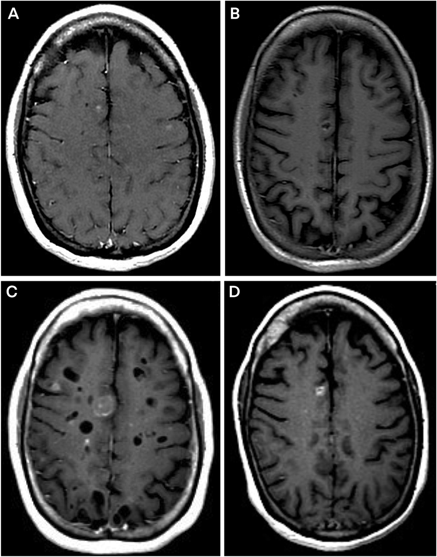
Imaging of the patient in CASE 6-1. Axial postcontrast T1-weighted MRIs show subcentimeter enhancing lesions at initial staging (A), radiographic response after initial treatment with pemetrexed and gemcitabine (B), recurrent disease after treatment with alectinib (C), and improvement of the largest metastasis and resolution of smaller metastases after treatment with lorlatinib (D).
FIGURE 6-4.

Images of the patient presented in CASE 6-2. Initial axial postcontrast T1-weighted (A)and fluid-attenuated inversion recovery (FLAIR) (B) MRIs show a single parenchymal brain metastasis with abundant surrounding vasogenic edema. C, Axial postcontrast MRI attwo months after treatment with stereotactic radiosurgery. New contrast enhancement is apparent (arrow). D, Axial brain positron emission tomography (PET) shows hypometabolism (arrow) at the sites of T1-enhancing disease, consistent with radiation necrosis. E, Sagittal postcontrast T1-weighted MRI shows linear contrast-enhancing deposits over the cerebellar folia and pons. F, Sagittal postcontrast T1-weighted MRI shows resolution of the enhancing deposits after whole-brain radiation therapy and carboplatin.
KEY POINTS.
Central nervous system metastases are common in patients with cancer and may occur in the brain, spinal cord, leptomeninges, epidural space, or dura.
Patients may harbor metastases in the brain parenchyma, spinal cord, leptomeninges, and epidural and dural spaces either singly or, more commonly, in combination. If a single discovered metastasis cannot adequately explain a patient’s symptoms or signs, imaging of additional sites is warranted.
Treatment of symptomatic lesions is the initial primary focus of care in patients with metastases to the central nervous system.
Parenchymal brain metastases grow within the confines of the blood-brain barrier, and systemic therapies must penetrate this space to be effective.
Leptomeningeal metastasis growth is bound by the blood-CSF barrier; treatments for malignancy within this space include intrathecal therapy and CSF-penetrant systemic therapies.
Central nervous system metastases are the result of selective genetic pressures and may therefore harbor mutations unlike those of the primary cancer.
When feasible, surgical resection of the symptomatic lesion(s) is preferred as it enables molecular and genetic characterization of the lesion.
When not surgically accessible, symptomatic central nervous system metastases may be treated with radiation therapy.
After surgical resection of central nervous system metastases, radiation is generally applied to the surgical bed to reduce the likelihood of recurrence.
The indications for the use of whole-brain radiation therapy are more restricted now than in the past.
Dural and epidural metastases reside outside the blood-brain and blood-CSF barriers and thus can be treated with standard chemotherapies, immunotherapies, and targeted therapies.
Intrathecal therapy is inappropriate for patients with elevated intracranial pressure or bulky leptomeningeal deposits.
Delivery of intrathecal therapy through an intraventricular catheter, such as an Ommaya reservoir, is preferable to delivery of the drug into the lumbar cistern.
Many new central nervous system–penetrating targeted therapies and immunotherapies have emerged that may be used in select cases to treat central nervous system metastases in lieu of radiation therapy.
Immunotherapy shows promise for parenchymal brain metastases from melanoma and non–small cell lung cancer in certain patient subpopulations.
RELATIONSHIP DISCLOSURE:
Dr Boire has received research/grant support from the Damon Runyon Cancer Research Foundation, the Pershing Square Sohn Cancer Research Alliance, The Pew Charitable Trusts, and theW. M. Keck Foundation.
Footnotes
UNLABELED USE OF PRODUCTS/INVESTIGATIONAL USE DISCLOSURE: Dr Boire discusses the unlabeled/investigational intrathecal use of trastuzumab to treat leptomeningeal metastasis.
REFERENCES
- 1.Lin J, Jandial R, Nesbit A, et al. Current and emerging treatments for brain metastases. Oncology (Williston Park) 2015;29(4):250–257. [PubMed] [Google Scholar]
- 2.Valiente M, Ahluwalia MS, Boire A, et al. The evolving landscape of brain metastasis. Trends Cancer 2018;4(3):176–196. doi: 10.1016/j.trecan.2018.01.003 [DOI] [PMC free article] [PubMed] [Google Scholar]
- 3.Gaspar L, Scott C, Rotman M, et al. Recursive partitioning analysis (RPA) of prognostic factors in three Radiation Therapy Oncology Group (RTOG) brain metastases trials. Int J Radiat Oncol Biol Phys 1997;37(4):745–751. doi: 10.1016/s0360-3016(96)00619-0 [DOI] [PubMed] [Google Scholar]
- 4.Stelzer KJ. Epidemiology and prognosis of brain metastases. Surg Neurol Int 2013;4(suppl 4): S192–S202. doi: 10.4103/2152-7806.111296 [DOI] [PMC free article] [PubMed] [Google Scholar]
- 5.Park JH, Kim YJ, Lee JO, et al. Clinical outcomes of leptomeningeal metastasis in patients with non-small cell lung cancer in the modern chemotherapy era. Lung Cancer 2012;76(3): 387–392. doi: 10.1016/j.lungcan.2011.11.022 [DOI] [PubMed] [Google Scholar]
- 6.Harstad L, Hess KR, Groves MD. Prognostic factors and outcomes in patients with leptomeningeal melanomatosis. Neuro Oncol 2008;10(6):1010–1018. doi: 10.1215/15228517-2008-062 [DOI] [PMC free article] [PubMed] [Google Scholar]
- 7.Kingston B, Kayhanian H, Brooks C, et al. Treatment and prognosis of leptomeningeal disease secondary to metastatic breast cancer: a single-centre experience. Breast 2017;36: 54–59. doi: 10.1016/j.breast.2017.07.015 [DOI] [PubMed] [Google Scholar]
- 8.Morikawa A, Jordan L, Rozner R, et al. Characteristics and outcomes of patients with breast cancer with leptomeningeal metastasis. Clin Breast Cancer 2017;17(1):23–28. doi: 10.1016/j.clbc.2016.07.002 [DOI] [PMC free article] [PubMed] [Google Scholar]
- 9.Schumacher M, Orszagh M. Imaging techniques in neoplastic meningiosis. J Neurooncol 1998; 38(2–3):111–120. doi: 10.1023/a:1005914321578 [DOI] [PubMed] [Google Scholar]
- 10.Sze G, Soletsky S, Bronen R, Krol G. MR imaging of the cranial meninges with emphasis on contrast enhancement and meningeal carcinomatosis. AJNR Am J Neuroradiol 1989;10(5):965–975. [PMC free article] [PubMed] [Google Scholar]
- 11.Glantz MJ, Cole BF, Glantz LK, et al. Cerebrospinal fluid cytology in patients with cancer: minimizing false-negative results. Cancer 1998;82(4):733–739. doi: 10.1002/(sici)1097-0142(19980215)82:4<733::aid-cncr17>3.0.co;2-z [DOI] [PubMed] [Google Scholar]
- 12.Wasserstrom WR, Glass JP, Posner JB. Diagnosis and treatment of leptomeningeal metastases from solid tumors: experience with 90 patients. Cancer 1982;49(4):759–772. doi: 10.1002/1097-0142(19820215)49:4<759∷aid-cncr2820490427>3.0.co;2-7 [DOI] [PubMed] [Google Scholar]
- 13.Kumar A, Weber MH, Gokaslan Z, et al. Metastatic spinal cord compression and steroid treatment: a systematic review. Clin Spine Surg 2017;30(4):156–163. doi: 10.1097/BSD.0000000000000528 [DOI] [PubMed] [Google Scholar]
- 14.Laufer I, Zuckerman SL, Bird JE, et al. Predicting neurologic recovery after surgery in patients with deficits secondary to MESCC: systematic review. Spine (Phila Pa 1976) 2016;41(suppl 20): S224–S230. doi: 10.1097/BRS.0000000000001827 [DOI] [PMC free article] [PubMed] [Google Scholar]
- 15.Brastianos PK, Carter SL, Santagata S, et al. Genomic characterization of brain metastases reveals branched evolution and potential therapeutic targets. Cancer Discov 2015;5: 1164–1177. doi: 10.1158/2159-8290.CD-15-0369 [DOI] [PMC free article] [PubMed] [Google Scholar]
- 16.De Mattos-Arruda L, Ng CKY, Piscuoglio S, et al. Genetic heterogeneity and actionable mutations in HER2-positive primary breast cancers and their brain metastases. Oncotarget 2018;9(29): 20617–20630. doi: 10.18632/oncotarget.25041 [DOI] [PMC free article] [PubMed] [Google Scholar]
- 17.Swinkels DW, de Kok JB, Hanselaar A, et al. Early detection of leptomeningeal metastasis by PCR examination of tumor-derived K-ras DNA in cerebrospinal fluid. Clin Chem 2000;46(1):132–133. doi: 10.1093/clinchem/46.1.132 [DOI] [PubMed] [Google Scholar]
- 18.Li Y, Pan W, Connolly ID, et al. Tumor DNA in cerebral spinal fluid reflects clinical course in a patient with melanoma leptomeningeal brain metastases. J Neurooncol 2016;128(1):93–100. doi: 10.1007/s11060-016-2081-5 [DOI] [PMC free article] [PubMed] [Google Scholar]
- 19.Pentsova EI, Shah RH, Tang J, et al. Evaluating cancer of the central nervous system through next-generation sequencing of cerebrospinal fluid. J Clin Oncol 2016;34(20):2404–2415. doi: 10.1200/JCO.2016.66.6487 [DOI] [PMC free article] [PubMed] [Google Scholar]
- 20.Pan W, Gu W, Nagpal S, et al. Brain tumor mutations detected in cerebral spinal fluid. Clin Chem 2015;61(3):514–522. doi: 10.1373/clinchem.2014.235457 [DOI] [PMC free article] [PubMed] [Google Scholar]
- 21.Li YS, Jiang BY, Yang JJ, et al. Unique genetic profiles from cerebrospinal fluid cell-free DNA in leptomeningeal metastases of EGFR-mutant non-small-cell lung cancer: a new medium of liquid biopsy. Ann Oncol 2018;29(4):945–952. doi: 10.1093/annonc/mdy009 [DOI] [PubMed] [Google Scholar]
- 22.Momtaz P, Pentsova E, Abdel-Wahab O, et al. Quantification of tumor-derived cell free DNA (cfDNA) by digital PCR (DigPCR) in cerebrospinal fluid of patients with BRAFV600 mutated malignancies. Oncotarget 2016;7(51): 85430–85436. doi: 10.18632/oncotarget.13397 [DOI] [PMC free article] [PubMed] [Google Scholar]
- 23.Boire A, Brandsma D, Brastianos PK, et al. Liquid biopsy in central nervous system metastases:a RANO review and proposals for clinical applications. Neuro Oncol 2019;21(5):571–584. doi: 10.1093/neuonc/noz012 [DOI] [PMC free article] [PubMed] [Google Scholar]
- 24.Patchell RA, Tibbs PA, Walsh JW, et al. A randomized trial of surgery in the treatmentof single metastases to the brain. N Engl J Med 1990;322(8):494–500. doi: 10.1056/NEJM199002223220802 [DOI] [PubMed] [Google Scholar]
- 25.Vecht CJ, Haaxma-Reiche H, Noordijk EM, et al. Treatment of single brain metastasis: radiotherapy alone or combined with neurosurgery? Ann Neurol 1993;33(6):583–590. doi: 10.1002/ana.410330605 [DOI] [PubMed] [Google Scholar]
- 26.Patchell RA, Tibbs PA, Regine WF, et al. Postoperative radiotherapy in the treatment of single metastases to the brain: a randomized trial. JAMA 1998;280(17):1485–1489. doi: 10.1001/jama.280.17.1485 [DOI] [PubMed] [Google Scholar]
- 27.Brown PD, Ballman KV, Cerhan JH, et al. Postoperative stereotactic radiosurgery compared with whole brain radiotherapy for resected metastatic brain disease (NCCTG N107C/CEC·3): a multicentre, randomised, controlled, phase 3 trial. Lancet Oncol 2017;18(8): 1049–1060. doi: 10.1016/S1470-2045(17)30441-2 [DOI] [PMC free article] [PubMed] [Google Scholar]
- 28.McTyre E, Ayala-Peacock D, Contessa J, et al. Multi-institutional competing risks analysis of distant brain failure and salvage patterns after upfront radiosurgery without whole brain radiotherapy for brain metastasis. Ann Oncol 2018;29(2):497–503. doi: 10.1093/annonc/mdx740 [DOI] [PubMed] [Google Scholar]
- 29.Atkins KM, Pashtan IM, Bussière MR, et al. Proton stereotactic radiosurgery for brain metastases: a single-institution analysis of 370 patients. Int J Radiat Oncol Biol Phys 2018;101(4):820–829. doi: 10.1016/j.ijrobp.2018.03.056 [DOI] [PubMed] [Google Scholar]
- 30.Mehta MP, Rodrigus P, Terhaard CH, et al. Survival and neurologic outcomes in a randomized trial of motexafin gadolinium and whole-brain radiation therapy in brain metastases. J Clin Oncol 2003;21(13):2529–2536. doi: 10.1200/JCO.2003.12.122 [DOI] [PubMed] [Google Scholar]
- 31.Suh JH, Stea B, Nabid A, et al. Phase III study of efaproxiral as an adjunct to whole-brain radiation therapy for brain metastases. J Clin Oncol 2006; 24(1):106–114. doi: 10.1200/JCO.2004.00.1768 [DOI] [PubMed] [Google Scholar]
- 32.Borgelt B, Gelber R, Kramer S, et al. The palliation of brain metastases: final results of the first two studies by the Radiation Therapy Oncology Group. Int J Radiat Oncol Biol Phys 1980;6(1):1–9. doi: 10.1016/0360-3016(80)90195-9 [DOI] [PubMed] [Google Scholar]
- 33.Brown PD, Jaeckle K, Ballman KV, et al. Effect of radiosurgery alone vs radiosurgery with whole brain radiation therapy on cognitive function in patients with 1 to 3 brain metastases: a Randomized Clinical Trial. JAMA 2016;316(4): 401–409. doi: 10.1001/jama.2016.9839 [DOI] [PMC free article] [PubMed] [Google Scholar]
- 34.Day J, Zienius K, Gehring K, et al. Interventions for preventing and ameliorating cognitive deficits in adults treated with cranial irradiation. Cochrane Database Syst Rev 2014;(12):CD011335. doi: 10.1002/14651858.CD011335.pub2 [DOI] [PMC free article] [PubMed] [Google Scholar]
- 35.Brown PD, Pugh S, Laack NN, et al. Memantine for the prevention of cognitive dysfunction in patients receiving whole-brain radiotherapy: a randomized, double-blind, placebo-controlled trial. Neuro Oncol 2013;15(10):1429–1437. doi: 10.1093/neuonc/not114 [DOI] [PMC free article] [PubMed] [Google Scholar]
- 36.Mulvenna P, Nankivell M, Barton R, et al. Dexamethasone and supportive care with or without whole brain radiotherapy in treating patients with non-small cell lung cancer with brain metastases unsuitable for resection or stereotactic radiotherapy (QUARTZ): results from a phase 3, non-inferiority, randomised trial. Lancet 2016;388 (10055):2004–2014. doi: 10.1016/S0140-6736(16)30825-X [DOI] [PMC free article] [PubMed] [Google Scholar]
- 37.Fisher CG, DiPaola CP, Ryken TC, et al. A novel classification system for spinal instability in neoplastic disease: an evidence-based approach and expert consensus from the Spine Oncology Study Group. Spine (Phila Pa 1976) 2010;35 (22): E1221–E1229. doi: 10.1097/BRS.0b013e3181e16ae2 [DOI] [PubMed] [Google Scholar]
- 38.Wang JC, Boland P, Mitra N, et al. Single-stage posterolateral transpedicular approach for resection of epidural metastatic spine tumors involving the vertebral body with circumferential reconstruction: results in 140 patients. Invited submission from the Joint Section Meeting on Disorders of the Spine and Peripheral Nerves, March 2004. J Neurosurg Spine 2004;1(3): 287–298. doi: 10.3171/spi.2004.1.3.0287 [DOI] [PubMed] [Google Scholar]
- 39.Mendel E, Bourekas E, Gerszten P, Golan JD. Percutaneous techniques in the treatment of spine tumors: what are the diagnostic and therapeutic indications and outcomes? Spine (Phila Pa 1976) 2009;34(22 suppl):S93–S100. doi: 10.1097/BRS.0b013e3181b77895 [DOI] [PubMed] [Google Scholar]
- 40.Patchell RA, Tibbs PA, Regine WF, et al. Direct decompressive surgical resection in the treatment of spinal cord compression caused by metastatic cancer: a randomised trial. Lancet 2005;366(9486):643–648. doi: 10.1016/S0140-6736(05)66954-1 [DOI] [PubMed] [Google Scholar]
- 41.Maranzano E, Trippa F, Casale M, et al. 8Gy single-dose radiotherapy is effective in metastatic spinal cord compression: results of a phase III randomized multicentre Italian trial. Radiother Oncol 2009;93(2):174–179. doi: 10.1016/j.radonc.2009.05.012 [DOI] [PubMed] [Google Scholar]
- 42.Rades D, Fehlauer F, Schulte R, et al. Prognostic factors for local control and survival after radiotherapy of metastatic spinal cord compression. J Clin Oncol 2006;24(21): 3388–3393. doi: 10.1200/JCO.2005.05.0542 [DOI] [PubMed] [Google Scholar]
- 43.Laufer I, Rubin DG, Lis E, et al. The NOMS framework: approach to the treatment of spinal metastatic tumors. Oncologist 2013;18(6):744–751. doi: 10.1634/theoncologist.2012-0293 [DOI] [PMC free article] [PubMed] [Google Scholar]
- 44.Lyndon D, Lansley JA, Evanson J, Krishnan AS. Dural masses: meningiomas and their mimics. Insights Imaging 2019;10(1):11. doi: 10.1186/s13244-019-0697-7 [DOI] [PMC free article] [PubMed] [Google Scholar]
- 45.Scherer K, Johnston J, Panda M. Dural based mass: malignant or benign. J Radiol Case Rep 2009;3(11):1–12. doi: 10.3941/jrcr.v3i11.189 [DOI] [PMC free article] [PubMed] [Google Scholar]
- 46.Pan Z, Yang G, He H, et al. Leptomeningeal metastasis from solid tumors: clinical features and its diagnostic implication. Sci Rep 2018;8: 10445. doi: 10.1038/s41598-018-28662-w [DOI] [PMC free article] [PubMed] [Google Scholar]
- 47.Mehta M, Bradley K. Radiation therapy for leptomeningeal cancer. Cancer Treat Res 2005; 125:147–158. doi: 10.1007/0-387-24199-x_9 [DOI] [PubMed] [Google Scholar]
- 48.Morris PG, Reiner AS, Szenberg OR, et al. Leptomeningeal metastasis from non-small cell lung cancer: survival and the impact of whole brain radiotherapy. J Thorac Oncol 2012;7(2): 382–385. doi: 10.1097/JTO.0b013e3182398e4f [DOI] [PubMed] [Google Scholar]
- 49.Gani C, Müller AC, Eckert F, et al. Outcome after whole brain radiotherapy alone in intracranial leptomeningeal carcinomatosis from solid tumors. Strahlenther Onkol 2012;188(1):148–153. doi: 10.1007/s00066-011-0025-8 [DOI] [PubMed] [Google Scholar]
- 50.Omuro AM, Lallana EC, Bilsky MH, DeAngelis LM. Ventriculoperitoneal shunt in patients with leptomeningeal metastasis. Neurology 2005; 64(9):1625–1627. doi: 10.1212/01.WNL.0000160396.69050.DC [DOI] [PubMed] [Google Scholar]
- 51.Nigim F, Critchlow JF, Kasper EM. Role of ventriculoperitoneal shunting in patients with neoplasms of the central nervous system: an analysis of 59 cases. Mol Clin Oncol 2015;3(6): 1381–1386. doi: 10.3892/mco.2015.627 [DOI] [PMC free article] [PubMed] [Google Scholar]
- 52.Glantz MJ, Cole BF, Recht L, et al. High-dose intravenous methotrexate for patients with nonleukemic leptomeningeal cancer: is intrathecal chemotherapy necessary? J Clin Oncol 1998;16(4): 1561–1567. doi: 10.1200/JCO.1998.16.4.1561 [DOI] [PubMed] [Google Scholar]
- 53.Grossman SA, Finkelstein DM, Ruckdeschel JC, et al. Randomized prospective comparison of intraventricular methotrexate and thiotepa in patients with previously untreated neoplastic meningitis. Eastern Cooperative Oncology Group. J Clin Oncol 1993;11(3):561–569. doi: 10.1200/JCO.1993.11.3.561 [DOI] [PubMed] [Google Scholar]
- 54.Pietanza MC, Kadota K, Huberman K, et al. Phase II trial of temozolomide in patients with relapsed sensitive or refractory small cell lung cancer, with assessment of methylguanine-DNA methyltransferase as a potential biomarker. Clin Cancer Res 2012;18(4):1138–1145. doi: 10.1158/1078-0432.CCR-11-2059 [DOI] [PubMed] [Google Scholar]
- 55.Trudeau ME, Crump M, Charpentier D, et al. Temozolomide in metastatic breast cancer (MBC): a phase II trial of the National Cancer Institute of Canada – Clinical Trials Group (NCIC-CTG). Ann Oncol 2006;17(6):952–956. doi: 10.1093/annonc/mdl056 [DOI] [PubMed] [Google Scholar]
- 56.Lee DH, Han JY, Kim HT, et al. Primary chemotherapy for newly diagnosed nonsmall cell lung cancer patients with synchronous brain metastases compared with whole-brain radiotherapy administered first: result of a randomized pilot study. Cancer 2008;113(1): 143–149. doi: 10.1002/cncr.23526 [DOI] [PubMed] [Google Scholar]
- 57.Rivera E, Meyers C, Groves M, et al. Phase I study of capecitabine in combination with temozolomide in the treatment of patients with brain metastases from breast carcinoma. Cancer 2006;107(6):1348–1354. doi: 10.1002/cncr.22127 [DOI] [PubMed] [Google Scholar]
- 58.Christodoulou C, Bafaloukos D, Linardou H, et al. Temozolomide (TMZ) combined with cisplatin (CDDP) in patients with brain metastases from solid tumors: a Hellenic Cooperative Oncology Group (HeCOG) Phase II study. J Neurooncol 2005;71(1):61–65. doi: 10.1007/s11060-004-9176-0 [DOI] [PubMed] [Google Scholar]
- 59.Ekenel M, Hormigo AM, Peak S, et al. Capecitabine therapy of central nervous system metastases from breast cancer. J Neurooncol 2007;85(2):223–227. doi: 10.1007/s11060-007-9409-0 [DOI] [PubMed] [Google Scholar]
- 60.Groen HJ, Smit EF, Haaxma-Reiche H, Postmus PE. Carboplatin as second line treatment for recurrent or progressive brain metastases from small cell lung cancer. Eur J Cancer 1993;29A(12): 1696–1699. doi: 10.1016/0959-8049(93)90107-q [DOI] [PubMed] [Google Scholar]
- 61.Chen G, Huynh M, Fehrenbacher L, et al. Phase II trial of irinotecan and carboplatin for extensive or relapsed small-cell lung cancer. J Clin Oncol 2009;27(9):1401–1404. doi: 10.1200/JCO.2008.20.2127 [DOI] [PMC free article] [PubMed] [Google Scholar]
- 62.Bailon O, Chouahnia K, Augier A, et al. Upfront association of carboplatin plus pemetrexed in patients with brain metastases of lung adenocarcinoma. Neuro Oncol 2012; 14(4):491–495. doi: 10.1093/neuonc/nos004 [DOI] [PMC free article] [PubMed] [Google Scholar]
- 63.Korfel A, Oehm C, von Pawel J, et al. Response to topotecan of symptomatic brain metastases of small-cell lung cancer also after whole-brain irradiation. A multicentre phase II study. Eur J Cancer 2002;38(13):1724–1729. doi: 10.1016/s0959-8049(02)00140-5 [DOI] [PubMed] [Google Scholar]
- 64.Beauchesne P Intrathecal chemotherapy for treatment of leptomeningeal dissemination of metastatic tumours. Lancet Oncol 2010(9);11: 871–879. doi: 10.1016/S1470-2045(10)70034-6 [DOI] [PubMed] [Google Scholar]
- 65.Siegal T, Lossos A, Pfeffer MR. Leptomeningeal metastases: analysis of 31 patients with sustained off-therapy response following combined-modality therapy. Neurology 1994; 44(8):1463–1469. doi: 10.1212/wnl.44.8.1463 [DOI] [PubMed] [Google Scholar]
- 66.Hitchins RN, Bell DR, Woods RL, Levi JA. A prospective randomized trial of single-agent versus combination chemotherapy in meningeal carcinomatosis. J Clin Oncol 1987;5(10):1655–1662. doi: 10.1200/JCO.1987.5.10.1655 [DOI] [PubMed] [Google Scholar]
- 67.Zagouri F, Sergentanis TN, Bartsch R, et al. Intrathecal administration of trastuzumab for the treatment of meningeal carcinomatosis in HER2-positive metastatic breast cancer: a systematic review and pooled analysis. Breast Cancer Res Treat 2013;139(1):13–22. doi: 10.1007/s10549-013-2525-y [DOI] [PubMed] [Google Scholar]
- 68.Costa DB, Shaw AT, Ou SH, et al. Clinical experience with crizotinib in patients with advanced ALK-rearranged non-small-cell lung cancer and brain metastases. J Clin Oncol 2015; 33(17):1881–1888. doi: 10.1200/JCO.2014.59.0539 [DOI] [PMC free article] [PubMed] [Google Scholar]
- 69.Gadgeel SM, Gandhi L, Riely GJ, et al. Safety and activity of alectinib against systemic disease and brain metastases in patients with crizotinib-resistant ALK-rearranged non-small-cell lung cancer (AF-002JG): results from the dose-finding portion of a phase 1/2 study. Lancet Oncol 2014; 15(10):1119–1128. doi: 10.1016/S1470-2045(14)70362-6 [DOI] [PubMed] [Google Scholar]
- 70.Kim DW, Tiseo M, Ahn MJ, et al. Brigatinib in patients with crizotinib-refractory anaplastic lymphoma kinase-positive non-small-cell lung cancer: a randomized, multicenter phase II trial. J Clin Oncol 2017;35(22):2490–2498. doi: 10.1200/JCO.2016.71.5904 [DOI] [PubMed] [Google Scholar]
- 71.Shaw AT, Solomon BJ, Chiari R, et al. Lorlatinib in advanced ROS1-positive non-small-cell lung cancer: a multicentre, open-label, single-arm, phase 1–2 trial. Lancet Oncol 2019;20(12):1691–1701. doi: 10.1016/S1470-2045(19)30655-2 [DOI] [PubMed] [Google Scholar]
- 72.Solomon BJ, Besse B, Bauer TM, et al. Lorlatinib in patients with ALK-positive non-small-cell lung cancer: results from a global phase 2 study. Lancet Oncol 2018;19(12):1654–1667. doi: 10.1016/S1470-2045(18)30649-1 [DOI] [PubMed] [Google Scholar]
- 73.Long GV, Flaherty KT, Stroyakovskiy D, et al. Dabrafenib plus trametinib versus dabrafenib monotherapy in patients with metastatic BRAF V600E/K-mutant melanoma: long-term survival and safety analysis of a phase 3 study. Ann Oncol 2019;30(11):1848. doi: 10.1093/annonc/mdz221 [DOI] [PMC free article] [PubMed] [Google Scholar]
- 74.Bai H, Han B. The effectiveness of erlotinib against brain metastases in non-small cell lung cancer patients. Am J Clin Oncol 2013;36(2): 110–115. doi: 10.1097/COC.0b013e3182438c91 [DOI] [PubMed] [Google Scholar]
- 75.Sperduto PW, Wang M, Robins HI, et al. A phase 3 trial of whole brain radiation therapy and stereotactic radiosurgery alone versus WBRT and SRS with temozolomide or erlotinib for non-small cell lung cancer and 1 to 3 brain metastases: Radiation Therapy Oncology Group 0320. Int J Radiat Oncol Biol Phys 2013;85(5):1312–1318. doi: 10.1016/j.ijrobp.2012.11.042 [DOI] [PMC free article] [PubMed] [Google Scholar]
- 76.Lee SM, Lewanski CR, Counsell N, et al. Randomized trial of erlotinib plus whole-brain radiotherapy for NSCLC patients with multiple brain metastases. J Natl Cancer Inst 2014;106(7). Pii:dju151. doi: 10.1093/jnci/dju151 [DOI] [PMC free article] [PubMed] [Google Scholar]
- 77.Iuchi T, Shingyoji M, Sakaida T, et al. Phase II trial of gefitinib alone without radiation therapy for Japanese patients with brain metastases from EGFR-mutant lung adenocarcinoma. Lung Cancer 2013;82(2):282–287. doi: 10.1016/j.lungcan.2013.08.016 [DOI] [PubMed] [Google Scholar]
- 78.Ceresoli GL, Cappuzzo F, Gregorc V, et al. Gefitinib in patients with brain metastases from non-small-cell lung cancer: a prospective trial. Ann Oncol 2004;15(7):1042–1047. doi: 10.1093/annonc/mdh276 [DOI] [PubMed] [Google Scholar]
- 79.Soria JC, Ohe Y, Vansteenkiste J, et al. Osimertinib in untreated EGFR-mutated advanced non-small-cell lung cancer. N Engl J Med 2018; 378(2):113–125. doi: 10.1056/NEJMoa1713137 [DOI] [PubMed] [Google Scholar]
- 80.Wu YL, Ahn MJ, Garassino MC, et al. CNS efficacy of osimertinib in patients with T790M-positive advanced non-small-cell lung cancer: data from a randomized phase III trial (AURA3). J Clin Oncol 2018;36(26):2702–2709. doi: 10.1200/JCO.2018.77.9363 [DOI] [PubMed] [Google Scholar]
- 81.Yang JC, Kim SW, Kim DW, et al. Osimertinib in patients with epidermal growth factor receptor mutation–positive non–small-cell lung cancer and leptomeningeal metastases: the BLOOM Study. J Clin Oncol 2020;38(6):538–547. doi: 10.1200/JCO.19.00457 [DOI] [PMC free article] [PubMed] [Google Scholar]
- 82.Lin NU, Diéras V, Paul D, et al. Multicenter phase II study of lapatinib in patients with brain metastases from HER2-positive breast cancer. Clin Cancer Res 2009;15(4):1452–1459. doi: 10.1158/1078-0432.CCR-08-1080 [DOI] [PubMed] [Google Scholar]
- 83.Bachelot T, Romieu G, Campone M, et al. Lapatinib plus capecitabine in patients with previously untreated brain metastases from HER2-positive metastatic breast cancer (LANDSCAPE): a single-group phase 2 study. Lancet Oncol 2013;14(1):64–71. doi: 10.1016/S1470-2045(12)70432-1 [DOI] [PubMed] [Google Scholar]
- 84.Cortés J, Dieras V, Ro J, et al. Afatinib alone or afatinib plus vinorelbine versus investigator’s choice of treatment for HER2-positive breast cancer with progressive brain metastases after trastuzumab, lapatinib, or both (LUX-Breast 3): a randomised, open-label, multicentre, phase 2 trial. Lancet Oncol 2015;16(16):1700–1710. doi: 10.1016/S1470-2045(15)00373-3 [DOI] [PubMed] [Google Scholar]
- 85.Freedman RA, Gelman RS, Anders CK, et al. TBCRC 022: a phase II trial of neratinib and capecitabine for patients with human epidermal growth factor receptor 2-positive breast cancer and brain metastases. J Clin Oncol 2019;37(13): 1081–1089. doi: 10.1200/JCO.18.01511 [DOI] [PMC free article] [PubMed] [Google Scholar]
- 86.Lin NU, Borges V, Anders C, et al. Intracranial efficacy and survival with tucatinib plus trastuzumab and capecitabine for previously treated HER2-positive breast cancer with brain metastases in the HER2CLIMB trial. J Clin Oncol 2020;38(23):2610–2619. doi: 10.1200/JCO.20.00775 [DOI] [PMC free article] [PubMed] [Google Scholar]
- 87.Davies MA, Saiag P, Robert C, et al. Dabrafenib plus trametinib in patients with BRAFV600-mutant melanoma brain metastases (COMBI-MB): a multicentre, multicohort, open-label, phase 2 trial. Lancet Oncol 2017;18(7):863–873. doi: 10.1016/S1470-2045(17)30429-1 [DOI] [PMC free article] [PubMed] [Google Scholar]
- 88.Kim KB, Kefford R, Pavlick AC, et al. Phase II study of the MEK1/MEK2 inhibitor trametinib in patients with metastatic BRAF-mutant cutaneous melanoma previously treated with or without a BRAF inhibitor. J Clin Oncol 2013;31(4):482–489. doi: 10.1200/JCO.2012.43.5966 [DOI] [PMC free article] [PubMed] [Google Scholar]
- 89.Shapiro WR, Young DF, Mehta BM. Methotrexate: distribution in cerebrospinal fluid after intravenous, ventricular and lumbar injections. N Engl J Med 1975;293(4):161–166. doi: 10.1056/NEJM197507242930402 [DOI] [PubMed] [Google Scholar]
- 90.Larson SM, Schall GL, Di Chiro G. Theinfluence of previous lumbar puncture and pneumoencephalography on the incidence of unsuccessful radioisotope cisternography. J Nucl Med 1971;12(8):555–557. [PubMed] [Google Scholar]
- 91.Grommes C, Oxnard GR, Kris MG, et al. “Pulsatile” high-dose weekly erlotinib for CNS metastases from EGFR mutant non-small cell lung cancer. Neuro Oncol 2011;13(12):1364–1369. doi: 10.1093/neuonc/nor121 [DOI] [PMC free article] [PubMed] [Google Scholar]
- 92.Hendriks LEL, Henon C, Auclin E, et al. Outcome of patients with non-small cell lung cancer and brain metastases treated with checkpoint inhibitors. J Thorac Oncol 2019;14(7):1244–1254. doi: 10.1016/j.jtho.2019.02.009 [DOI] [PubMed] [Google Scholar]
- 93.Goldberg SB, Gettinger SN, Mahajan A, et al. Pembrolizumab for patients with melanoma or non-small-cell lung cancer and untreated brain metastases: early analysis of a non-randomised, open-label, phase 2 trial. Lancet Oncol 2016;17(7): 976–983. doi: 10.1016/S1470-2045(16)30053-5 [DOI] [PMC free article] [PubMed] [Google Scholar]
- 94.Crino L, Bronte G, Bidoli P, et al. Nivolumab and brain metastases in patients with advanced non-squamous non-small cell lung cancer. Lung Cancer 2019;129:35–40. doi: 10.1016/j.lungcan.2018.12.025 [DOI] [PubMed] [Google Scholar]
- 95.Gadgeel SM, Lukas RV, Goldschmidt J, et al. Atezolizumab in patients with advanced non-small cell lung cancer and history of asymptomatic, treated brain metastases: Exploratory analyses of the phase III OAK study. Lung Cancer 2019;128:105–112. doi: 10.1016/j.lungcan.2018.12.017 [DOI] [PubMed] [Google Scholar]
- 96.Rulli E, Legramandi L, Salvati L, Mandala M. The impact of targeted therapies and immunotherapy in melanoma brain metastases: a systematic review and meta-analysis. Cancer 2019;125(21): 3776–3789. doi: 10.1002/cncr.32375 [DOI] [PubMed] [Google Scholar]
- 97.Tawbi HA, Forsyth PA, Algazi A, et al. Combined nivolumab and ipilimumab in melanoma metastatic to the brain. N Engl J Med 2018;379(8): 722–730. doi: 10.1056/NEJMoa1805453 [DOI] [PMC free article] [PubMed] [Google Scholar]
- 98.Long GV, Atkinson V, Lo S, et al. Combination nivolumab and ipilimumab or nivolumab alone in melanoma brain metastases: a multicentre randomised phase 2 study. Lancet Oncol 2018; 19(5):672–681. doi: 10.1016/S1470-2045(18)30139-6 [DOI] [PubMed] [Google Scholar]