



Abstract
Background
Seroprevalence studies, to estimate the proportion of people that has been infected by SARS-CoV-2 are importance in African countries, where incidence is among the lowest in the world.
Objective
This study aimed at evaluating the exposure to SARS-CoV-2 within a university setting of Cameroon.
Methods
A cross-sectional study performed in December 2020 - December 2021, among students and staffs of the Evangelical University of Cameroon. COVID-19 antigen rapid detection test (RDT) was performed using Standard Q Biosensor, and one year after SARS-CoV-2 antibody-test was performed within the same population using RDT and chemiluminescence immunoassay (CLIA).
Results
106 participants were enrolled (80% students), female sex was the most represented. Positivity to SARS-CoV-2 was 0.0% based on antigen RDTs. The seroprevalence of SARSCoV- 2 antibodies was estimated at 73.6% (95% CI. 64.5-81.0) for IgG and 1.9% (95% CI. 0.2-6.8) for IgM/IgG with RDTs, and 91.9% (95% CI. 84.7-96.4) for anti-nucleocapsid with CLIA. 95.3% (101) reported having developed at least one of the known COVID-19 symptoms (cough and headache being the most common). 90.3% (28) of people who experienced at least one of these symptoms developed IgG antibodies. 40.6% (43) of participants took natural herbs, whereas 55.7% (59) took conventional drugs. The most used herb was Zingiber officinale, while the most used drugs were antibiotics.
Conclusion
In this Cameroonian University community, SARS-CoV-2 seroprevalence is high, with a greater detection using advanced serological assays. This indicates a wide viral exposure, and the need to adequate control measures especially for those experiencing any related COVID-19 symptoms.
Key words: SARS-CoV-2, antibodies, seroprevalence, University of Cameroon, anti-N protein
Introduction
The world has experienced several major pandemics and epidemics since time ranging from the plague, Spanish flu, Asian flu, AIDS which is still present, to most recent the COVID-19 pandemic. 1 The latter has appeared since December 2019, and remains a concern for the scientific community, although there are different vaccines and treatments to prevent and manage the disease.2,3 Nevertheless, the immunological and inflammatory effects associated with its pathophysiology can be controlled with known drugs depending on the patient’s immunocompetence and the severity of the symptoms.4–7 A fundamental challenge in achieving COVID- 19 control is understanding transmission dynamics and regional disease prevalence and knowing what proportion of the population has been infected with the virus is an important step to optimize public health interventions, e.g., distancing measures and plan vaccination strategies. The first case of COVID-19 in Cameroon was diagnosed on 6 March 2020. As of 31st of March, and so far, there have been 119 947 confirmed cases of COVID-19 with 1 930 deaths.8 Given the difficulty and cost of RT-PCR based testing in resource-limited countries like Cameroon mildly affected or asymptomatic individuals are not usually screened, the number of confirmed SARS-CoV-2 infections is likely vastly underestimated.9
In this context, seroprevalence surveys are of the most importance to assess the proportion of the population that have already developed antibodies following a previous exposure to SARSCoV- 2 and the immune responses to the virus.10–12 The spike glycoprotein (S) and the nucleocapsid phosphoprotein (N) are the main viral antigenic targets against which antibodies are detected. 13,14 This Study was to evaluate exposure to SARS-CoV-2 and associated clinical factors within the University community of Cameroon.
Materials and methods
Ethical considerations
The antigenic screening was conducted under the direction of the Bandjoun District Health Services, as part of the epidemiological surveillance of COVID-19 in the municipality of Bandjoun (Cameroon). For the seroprevalence part, authorization was obtained from the Bandjoun District Health Services. All necessary precautions were taken to ensure that the rights, privacy, and freedom of participants were respected. Each participant was informed about the objective and methods of the study and signed free and informed consent before participating to the study.
Study design and population
Following school reopening and classical teaching method resumption, a screening campaign based on rapid antigenic tests was conducted on December 2020 at the Evangelical University of Cameroon (UEC) campus. A year later, a sero-surveillance campaign for anti-SARS-CoV-2 detection was carried out to assess the level of virus circulation within the same student community. The interval time between the two investigations (December 2020- February 2022) has been characterized by 2 successive waves of COVID-19.
Description of the study site
The Evangelical University of Cameroon is located in the Mbouo district of Bandjoun, Pomougne district, Koung-Khi department, Western Cameroon region. The town of Bandjoun is located on the right bank of the South Mifi, 17 km south of the regional capital Bafoussam.15 The district in which the UEC is located is one of the most popular and busiest because, in the same area, there is the large Protestant hospital of Mbouo, a nursery and primary school, a secondary school, as well as the evangelical church.
Enrolment of study participants
This study took place at the UEC campus at two different periods, Staff as well as students, were included. Antigenic RDTs (nasopharyngeal swabs) were performed in December 2020, while blood collection for seroprevalence testing took place between December 2021 and February 2022.The questionnaire was the main instrument for data collection. Whole blood was collected in BD VACUTAINER K3 EDTA tubes, plasma was obtained by centrifugation at 3000 rpm for 10 minutes at room temperature and stored at -20°C until use.
Laboratory procedures
Detection of SARS-CoV-2 antigen by rapid antigen test
The SARS-CoV-2 RDTs in nasopharyngeal specimens was performed with the STANDARD Q COVID-19 Ag Test (SD Biosensor Inc, KR). It is a Lateral-flow Immunochromatographic Assay (LFIA) for the qualitative detection of specific antigens to SARS-CoV-2 present in the human nasopharynx. The analysis was performed following the recommendation of the manufacturer. The result was considered negative when only the control line C was present. It was considered positive when there was presence of both test line T and control line C in the window result. It was invalid when the control line did not appear. The sensitivity reported by manufacturer was 88.7% and the specificity 97.6%.
Detection of antibodies SARS-CoV-2 by rapid diagnostic test
The Pharmact SARS-CoV-2 IgM/IgG Rapid Test kit (Pharmact, GmbH, DE) was used to screen for IgG and IgM antibodies against the SARS-CoV-2 virus on serum/plasma, following the manufacturer’s instructions. It is a LFIA for the qualitative detection of IgG and IgM antibodies against SARS-CoV-2 nucleocapsid (N) protein. Test results were categorized into one of the five categories: IgM positive only (indicating that recent infection within the first 7 to 10 days), IgG positive only (indicating that the body has been exposed to the virus in the past), IgG and IgM positive (also indicating recent infection between 7 and 21 days), negative (indicating that there are no such antibodies in the blood) and invalid/inconclusive. Results were repeated and graded accordingly. The manufacturer rates the sensitivity for IgM at 98.1% and for IgG at 98.2%, IgM specificity estimated at 99.5% and IgG 99.7% IgM precision estimated at 99.0% and IgG 99.2%.
Anti- Nucleocapsid Protein (anti-N) detection
The determination of SARS-CoV-2 anti-N antibodies to the nucleocapsid antigen, was performed in the Virology and Immunology Laboratory of the Bambino Gesù Hospital in Rome. Chemiluminescence assay (CLIA) was used to identify total antibodies (IgA, IgM, or IgG) directed against the N protein of the SARS-CoV-2. This was the qualitative detection test.
The assays were performed according to the manufacturer’s recommendations of Elecsys® Anti SARS-CoV-2 immunoassay (Roche Diagnostics, CH) based on the Sandwich principle. The sensitivity reported by the manufacturer was 99.5% and the specificity 99.8%. The sample was considered anti-N positive if the cutoff index ≥1.
Statistical analysis
All data were analysed using SPSS.20. Pearson’s chi 2 Test (with 95% confidence interval) was used for the statistical analysis. A statistical level less than 0.05 was considered statistically significant. The graphs were performed with GraphPad Prism 6.0.
Results
Detection of SARS-CoV-2 antigen with Rapid antigen test
Of the 106 participants in December 2020, 84.9% (90) were students and 15.1% (16) were staff; 56.6% (60) were female while 43.4% (46) were male. The mean age of the participants was 24.0±1 years. The results of the antigen RDTs indicated the absence of SARS-CoV-2 in all participants (100%, 106) (Table 1).
Detection of SARS-CoV-2 antibodies
General characteristics of participants
Between December 2021 and February 2022, one year after the SARS-COV-2 antigen screening campaign within the UEC campus, a SARS-CoV-2 seroprevalence investigation was conducted on the same community. 106 volunteers (80% were students while 20% were staff) participated to the seroprevalence screening. The female gender was the most represented (62.3%, 66) and 29.2% (55) of the participants declared they have been in contact at least once with confirmed COVID-19 positive patient. 95.3% (101) of participants claimed to have developed at least one of the known symptoms of COVID-19 since the last campaign in December 2020 (Table 2).
To fight against these symptoms, 62.6% (66) of the participants took drugs such as analgesics, anti-inflammatories, antibiotics, and others, with antibiotics (46.2%, 49) as the most widely used drugs class. 50.9% (54) used natural and local plants including Zingiber officinale (Ginger), Artemisia afra Jacq (Artemisia), Cymbopogon flexuosus (Lemongrass), Combretum micranthum (Kinkeliba). Ginger was the most widely used plant followed by Lemongrass and Artemisia (Table 3).
The seroprevalence of anti-SARS-CoV-2 antibodies found with RDTs in our study population was 73.6% (95% CI 64.5-81.0) for IgG and 1.9% (95% CI 0.2-6.8) for IgM/IgG. About 90.3% (28) had contact with a COVID-19 case and were SARS-CoV-2 IgG positive (Table 3).


Student population
For this study, 80% (85) were UEC students, with the largest proportion (28.2%, 24) from biomedical sciences (BMS). Level 3 (L3) students were the most represented (38.8%, 33), as well as female gender (67.1%, 57). The age of the participants ranged between 18-29 years, and the average age was 22.0± 2.1 years (Table 4).
In this young population, 28.2% (24) indicated have being in contact with positive cases of COVID-19. 93% (79) of these students claimed to have experienced at least one of the known symptoms of COVID-19 during the year 2021. 56.5% (48) took medication for these symptoms while 40.0% (34) took natural local plants.
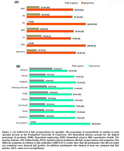
The results of rapid anti-SARS-CoV-2 antibody test showed that 80.0 % (CI 70.3-87.2) of students who participated in this study developed IgG antibodies against SARS-CoV-2 (Table 4). All biomedical engineering (BME) and reproductive health (RH) students tested positive of anti-SARS-CoV-2 and Agronomy and biomedical science (BMS) students showed the highest positive percentage 83.3% (05) and 79.2% (19) respectively (Figure 1A).
Clinical factors associated with the seroprevalence of SARSCoV- 2
More than half of the participants (75.2%, 76) who experienced symptoms were positive for the detection of IgG antibodies SARS-COV-2, while all individuals who had no symptoms tested positive for IgG detection (100.0%, 5). The most common symptoms reported among the IgG seropositive participants were headache, cough, rhinitis, and fever (Figure 1B).
The comparison between the results obtained from SARSCOV- 2 antigen detection in 2020 and seroprevalence investigation after one year clearly shows that, between December 2020 and December 2021 the virus strongly circulated through this community. In December 2020 there were no antigen positive cases in this population. However, the antibodies detection one year later shows that these people have indeed been in contact with the virus, although there have been no cases of serious symptoms or hospitalization (Table 5).
Detection of anti-N proteins
The detection of antibodies against protein N was done by the CLIA method, which is known as a gold standard compared to LFIA.16,17Analyses were performed of 99 samples previously tested for IgG detection by LFIA (the remaining 07 samples were excluded because the quality and quantity did not meet the requirements of the kit). The results showed that of these selected participants, 78.8% (95% CI 69.4-86.4) were positive for IgG by LFIA while 91.9% (95% CI 84.7-96.4) were positive for anti-N (Table 5). Of the 78.8% positive, 2% (2/99) had not detected for anti-N, while 76.8% (76/99) were detected for total anti-N against SARSCoV- 2, and 15.5% (15/99) of the IgG negative samples were detectable by CLIA. When the two types of analysis (LFIA and CLIA) were combined, 6.1% (6/99) of the participants had no antibodies against N protein at the time of the study.
Discussion
This study aimed to evaluate the circulation of SARS-CoV-2 within the community of the UEC.
Indeed, the perception of action in relation to COVID-19 is still mixed and dominated by hesitation in the Cameroonian community, 18 as we observed a low rate of acceptance of participation by students from other departments, except biomedical sciences.


The SARS-CoV-2 rapid antigen detection campaign showed no positive results among the participants. For COVID-19 screening, several rapid and molecular tests have been recently developed and quickly adopted in several countries across the globe including Cameroon.19,20 Availability and low cost guided the choice of Ag RDTs for this campaign, although it would have been best to combine them with molecular tests for SARS-CoV-2 to avoid false negatives.21
Since the beginning of COVID-19, low contaminations prevalence are declared in many countries, especially in Africa.22–24 In contrast to that, many seroprevalence studies showed important circulation of the virus although asymptomatic forms are recurrent.25 In this study, the seroprevalence of IgG anti-SARSCoV- 2 was 73.6% with 80% in young people aged 18-29 years. Similar studies were conducted in general population of the central part of Cameroon (Yaoundé) between June-August 2020 and October-November 2020, with an IgG seroprevalence of 24% in the first study and 29.2% in the second.26,27 The seroprevalences obtained in these studies are lower than those found in our study, this difference can be justified by the fact that our study was conducted in a young and student environment unlike theirs. In the same region, a more recent study (January-February and April- May 2021) showed an SARS-CoV-2 IgG seroprevalence increased significantly from 18.6% in the first survey to 51.3% in the second, illustrating high community transmission during the second wave of COVID-19.28 These results show that from one wave of COVID-19 to the next in Cameroon, the immune response is higher in the community, proving that the SARS-CoV-2 virus has circulated widely in this population, despite the low morbidity and mortality observed. Seroprevalence was highest in studies conducted in Central Africa, as well as among African-Americans, and antibody prevalence was highest among those under 30 years of age.13,29 It has been shown that LFIA assays may have a lower performance than enzyme-linked immunosorbent assays (ELISA) as well as chemiluminescent immunosorbent assays (CLIA), and the sensitivity of LFIA tests may be related to the profile of the infected patients.30–32 To address this gap, anti-N was also performed by the CLIA method, and the results revealed that 91.9% of the participants developed antibodies against protein N. This value was higher than those found by LFIA which could be justified as the CLIA method is more efficient and it has been shown that the sensitivity of LFIA is higher in symptomatic hospitalised patients than in asymptomatic patients,30 which constitutes the population of this study.
The SARS-CoV-2 N protein is one of the major immunogenic elements of the virus,33 and anti-nucleocapsid protein IgG antibodies are the best markers of infection and exposition.34,35 Furthermore, it has been shown that responses against N protein seem to attenuate over time, 36–38 and the high level of this antibody found in our study population would therefore indicate exposure to SARS-CoV-2 infection.
West Cameroon and precisely in Bandjoun where this study was conducted is a cold and dusty area with high prevalence of infectious diseases (respiratory and gut infections, parasitosis, and other) among inhabitants.39 This situation could have conferred to the population. a high adaptability for inflammation, one of the symptoms implicated in COVID-19 physiopathology in the lungs,4,7explaining the high circulation of SARS-CoV-2 without serious forms. In addition, our study population was young people, and their strong immune system could have prevented serious forms development.
At the start of the pandemic, many people rushed to self-medication, 40 and took, among others, natural plants,41–43 to prevent or treat symptoms of COVID-19. Several combinations of natural plants were used, perhaps these aspects could also justify the asymptomatic forms observed in this study, as several reports reviewed by Attah et al,44 confirmed antiviral properties of some of these plants to SARS-CoV-2 with cross-kingdom activity.45 The regulation of plant across kingdoms is a novel concept confirmed in other studies in which anti-inflammatory and anti-tumor activity was observed due to epigenetic alterations.46–49 Nevertheless, the excessive consumption of drugs in our study, like antibiotics, could soon raise another pandemic linked to antibiotic resistance.
More than half of the participants (52.0%) said they no longer respect social distancing, this observation had also been made previously by Metchum et al in the same setting.50 This non-respect of social distancing could be one of the factors that favoured the circulation of the virus among these students. In addition, most of the participants were health science students who, since December 2020 have completed several internships in a hospital environment. This undoubtedly increased their contamination risk as it is well known that the prevalence of COVID-19 is high among healthcare workers.51
Limitations of the study
The main limitation of this study was the small number of participants and the poor completing of questionnaire administered to each participant. The lack of adjusted seroprevalence data based on the pre-pandemic antibody survey. The need to perform the Ag detection by RDT in 2021 to strengthen the antibody findings.
Conclusions
Our finding shows high seroprevalence of SARS-CoV-2 in UEC young students and staff. These results may signify that after the first wave of COVID-19 with lockdown, the resumption of face-to-face school activities would have facilitated collective immunity within populations. SARS-CoV-2 already circulated widely among staff and students at a university in Cameroon. Despite a low level of COVID-19 vaccination there were no serious symptoms or hospitalisations reported. Several participants resorted to self-medication and local natural plants as a means of prevention from serious forms.
Acknowledgements
We thank all the students who participated in the data collection for this study. We would like to express our gratitude to the Charité Universital Medical Centre Berlin, the German Hospital Partnerships program and to the collaborators of the major tropical epidemic’s laboratory in Ndjamena. We are grateful to the Italian Agency for Development Cooperation for the biological analysis of our samples and the EDCTP, the European and Developing Countries Clinical Trial Partnership - PERFECT STUDY (RIA2020EF-3000) for financial support. Contributions: ALDW wrote the original draft, prepared, and designed the study. Data collection and lab analysis were done by BTN, AB, BMT, GL and ALDW. Writing review and editing by ACM, AMNS, CD, ATT, HKT, GC, CR, and JRK. Validation, interpretation and writing of the manuscript were done by all authors led by CFP, LS, and AG. All authors approved the final version and made the decision to submit for publication.
Funding Statement
Funding: This study received the support of Italian Agency for Development Cooperation and the EDCTP, the European and Developing Countries Clinical Trial Partnership - PERFECT STUDY (RIA2020EF- 3000).
References
- 1.History.com. Pandemics that changed history: timeline - history. History 2020. Available from: https://www.history.com/topics/middle-ages/pandemics-timeline [Google Scholar]
- 2.CDC. Stay up to date with your vaccines centers for disease control and prevention. 2022. Available from: https://www.cdc.gov/coronavirus/2019-ncov/vaccines/stayup-todate.html?CDC_AA_refVal=https%3A%2F%2Fwww.cdc.gov%2Fcoronavirus%2F2019-ncov%2Fvaccines%2Fdifferentvaccines.html [Google Scholar]
- 3.WHO. Therapeutics and COVID-19: living guideline 2022. Available from: https://www.who.int/publications/i/item/WHO-2019-nCoVtherapeutics-2022.4 [PubMed] [Google Scholar]
- 4.Tay MZ, Poh CM, Rénia L, et al. The trinity of COVID-19: immunity, inflammation and intervention. Nat Rev Immunol 2020;20:363–74. Available from: 10.1038/s41577-020-0311-8 [DOI] [PMC free article] [PubMed] [Google Scholar]
- 5.Marik PE, Iglesias J, Varon J, Kory P. A scoping review of the pathophysiology of COVID-19. Int J Immunopathol Pharmacol 2021;35:1–16. [DOI] [PMC free article] [PubMed] [Google Scholar]
- 6.García LF. Immune response, inflammation, and the clinical spectrum of COVID-19. Frontiers in Immunology. 2020: 4–8. [DOI] [PMC free article] [PubMed] [Google Scholar]
- 7.Song JW, Zhang C, Fan X, et al. Immunological and inflammatory profiles in mild and severe cases of COVID-19. Nat Commun. 2020;11. [DOI] [PMC free article] [PubMed] [Google Scholar]
- 8.Cameroon: WHO Coronavirus Disease (COVID-19) dashboard with vaccination data. Available from: https://covid19.who.int/region/afro/country/cm [Google Scholar]
- 9.Gelanew T, Seyoum B, Mulu A, et al. High seroprevalence of Anti-SARS-CoV-2 antibodies among ethiopian healthcare workers. Res Sq 2021; 22:261.Available from: /pmc/articles/PMC8312903/ [DOI] [PMC free article] [PubMed] [Google Scholar]
- 10.Long QX, Liu BZ, Deng HJ, et al. Antibody responses to SARS-CoV-2 in patients with COVID-19. Nat Med 2020;26:845–8. Available from: https://pubmed.ncbi.nlm.nih.gov/32350462/ [DOI] [PubMed] [Google Scholar]
- 11.Amanat F, Stadlbauer D, Strohmeier S, et al. A serological assay to detect SARS-CoV-2 seroconversion in humans. Nat Med 2020;26:1033. Available from: /pmc/articles/PMC8183627/ [DOI] [PMC free article] [PubMed] [Google Scholar]
- 12.Stadlbauer D, Amanat F, Chromikova V, et al. SARS-CoV-2 Seroconversion in Humans: a detailed protocol for a serological assay, antigen production, and test setup. Curr Protoc Microbiol 2020;57. Available from: https://pubmed.ncbi.nlm.nih.gov/32302069/ [DOI] [PMC free article] [PubMed] [Google Scholar]
- 13.Dwyer CJ, Cloud CA, Wang C, et al. Comparative analysis of antibodies to SARS-CoV-2 between asymptomatic and convalescent patients. iScience 2021;24:102489. Available from: /pmc/articles/PMC8087581/ [DOI] [PMC free article] [PubMed] [Google Scholar]
- 14.Jaimes JA, André NM, Chappie JS, et al. Phylogenetic analysis and structural modeling of SARS-CoV-2 spike protein reveals an evolutionary distinct and proteolytically sensitive activation loop. J Mol Biol 2020;432:3309–25. Available from: https://pubmed.ncbi.nlm.nih.gov/32320687/ [DOI] [PMC free article] [PubMed] [Google Scholar]
- 15.Pète-Bandjoun-Wikipédia. Available from: https://fr.wikipedia.org/wiki/Pète-Bandjoun [Google Scholar]
- 16.Mahajan S, Agarwal R, Rawat V, et al. Comparative evaluation of three rapid immunochromatographic test assays with chemiluminescent microparticle immunoassay for the detection of hepatitis C virus antibody. Virus Disease 2019;30:373–9. Available from: https://link.springer.com/article/10.1007/s13337-019-00542-5 [DOI] [PMC free article] [PubMed] [Google Scholar]
- 17.Mylemans M, Van Honacker E, Nevejan L, et al. Diagnostic and analytical performance evaluation of ten commercial assays for detecting SARS-CoV-2 humoral immune response. J Immunol Methods 2021;493:113043. Available from: /pmc/articles/PMC7989098/ [DOI] [PMC free article] [PubMed] [Google Scholar]
- 18.Dinga JN, Sinda LK, Titanji VPK. Assessment of vaccine hesitancy to a covid-19 vaccine in cameroonian adults and its global implication. Vaccines (Basel). 2021;9:175. Available from: /pmc/articles/PMC7922050/ [DOI] [PMC free article] [PubMed] [Google Scholar]
- 19.Yusuf L, Appeaning M, Amole TG, et al. Rapid, cheap, and effective COVID-19 diagnostics for Africa. Diagnostics (Basel) 2021;11:2105. Available from: https://pubmed.ncbi.nlm.nih.gov/34829451/ [DOI] [PMC free article] [PubMed] [Google Scholar]
- 20.Jacobs J, Kühne V, Lunguya O, et al. Implementing COVID- 19 (SARS-CoV-2 ) rapid diagnostic tests in sub-Saharan Africa : a review. Front Med (Lausanne) 2020;7:557797. [DOI] [PMC free article] [PubMed] [Google Scholar]
- 21.Mouliou DS, Gourgoulianis KI. False-positive and false-negative COVID-19 cases: respiratory prevention and management strategies, vaccination, and further perspectives. Expert Rev Respir Med 2021;15:993–1002. [DOI] [PMC free article] [PubMed] [Google Scholar]
- 22.Alex Ezeh, Michael Silverman SS. The impact of COVID-19 has been lower in Africa. We explore the reasons 2021. 2021;19. Available from: https://theconversation.com/theimpact-of-covid-19-has-been-lower-in-africa-we-explore-thereasons-164955 [Google Scholar]
- 23.Buguzi S. Reasons for Africa’s low COVID-19 rates revealed - sub-Saharan Africa 2020. Available from: https://medicalxpress.com/news/2020-10-africa-covid-revealed.html [Google Scholar]
- 24.Njenga MK, Dawa J, Nanyingi M, et al. Why is there low morbidity and mortality of COVID-19 in Africa? Am J Trop Med Hyg 2020;103:564. Available from: /pmc/articles/PMC7410455/ [DOI] [PMC free article] [PubMed] [Google Scholar]
- 25.Chisale MRO, Ramazanu S, Mwale SE, et al. Seroprevalence of anti-SARS-CoV-2 antibodies in Africa: A systematic review and meta-analysis. Rev Med Virol 2022;32: e2271. Available from: /pmc/articles/PMC8420234/ [DOI] [PMC free article] [PubMed] [Google Scholar]
- 26.Cheng MP, Yansouni CP, Basta NE, et al. Serodiagnostics for Severe Acute respiratory syndrome-related coronavirus 2 : a narrative review. Ann Intern Med 2020;173:450–60. Available from: https://pubmed.ncbi.nlm.nih.gov/32496919/ [DOI] [PMC free article] [PubMed] [Google Scholar]
- 27.Nwosu K, Fokam J, Wanda F, et al. SARS-CoV-2 antibody seroprevalence and associated risk factors in an urban district in Cameroon. Nat Commun 2021;12. Available from: https://pubmed.ncbi.nlm.nih.gov/34615863/ [DOI] [PMC free article] [PubMed] [Google Scholar]
- 28.Ndongo FA, Guichet E, Mimbé ED, et al. Rapid increase of community SARS-CoV-2 seroprevalence during second wave of COVID-19, Yaoundé, Cameroon. Emerg Infect Dis 2022;28: 1233-1236. Available from: https://pubmed.ncbi.nlm.nih.gov/35470795/ [DOI] [PMC free article] [PubMed] [Google Scholar]
- 29.Hajissa K, Islam MA, Hassan SA, et al. Seroprevalence of SARS-CoV-2 antibodies in Africa: a aystematic review and meta-analysis. Int J Environ Res Public Health. 2022;19:7257. [DOI] [PMC free article] [PubMed] [Google Scholar]
- 30.Hantz S. Diagnostic biologique de l’infection à Sars-CoV-2 : stratégies et interprétation des résultats. Rev Francoph Lab 2020:48-56. Available from: /pmc/articles/PMC7604167/ [DOI] [PMC free article] [PubMed] [Google Scholar]
- 31.Makoah NA, Tipih T, Litabe MM, et al. A systematic review and meta-analysis of the sensitivity of antibody tests for the laboratory confirmation of COVID-19. Future Virol 2021;17:119–39. Available from: /pmc/articles/PMC8686841/ [DOI] [PMC free article] [PubMed] [Google Scholar]
- 32.Montesinos I, Gruson D, Kabamba B, et al. Evaluation of two automated and three rapid lateral flow immunoassays for the detection of anti-SARS-CoV-2 antibodies. J Clin Virol 2020;128:104413. Available from: /pmc/articles/PMC7198434/ [DOI] [PMC free article] [PubMed] [Google Scholar]
- 33.Bai Z, Cao Y, Liu W, Li J. The SARS-CoV-2 nucleocapsid protein and its role in viral structure, biological functions, and a potential target for drug or vaccine mitigation. Viruses 2021;13:1115. Available from: /pmc/articles/PMC8227405/ [DOI] [PMC free article] [PubMed] [Google Scholar]
- 34.Qiu M, Shi Y, Guo Z, et al. Antibody responses to individual proteins of SARS coronavirus and their neutralization activities. Microbes Infect 2005;7:882–9. Available from: https://pubmed.ncbi.nlm.nih.gov/15878679/ [DOI] [PMC free article] [PubMed] [Google Scholar]
- 35.Szymczak A, Jędruchniewicz N, Torelli A, et al. Antibodies specific to SARS-CoV-2 proteins N, S and E in COVID-19 patients in the normal population and in historical samples. J Gen Virol 2021;102:1692. Available from: /pmc/articles/PMC8742988/ [DOI] [PMC free article] [PubMed] [Google Scholar]
- 36.Xiaojie S, Yu L, lei Y, et al. Neutralizing antibodies targeting SARS-CoV-2 spike protein. Stem Cell Res 2021;50:102125. Available from: /pmc/articles/PMC7737530/ [DOI] [PMC free article] [PubMed] [Google Scholar]
- 37.Fenwick C, Croxatto A, Coste AT, et al. Changes in SARSCoV- 2 Spike versus nucleoprotein antibody responses impact the estimates of infections in population-based seroprevalence studies. J Virol 2021;95:1–12. Available from: https://pubmed.ncbi.nlm.nih.gov/33144321/ [DOI] [PMC free article] [PubMed] [Google Scholar]
- 38.Weisblum Y, Schmidt F, Zhang F, et al. Escape from neutralizing antibodies by SARS-CoV-2 spike protein variants. Elife 2020;9:1. Available from: https://pubmed.ncbi.nlm.nih.gov/33112236/ [DOI] [PMC free article] [PubMed] [Google Scholar]
- 39.Monamele CG, Kengne-Nde C, Njifon HLM, et al. Clinical signs predictive of influenza virus infection in Cameroon. PLoS One. 2020;15:e0236267. [DOI] [PMC free article] [PubMed] [Google Scholar]
- 40.Quincho-Lopez A, Benites-Ibarra CA, Hilario-Gomez MM, et al. Self-medication practices to prevent or manage COVID-19: A systematic review. PLoS ONE. 2021; 16:e0259317. [DOI] [PMC free article] [PubMed] [Google Scholar]
- 41.Lim XY, Teh BP, Tan TYC. Medicinal plants in COVID-19: potential and limitations. Front Pharmacol 2021;12:611408. [DOI] [PMC free article] [PubMed] [Google Scholar]
- 42.Omokhua-Uyi AG, Van Staden J. Natural product remedies for COVID-19: A focus on safety. S Afr J Bot 2021;139:386-398. [DOI] [PMC free article] [PubMed] [Google Scholar]
- 43.Thota SM, Balan V, Sivaramakrishnan V. Natural products as home-based prophylactic and symptom management agents in the setting of COVID-19. Phytother Res. 2020;34:3148-3167. [DOI] [PMC free article] [PubMed] [Google Scholar]
- 44.Attah AF, Fagbemi AA, Olubiyi O, et al. Therapeutic potentials of antiviral plants used in traditional african medicine with COVID-19 in Focus: a Nigerian perspective. Front Pharmacol. 2021;12:596855. Available from: /pmc/articles/PMC8108136/ [DOI] [PMC free article] [PubMed] [Google Scholar]
- 45.Fongnzossie Fedoung E, Biwole AB, Nyangono Biyegue CF, et al. A review of Cameroonian medicinal plants with potentials for the management of the COVID-19 pandemic. Adv Tradit Med. 2021; 26:1–26. [Google Scholar]
- 46.Minutolo A, Potestà M, Roglia V, et al. Plant microRNAs from Moringa oleifera regulate immune response and HIV infection. Front Pharmacol. 2021;11:620038. Available from: https://pubmed.ncbi.nlm.nih.gov/33643043/ [DOI] [PMC free article] [PubMed] [Google Scholar]
- 47.Potestà M, Roglia V, Fanelli M, et al. Effect of microvesicles from Moringa oleifera containing miRNA on proliferation and apoptosis in tumor cell lines. Cell death Discov 2020;6:43. Available from: https://pubmed.ncbi.nlm.nih.gov/32550010/ [DOI] [PMC free article] [PubMed] [Google Scholar]
- 48.Minutolo A, Potestà M, Gismondi A, et al. Olea europaea small RNA with functional homology to human miR34a in crosskingdom interaction of anti-tumoral response. Sci Rep 2018;8. Available from: https://pubmed.ncbi.nlm.nih.gov/30120339/ [DOI] [PMC free article] [PubMed] [Google Scholar]
- 49.Pirrò S, Minutolo A, Galgani A, et al. Bioinformatics prediction and experimental validation of microRNAs involved in crosskingdom interaction. J Comput Biol 2016;23:976–89. Available from: http://www.liebertpub.com/doi/10.1089/cmb.2016.0059 [DOI] [PubMed] [Google Scholar]
- 50.TALOM Béatrice M, TALOM Alaric T, Aimé Cesaire M, et al. COVID-19 Knowledge, attitudes, and practices in the community of the Evangelical University of Cameroon. Special Journal of Public Heatlh, Nutrition, and Dietetics 2022; 2:1-12. [Google Scholar]
- 51.Haft JW, Atluri P, Ailawadi G, et al. Seroprevalence of SARSCoV- 2 antibodies and associated factors in healthcare workers: a systematic review and meta-analysis. Ann Thorac Surg. 2020;110:697–700.32305286 [Google Scholar]