



Abstract
Arthrogryposis multiplex congenita (AMC) [also known as multiple joints contracture or Fetal Akinesia Deformation Sequence (FADS)] is etiologically a heterogeneous condition with an estimated incidence of approximately 1 in 3000 live births and much higher incidence when prenatally diagnosed cases are included. The condition can be acquired or secondary to fetal exposures and can also be caused by a variety of single-gene disorders affecting the brain, spinal cord, peripheral nerves, neuromuscular junction, muscle, and a variety of disorders affecting the connective tissues (Niles et al., Prenatal Diagnosis, 2019; 39:720-731). The introduction of next-generation gene sequencing uncovered many genes and causative variants of AMC but also identified genes that cause both dominant and recessive inherited conditions with the variability of clinical manifestations depending on the genes and variants. Molecular diagnosis in these cases is not only important for prognostication but also for the determination of recurrence risk and for providing reproductive options including preimplantation and prenatal diagnosis. TTN, the largest known gene in the human genome, has been known to be associated with autosomal dominant dilated cardiomyopathy. However, homozygote and compound heterozygote pathogenic variants with recessive inheritance have rarely been reported. We report the effect of recessive variants located within the fetal IC and/or N2BA isoforms in association with severe FADS in three families. All parents were healthy obligate carriers and none of them had cardiac or skeletal muscle abnormalities. This report solidifies FADS as an alternative phenotypic presentation associated with homozygote/compound heterozygous pathogenic variants in the TTN.
Keywords: cardiomyopathy, fetal akinesia deformation sequence syndrome, myopathy, myopathy arthrogryposis multiplex congenita, TTN
1 |. INTRODUCTION
Myopathies are an etiologically heterogeneous group of conditions with variability in the clinical manifestations with the most severe forms presenting prenatally with fetal akinesia deformation sequence syndrome (FADS)/arthrogryposis multiplex congenita (AMC). Prenatal ultrasound diagnosis is done by showing by showing decreased or no fetal movements and multiple joint contractures although this is frequently not identified prior to delivery (Niles et al., 2019). The etiology of prenatally presenting myopathies is especially difficult to diagnose in view of missing functional information and the lack of histopathological findings since the endpoint of many of them shows similar histopathological manifestations, mainly muscle fibrosis. Furthermore, when intrauterine demise occurs, the fetal maceration makes further investigation impossible. Thus, more than 50% of the cases with FADS remain undiagnosed (Hall, 2014).
“Molecular autopsy,” using next-generation sequencing became crucial in the diagnosis of prenatal myopathies with different clinical presentations and modes of inheritance. Some of these genes are associated with well-known conditions when presenting postnatally but result in FADS when presenting prenatally.
We report three families with pregnancies complicated with fetal FADS and subsequent molecular analysis identifying compound heterozygote variants in TTN (MIM#188840) with the heterozygote parents having neither myopathy nor cardiomyopathy.
2 |. CLINICAL REPORTS
2.1 |. Family A
The parents were healthy, non-consanguineous, and of Jamaican descent. Detailed family history was non-contributory for congenital anomalies, recurrent miscarriages, stillbirth, cardiomyopathy, arrhythmia, sudden deaths, muscular dystrophy, or myopathy.
The couple’s first pregnancy resulted in a daughter who is well, and their second pregnancy (Figure 1a, II-2) was terminated for fetal akinesia and multiple joints contractures. Autopsy was not done. Their third pregnancy resulted in a daughter who is well. The couple was referred to the prenatal diagnosis and Medical Genetics Program at Mount Sinai Hospital, Toronto, Canada in their fourth pregnancy (Figure 1a, II-4) regarding the maternal feeling of decreased fetal movements. At this stage, the mother was 29 years old G4P2TA1L2 and the father was 31 years old. Screening for Down syndrome was declined and fetal ultrasound at 17 weeks gestation showed a nuchal fold of 6.9 mm with a small right pleural effusion, markedly decreased fetal movements, and breech presentation. The upper limbs were persistently crossed in front of the chest with clenched fists. The couple was counseled and decided to have amniocentesis showed a normal male karyotype. A follow-up ultrasound at 19 weeks confirmed a lack of fetal movements and multiple joint contractures. The couple was counseled and decided to interrupt the pregnancy. This was done by dilation and evacuation and DNA analysis for spinal muscular atrophy (SMA) and Walker-Warburg syndrome failed to identify a causative variant. Whole exome sequencing (WES) was not clinically available now.
FIGURE 1.

Pedigree of the three reported families with TTN truncating variants. Pedigree of a family with unaffected individuals is shown in white and affected with FADS are shown in black. Molecular testing was performed on individuals II-1, II-4, II-5, and II-6. ⊡ –Unaffected male carrier. ☉ –Unaffected female carrier.
The couple was counseled regarding the possible autosomal recessive mode of inheritance with a male and female affected with the same condition and decided to embark on their fifth pregnancy (Figure 1a, II-5). During this pregnancy, the mother was 32 years old and the nuchal translucency measured at 12 weeks gestation was 1.1 mm (normal: < 3.0 mm). Maternal serum screening for Down syndrome showed low risk. A detailed fetal ultrasound at 19 weeks gestation showed abnormal fetal position with persistent limbs’ joint contractures, small jerky body movements, edema of the forehead skin, and normal fetal anatomy. A repeat ultrasound at 20 weeks confirmed multiple joint contractures and fetal akinesia. The couple was counseled and decided to interrupt the pregnancy at 22 weeks gestation by induction of labor. Microarray analysis was normal and female. DNA analysis for the Pena-Shokeir syndrome panel, including DOK7, RAPSN, FCMD, FKRP, POMGnT1, POMT1, and POMT2, failed to identify a causative variant.
In their 6th pregnancy (Figure 1a, II-6), the mother was 36 years old and the fetal nuchal translucency measured 1.3 mm. Maternal serum screening for Down syndrome showed low risk. An early detailed fetal ultrasound at 15.5 weeks showed persistently flexed elbows and knees with no joint movements. There was an abnormal position of both feet and possible rocker-bottom feet. There were also minimal flexion and extension movements on both hips and no wrists or finger movements. Follow-up ultrasound at 17.5 weeks showed rocker bottom feet, flexed hands bilaterally with clenched left fist, and micrognathia. The findings were consistent with FADS. The couple decided to continue the pregnancy which resulted in intrauterine fetal death in the late second trimester. Microarray analysis was normal and female. DNA analysis for SMA and genes associated with the Pena-Shokeir syndrome panel was repeated and was negative for causative variants.
2.2 |. Family B
The mother was 26 years old G5P1SA3L1 of African American and Jamaican descent, and the father was 30 years old and of Haitian descent. The couple was healthy and non-consanguineous and have a daughter who is well. The patient has a history of two first trimester miscarriages and a prior ectopic pregnancy. Detailed family history was non-contributory. The mother presented during her fifth pregnancy (Figure 1b, II-5), at 29 weeks gestation for fetal ultrasound findings of hydrops, growth restriction, and decreased fetal movement.
Fetal ultrasound showed a nuchal thickening of 11.3 mm (normal <6 mm in the second trimester), skin edema along the back, mild retrognathia with the upper lip overhanging the lower lip, and intraabdominal umbilical vein dilation that did not meet the criteria for an umbilical vein varix (Figure 2). There were reduced fetal movements and the biophysical profile was 8/10. There was limited excitability with vibroacoustic stimulation. The amniotic fluid index was 11.0 and the estimated fetal weight was 1238 g (11th centile). She decided to have amniocentesis which showed a normal male microarray analysis. She was followed with serial ultrasounds and underwent a planned C-section for breech presentation at 39 + 2/7 weeks gestation.
FIGURE 2.

Fetal ultrasound images from the affected (II-4), Family B: (a) fetal profile. Note the forehead edema/increased skin thickness (arrowhead) and protruding upper lip (arrow). (b) Maxilla width measurement compared to mandible width measurement (2.66/3.24) showing mandible wider than the maxilla inconsistent with micrognathia. (c) Inferior facial angle measuring 62° (within normal range) inconsistent with micrognathia.
The neonatal exam was notable for a head circumference of 39 cm (99th centile), prominent forehead, microretrognathia, axial and appendicular hypotonia with the absence of spontaneous movements of the upper limbs, and minimal movement of the lower limbs, no sucking or rooting, complete head lag, absent DTRs, plantars upgoing, bilateral clinodactyly of the fifth fingers, absent flexion creases at all joints in upper and lower limbs, including absent palmar and plantar creases, joint contractures over the upper and lower limbs most severe in the ankles, hirsutism with significant arm, elbow, and leg hair and a small phallus.
Postnatal echocardiography showed pulmonary hypertension (~80% systemic), moderate patent ductus arteriosus with a left to right shunt, vertical vein noted entering the leftward aspect of the innominate vein (the differential included hemiazygos vein, prominent chest wall vein, or anomalous pulmonary vein) and patent foramen ovale. Skeletal survey showed thin bones and fractured left humerus. He initially took spontaneous breaths but had poor oxygen saturation resulting in intubation and assisted ventilation for the duration of his life. He initially received parental nutrition and later transitioned to enteral feedings. His initial CK was 475 U/L and this decreased during his hospital course. The baby died at 51 days of life due to Serratia sepsis from a chronic PICC line.
The couple subsequently conceived with an anatomically normal fetus. Chorionic villus sampling (CVS) identified the fetus as a heterozygous for the paternal variant.
2.3 |. Family C
The parents were healthy, non-consanguineous, and of Western-European descent and their detailed family history was non-contributory. The couple’s first pregnancy (Figure 1c, II-1), was complicated with an increased nuchal translucency at 12 + 4/7 weeks gestation. Following counseling, she decided to have CVS which showed a normal female microarray analysis. Detailed ultrasound at 20 weeks showed no detectable abnormalities. A repeat ultrasound at 36 weeks gestation showed polyhydramnios. The baby was born via vacuum-assisted vaginal delivery at 38 + 2/7 weeks gestation due to failure to progress and non-reassuring fetal heart rate. The baby’s weight was 2299 g (third centile) with Apgar scores of 2, 3, 5, and 7 at 1, 5, 10, and 15 min, respectively. At delivery, the baby was cyanotic, with no spontaneous movements, and of the upper and lower limbs and minimal movements of the fingers, and no respiratory effort. She was intubated and ventilated. Further examination showed facial dysmorphism with triangular facies, retromicrognathia, tenting of the upper lip, high arched palate, down-slanting palpebral fissures, low-set posteriorly rotated ears, widely spaced nipples, long fingers, prominent heels (Figure 3a–c). The neurology exam was remarkable for generalized hypotonia, decreased muscle bulk, and absent deep tendon reflexes. She had contractures at both wrists, elbows, knees, and ankles. DNA analysis for SMN1 and IGHMBP2 showed no detectable variants and deletion/duplication analysis of the IGHMBP2 gene was negative. The patient continued to require mechanical ventilation until care was withdrawn at 2 months of age.
FIGURE 3.

(a–c) Affected patients from Family C. (a–c) Patient II-1 and (d–f) patient II-4.
The couple’s second pregnancy resulted in a healthy daughter. In their third pregnancy, the mother presented at 11 + 5/7 week gestation with fetal ultrasound findings of NT of 4.2 mm, micrognathia, and abnormally positioned legs with the feet together and genu varus. CVS revealed a normal female microarray analysis. The patient opted for termination of pregnancy due to concerns of recurrence of the condition affecting their first pregnancy.
The couple’s fourth pregnancy was uncomplicated and fetal ultrasounds showed no detectable abnormalities apart from some decreased fetal movements in the third trimester. Delivery was spontaneous and vaginal at 39 weeks gestation. The birth weight was 2970 g (50th centile) and Apgar scores were 4, 6, 6, and 6 at 1, 5, 10, and 15 min, respectively. On examination, she was noted to have low-set and posteriorly rotated ears, widely spaced nipples, long fingers, retromicrognathia with tenting upper lip, and high arched palate (Figure 3d–f). The baby had a weak cry, generalized decreased muscle bulk, and poor tone. He was intubated and transferred to the NICU. The parents had genetic counseling and were informed of the similarities to their first two affected pregnancies and decided to withdraw care.
2.4 |. Materials and methods
WES was performed on the last three affected probands (II-4,5,6) from Family A, along with the biological parents (quint analysis) under the Care4Rare Canada initiative. The affected patients from Family B (Figure 1b, II-4) had clinical trio exome sequencing by a commercial laboratory. The affected patients from Family C (Figure 1c, II-1) had DNA analysis using a comprehensive muscular dystrophy/myopathy panel initially (183 genes) and later trio WES. Other individuals from Family C (Figure 1c, II-3) had testing of the familial variant, and (II-4) had rapid WES. Informed consent was obtained for WES, photographs, and publications from the parents according to the protocol approved by the patients’ hospitals [Mount Sinai Hospital in Toronto, Canada for Family A, The Johns Hopkins Center for Fetal Therapy for Family B, and University of Rochester Medical Center for Family C]. All the three molecular diagnostic laboratories have bioinformatic pipelines based on the general guidelines for variant interpretation by the American College of Medical Genetics and Genomics (Richards et al., 2015). Additionally, the bioinformatic pipelines incorporate gene-specific evidence on functional impact and genotype-phenotype correlation of different variant types. For example, truncating variants in highly spliced exons of TTN are more likely to have significant biological impact (Deo, 2016; Schafer et al., 2015) and their location within the gene is reported in association with disease severity (Savarese et al., 2020). Furthermore, when available, data on RNA molecular analyses, western blot, and functional studies are used to help decipher clinical significance of TTN variants (Perrin et al., 2020).
2.5 |. Histopathology
In Family A, a post-mortem examination was performed on the fragmented fetal tissue of the fourth pregnancy, and a complete examination of the intact fetus from pregnancy 5. Muscle tissue was submitted for histological examination and electron microscopy (EM) on the affected from Family A (II-5 and II-6) and Family C (II-1, 3, 4) and were examined using routine muscle biopsy and EM protocols. For the EM the tissue was fixed in buffered glutaraldehyde, post-fixed with uranium tetroxide and lead acetate, embedded in Epon Araldyte, and examined on a JEM-1400 transmission electron microscope. No muscle pathological examination was performed on the affected patient from Family B as MRI showed a paucity of intact muscle amenable for studies.
3 |. RESULTS
3.1 |. Molecular analysis
Summary of the molecular analysis results done on the three families is summarized in Table 1. In Family A, quint WES showed biallelic likely pathogenic variants in the three examined probands. Sanger sequencing confirmed the causative variants in all three affected siblings. None of the heterozygous parents showed cardiac dysfunction and echocardiography and ECG done on the parents were normal.
TABLE 1.
TTN variants identified in our patients
| Family | Number of tested probands | Decrease fetal movement | Contractures/fetal akinesia | Nucleotide change | Amino acid change | Ref Seq ID | Location | Region |
|---|---|---|---|---|---|---|---|---|
| Family A | 3 | + | + | c.38949delT | p.P12983fs*94 | NM_001267550 | Exon 200 | Metatranscript only |
| c.52601T>A | p.L17534* | NM_001267550 | Exon 275 | A-band | ||||
|
| ||||||||
| Family B | 2 | + | + | c.38442dupA | p.P12815Tfs*37 | NM_001267550 | Exon 194 | Metatranscript only |
| c.826C>T | p.Q276* | NM_001267550 | Exon 6 | Z-disk | ||||
|
| ||||||||
| Family C | 3 | + | + | c.15922C>T | p.R5308* | NM_001267550 | Exon 54 | I-band |
| c.85328T>A | p.V28443D | NM_001267550 | Exon 326 | A-band | ||||
In Family B, a prenatal WES showed biallelic compound heterozygote variants in TTN. These variants were both initially classified as VUS. The lab was recontacted for re-interpretation following the delivery of a subsequent healthy female who was a heterozygous carrier of the paternal allele. At this time, the maternal variant c.38442dupA (p.P12815Tfs*37) was reclassified as pathogenic and the paternal variant c.826C>T (p.Q276*) as likely pathogenic.
In Family C, DNA analysis of a comprehensive muscular dystrophy/myopathy panel (183 genes) identified biallelic variants in TTN: c.15922C>T (p.R5308*) classified as pathogenic and c.85328T>A (p.V28443D) classified as a variant of unknown significance (VUS). Follow-up WES on (CII-1) identified both TTN variants with the pathogenic being maternally inherited and the VUS being paternally inherited. No additional significant variants were identified. Other affected individuals from Family C (II-3) had targeted DNA analysis via Sanger sequencing for the familial variant which identified the familial biallelic variants in TTN. Familial testing was also performed on (II-2) and identified her as a carrier of one of the TTN variants. Moreover, another affected individual from Family C (II-4) had rapid WES analysis which identified the familial biallelic variants in TTN.
3.2 |. Autopsy and histopathology
Family A (II-5 and II-6): post-mortem examination of fetal remains from the fourth pregnancy demonstrated nuchal webbing, multiple joint contractures, pterygia at the axillae, elbows and knees, rocker-bottom feet, posteriorly placed anus and absent palmar creases. The fifth pregnancy (Figure 4a) demonstrated pterygia at the elbows, overlapping fingers with the second fingers overlapping the third and the third fingers overlapping the fourth. The lower limbs showed hips and knees contractures with pterygia and bilateral talipes equinovarus and rocker bottom feet. The muscle bulk was markedly reduced. In II-5, a complete autopsy enabled the examination of the viscera. The heart was normal in size and was developed appropriately for gestational age. The myocardium was histologically normal. Other internal organs were within normal limits and the brain and spinal cord showed no histopathological abnormalities. Examination of muscle tissue from both pregnancies demonstrated diffuse loss of muscle bulk with atrophic myofascicles, a marked excess of endomysial fibromyxoid tissue, and increased myofibers variation in size and maturational delay (Figure 4b). Muscle EM demonstrated myofibers with areas of myofibrillary disarray and in some areas, aggregates of electron-dense Z band material that resembled early nemaline body formation (Figure 4b).
FIGURE 4.
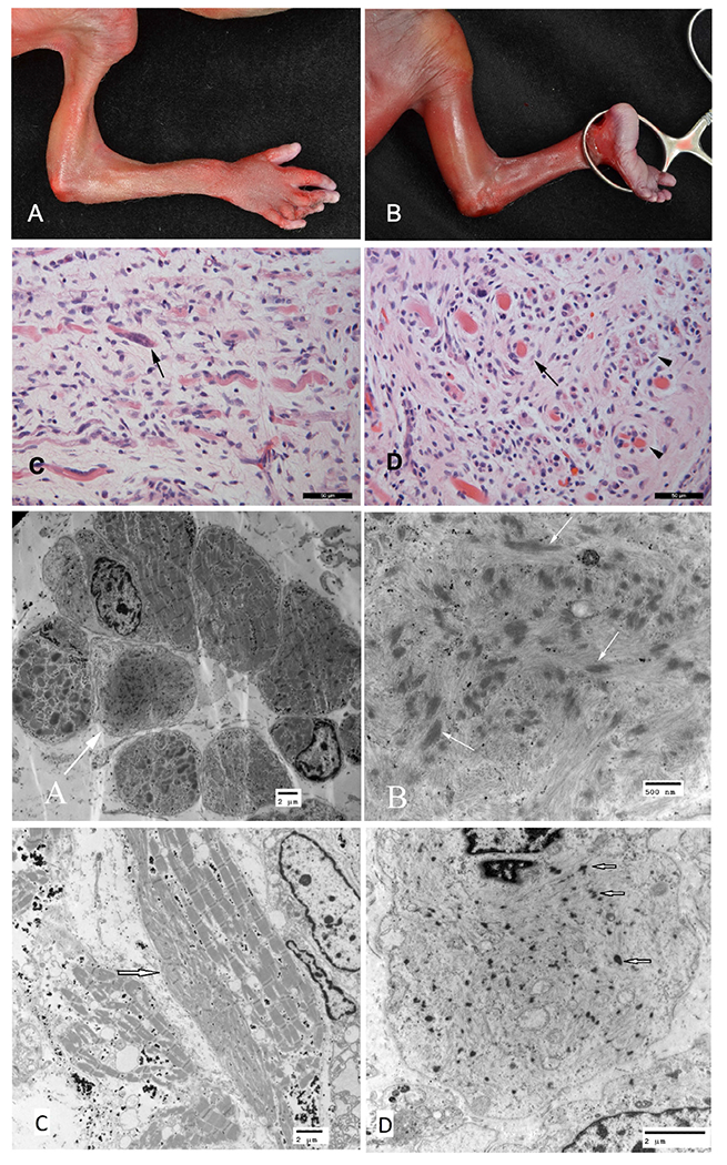
Family a, 5th pregnancy, demonstrating severe loss of muscle bulk with pterygia in axillae and elbows (a) and hips and knees (b). (b) Histology of fetal quadriceps in the same fetus with (c) longitudinal section demonstrating marked excess of intrafascicular fibromyxoid tissue with occasional fibers demonstrating clumped nuclei (arrow). (d) Cross section demonstrating individual fibers (arrow) and small hypoplastic fascicles containing poorly developed myocytes (arrowheads) surrounded by concentric loose connective tissue. (e) EM of muscle from the fifth pregnancy in the Family a demonstrates scattered fibers with myofibrillary disarray (arrow). (f) On higher magnification, some of the Z band material is forming electron-dense bodies (arrows) that are elongated in parallel to the thick filaments, strongly suggestive of early nemaline body formation. The muscle of pregnancy 4, although at an earlier gestational age, shows similar features with (g) many fibers have areas of sarcoplasmic disarray (arrow) adjacent to normal architecture, and in some (h) there are accumulations of Z band material similar to those shown in the sibling
Family C (II-1, 3, 4): muscle biopsy showed similar findings in all affected patients that suggested marked chronic denervation, characterized by groups of hypertrophic type 1 fibers and sheets of atrophic type 2 fibers. Autopsy performed on CII-4 showed flexion contractures of upper and lower limbs, symmetrical peripheral and axial muscle atrophy, and abnormal variation in fiber size due to muscle fiber atrophy and hypertrophy. EM demonstrated some myofibers with central nuclei and marginated organelles. The peripheral nerve was unremarkable (Figure 5).
FIGURE 5.

Family C—histology and ultrastructure of muscle, with histology suggestive of chronic denervation with re-innervation. (a) Muscle demonstrating clusters of hypertrophic fibers and groups of severely atrophic fibers (arrow). (b) Myofascicle composed largely of markedly atrophic fibers with occasional larger fibers. (c) NADH-tetrazolium reductase: Numerous small, darkly staining angulated fibers. (d) (ATPase, pH 4.2) Large groups of fibers with the same fiber type. EM (e) demonstrating rounded fibers with central nuclei (arrows), and (f) demonstrates myofibers with centralized sarcoplasm and peripheral organelles (arrow).
4 |. DISCUSSION
Titin sarcomere protein (TTN) plays an essential role in the sarcomere organization and elasticity of the cardiac and skeletal muscle cells (Tskhovrebova & Trinick, 2010). It is a giant muscle protein (also known as connectin; 4.2 MDa) which consists of four main regions, including Z-line, I-band, A-band, and M-line, with extensive genetic variation (Gigli et al., 2016) and a major role in assembling sarcomeres and supporting their contraction and relaxation (Tskhovrebova & Trinick, 2010). The TTN protein extends from its N-terminus anchored in the Z-disc to its C-terminus bound to thick filaments in the M-band. TTN contains 363 exons that are alternatively spliced to form different transcripts of varying lengths (Roberts et al., 2015). The TTN metatranscript (NM_001267550.1), is the longest titin isoform that includes all 363 coding exons. Many of the meta-only exons are exclusively expressed in fetal muscles and some have reduced expression postnatally (Savarese et al., 2018). The N2A is a shorter TTN isoform that is mainly expressed in skeletal muscle, the N2B isoform is expressed mainly in adult myocardium and N2BA is an isoform containing segments of N2A and N2B (Ge et al., 2019; Roberts et al., 2015; Schafer et al., 2017).
Dominant pathogenic variants are mainly associated with cardiomyopathies but some cases are also reported with several forms of myopathies, muscular dystrophies, and rarely the adult presentation of hereditary myopathy with early respiratory failure (Pfeffer et al., 2014). While the former remains a known disease-gene association, the improvement in next-generation sequencing technologies allowed us to discover new mutations and revealed an association with previously undiagnosed lethal FADS, caused by biallelic variants in the TTN with a recessive mode of inheritance (Chauveau et al., 2014).
Chauveau et al., (Chauveau et al., 2014) were the first to propose an association between biallelic TTN truncating variants and multi-mini core disease with arthrogryposis. Although many studies have described the association of recessive TTN variants with congenital myopathies (Ceyhan-Birsoy et al., 2013; Chauveau et al., 2014; Evilä et al., 2014, 2017; Harris et al., 2017; Savarese et al., 2018), only recently a few reports were published regarding the association of biallelic TTN variant and FADS (Bryen et al., 2020; Chervinsky et al., 2018; Fernández-Marmiesse et al., 2017; McDermott et al., 2021; Oates et al., 2018; Rees et al., 2021; Savarese et al., 2020). These biallelic variants are mainly metatranscript-only that affect the developmental isoforms, leading to a congenital phenotype of FADS with variable expression. Other reports on proximal recessive truncating variants in canonical exons expressed in adult isoforms lead to a prematurely truncated protein with nonsense-mediated decay and are associated with prenatal lethality (Savarese et al., 2018).
In this report, we identified three families with FADS and using WES were able to show compound heterozygous truncating variants in TTN, inherited from asymptomatic parents with a variety of ethnic backgrounds. In Family A, we identified the truncating variants p.P12984fs*94A and p.L17534* in trans, not previously reported in healthy control databases (EVS, 1000Genomes, gnomAD) or HGMD. Interestingly, the p.P12984fs*94A truncating variant presented in an alternate metatranscript, within the fetal IC isoform.
Variants located within the fetal IC isoform of TTN were reported in association with severe fetal FADS (Bryen et al., 2020; Chervinsky et al., 2018; Fernández-Marmiesse et al., 2017; McDermott et al., 2021; Oates et al., 2018; Savarese et al., 2020). The variants from Family B showed that the p.P12815Tfs*37 truncating variant presents solely in the alternate metatranscript, within the fetal IC isoform and is inherited in trans with a second truncating variant, p.Q276*. These variants were reclassified into pathogenic for the maternal allele (p.P12815Tfs*37) and likely pathogenic for the paternal allele (p.Q276*) after delivering a healthy female fetus who was found to be a heterozygous carrier of the paternal allele (Figure 1b, II-5). This added evidence of recessive inheritance. Both variants have not previously been reported in presumed healthy control databases, such as EVS or gnomAD (Lek et al., 2016).
In Family C, the p.R5308* and p.V28443D have been previously reported at a low level in the healthy control databases (EVS, 1000Genomes, gnomAD), and both are present within the alternate metatranscript but also within other coding transcripts of the TTN.
In Families A and B, one of the truncating variants was a TTN metatranscript-only variant, located within the fetal IC isoform. We thus conclude that having at least one of the recessive variants in the fetal IC isoform is likely to result in FADS providing a genotype-phenotype correlation. In contrast, Family C presented with two variants in the metatranscript but also in other coding portions of Titin primary transcripts (NM_133378.4 and NM_001256850.1) with the p.R5308* located within exon 54 and affecting N2BA and N2A elements. In reviewing the literature, we noted reports of one variant affecting these isoforms associated with generalized muscle weakness (Ge et al., 2019; Huang et al., 2021). Huang et al. (2021) reported an infant with generalized muscle weakness and a compound heterozygous splicing site variant that is predicted to affect the N2BA and N2A protein isoforms, within the I-band. The variant was in trans with another pathogenic nonsense variant. Ge et al. (2019) identified twins with generalized muscle weakness and compound heterozygous variants affecting also N2BA and N2A, within the I-band. Both Ge et al. (2019) and Huang et al. (2021) patients were reported mainly with congenital myopathy and not with contractures. The affected individuals from Family C in our report presented with congenital myopathy but were also found to have some joint contractures. None of the affected patients in our report had a cardiac complication which could be explained by the lack of N2B involvement. We thus conclude that the combination of variants either in metatranscript only or variants in the N2A or N2BA isoforms within the I-band would be associated with a spectrum of congenital myopathy and/or arthrogryposis.
Titinopathy has also been associated with different forms of cardiomyopathies, mostly dilated cardiomyopathy. Some studies also showed an association with arrhythmogenic right ventricular cardiomyopathy (Basso et al., 2009; Matthew et al., 2011). Although truncating dominant variants were found to be a major contributor to cardiomyopathy, some truncating variants were reported in asymptomatic individuals or parents of fetuses/newborns with autosomal recessive congenital myopathies/FADS (Chervinsky et al., 2018; Fernández-Marmiesse et al., 2017; Golbus et al., 2012; Herman et al., 2012). However, the non-cardiac effect of TTN variants has only been studied recently. In each of our families, the parents who carried the truncating variants were asymptomatic, had normal echocardiography and ECG, and had neither personal nor family history suggestive of cardiomyopathy. Some previous reports discussed the dissociation of some variants with the clinical phenotype in the dominantly inherited variants (Norton et al., 2013) raising the possibility of incomplete penetrance or variability in the clinical manifestations, known to be associated with autosomal dominant conditions (Peled et al., 2014). These studies expanded our knowledge of the molecular pathogenesis of titinopathies.
In Family A, the fetal muscle histopathology and EM at midgestation demonstrated severely underdeveloped skeletal muscle with sarcoplasmic disarray, and some features suggested early nemaline body formation with features, such as peripheral mitochondria, caps, and mini cores missing. The histopathological findings were dominated by severe atrophy of the developing muscle. Given the early stage of development, it is likely that the secondary changes in the myocytes simply have not developed. By contrast, the histology in Family C suggested denervation, with some features (myofiber group atrophy and hypertrophy) reminiscent of spinal muscular atrophy. However, central nuclei and marginated organelles were more obvious on the ultrastructural examination of the atrophic fibers. The severe muscle abnormalities, in the face of a normal central nervous system and peripheral nerves, point to a primary myopathic process.
In conclusion, our report confirms some of the previously reported genotype-phenotype correlations associated with TTN variants. As noted by us, pathogenic TTN variants with the fetal IC isoform, appear to represent the most severe end of the surviving spectrum of titinopathy associated with FADS. We thus recommend adding the TTN gene to the panel of genes included in the FADS/AMC DNA panel since identifying the TTN variants has a significant implication for the parents’ reproductive plans and can provide them with tools for prenatal and preimplantation genetic diagnosis.
ACKNOWLEDGMENTS
The authors thank the families for their participation in this project. No research funding was used for this research project.
Footnotes
CONFLICT OF INTEREST
The authors declare no conflict of interest.
DATA AVAILABILITY STATEMENT
Data sharing not applicable to this article as no datasets were generated or analysed during the current study.
REFERENCES
- Basso C, Corrado D, Marcus FI, Nava A, & Thiene G (2009). Arrhythmogenic right ventricular cardiomyopathy. The Lancet, 373, 1289–1300. [DOI] [PubMed] [Google Scholar]
- Bryen SJ, Ewans LJ, Pinner J, MacLennan SC, Donkervoort S, Castro D, Töpf A, O’Grady G, Cummings B, Chao KR, Weisburd B, Francioli L, Faiz F, Bournazos AM, Hu Y, Grosmann C, Malicki DM, Doyle H, Witting N, … Cooper ST (2020). Recurrent TTN metatranscript-only c.39974-11T>G splice variant associated with autosomal recessive arthrogryposis multiplex congenita and myopathy. Human Mutation, 41, 403–411. [DOI] [PMC free article] [PubMed] [Google Scholar]
- Ceyhan-Birsoy O, Agrawal PB, Hidalgo C, Schmitz-Abe K, DeChene ET, Swanson LC, Soemedi R, Vasli N, Iannaccone ST, Shieh PB, Shur N, Dennison JM, Lawlor MW, Laporte J, Markianos K, Fairbrother WG, Granzier H, & Beggs AH (2013). Recessive truncating titin gene, TTN, mutations presenting as centronuclear myopathy. Neurology, 81, 1205–1214. [DOI] [PMC free article] [PubMed] [Google Scholar]
- Chauveau C, Bonnemann CG, Julien C, Kho AL, Marks H, Talim B, Maury P, Arne-Bes MC, Uro-Coste E, Alexandrovich A, Vihola A, Schafer S, Kaufmann B, Medne L, Hübner N, Foley AR, Santi M, Udd B, Topaloglu H, … Ferreiro A (2014). Recessive TTN truncating mutations define novel forms of core myopathy with heart disease. Human Molecular Genetics, 23, 980–991. [DOI] [PMC free article] [PubMed] [Google Scholar]
- Chervinsky E, Khayat M, Soltsman S, Habiballa H, Elpeleg O, & Shalev S (2018). A homozygous TTN gene variant associated with lethal congenital contracture syndrome. American Journal of Medical Genetics. Part A, 176, 1001–1005. [DOI] [PubMed] [Google Scholar]
- Deo RC (2016). Alternative splicing, internal promoter, nonsense-mediated decay, or all three: Explaining the distribution of truncation variants in Titin. Circulation. Cardiovascular Genetics, 9, 419–425. [DOI] [PMC free article] [PubMed] [Google Scholar]
- Evilä A, Palmio J, Vihola A, Savarese M, Tasca G, Penttilä S, Lehtinen S, Jonson PH, De Bleecker J, Rainer P, Auer-Grumbach M, Pouget J, Salort-Campana E, Vilchez JJ, Muelas N, Olive M, Hackman P, & Udd B (2017). Targeted next-generation sequencing reveals novel TTN mutations causing recessive distal Titinopathy. Molecular Neurobiology, 54, 7212–7223. [DOI] [PubMed] [Google Scholar]
- Evilä A, Vihola A, Sarparanta J, Raheem O, Palmio J, Sandell S, Eymard B, Illa I, Rojas-Garcia R, Hankiewicz K, Negrão L, Löppönen T, Nokelainen P, Kärppä M, Penttilä S, Screen M, Suominen T, Richard I, Hackman P, & Udd B (2014). Atypical phenotypes in titinopathies explained by second titin mutations. Annals of Neurology, 75, 230–240. [DOI] [PubMed] [Google Scholar]
- Fernández-Marmiesse A, Carrascosa-Romero MC, Alfaro Ponce B, Nascimento A, Ortez C, Romero N, Palacios L, Jimenez-Mallebrera C, Jou C, Gouveia S, & Couce ML (2017). Homozygous truncating mutation in prenatally expressed skeletal isoform of TTN gene results in arthrogryposis multiplex congenita and myopathy without cardiac involvement. Neuromuscular Disorders, 27, 188–192. [DOI] [PubMed] [Google Scholar]
- Ge L, Fu X, Zhang W, Wang D, Wang Z, Yuan Y, Nonaka I, & Xiong H (2019). Recessive mutations in proximal I-band of TTN gene cause severe congenital multi-minicore disease without cardiac involvement. Neuromuscular Disorders, 29, 350–357. [DOI] [PubMed] [Google Scholar]
- Gigli M, Begay RL, Morea G, Graw SL, Sinagra G, Taylor MRG, Granzier H, & Mestroni L (2016). A review of the giant protein titin in clinical molecular diagnostics of cardiomyopathies. Frontiers in Cardiovascular Medicine, 3, 21. [DOI] [PMC free article] [PubMed] [Google Scholar]
- Golbus JR, Puckelwartz MJ, Fahrenbach JP, Dellefave-Castillo LM, Wolfgeher D, & McNally EM (2012). Population-based variation in cardiomyopathy genes. Circulation. Cardiovascular Genetics, 5, 391–399. [DOI] [PMC free article] [PubMed] [Google Scholar]
- Hall JG (2014). Arthrogryposis (multiple congenital contractures): Diagnostic approach to etiology, classification, genetics, and general principles. European Journal of Medical Genetics, 57, 464–472. [DOI] [PubMed] [Google Scholar]
- Harris E, Töpf A, Vihola A, Evilä A, Barresi R, Hudson J, Hackman P, Herron B, MacArthur D, Lochmüller H, Bushby K, Udd B, & Straub V (2017). A “second truncation” in TTN causes early onset recessive muscular dystrophy. Neuromuscular Disorders, 27, 1009–1017. [DOI] [PubMed] [Google Scholar]
- Herman DS, Lam L, Taylor MRG, Wang L, Teekakirikul P, Christodoulou D, Conner L, DePalma SR, McDonough B, Sparks E, Teodorescu DL, Cirino AL, Banner NR, Pennell DJ, Graw S, Merlo M, Di Lenarda A, Sinagra G, Bos JM, … Seidman CE (2012). Truncations of titin causing dilated cardiomyopathy. The New England Journal of Medicine, 366, 619–628. [DOI] [PMC free article] [PubMed] [Google Scholar]
- Huang S, Ma Y, Zhang Y, Xiong H, & Chang X (2021). Centronuclear myopathy due to a de novo nonsense variant and a maternally inherited splice-site variant in TTN: A case report. Clinical Case Reports, 9, e04478. [DOI] [PMC free article] [PubMed] [Google Scholar]
- Lek M, Karczewski KJ, Minikel EV, Samocha KE, Banks E, Fennell T, O’Donnell-Luria AH, Ware JS, Hill AJ, Cummings BB, Tukiainen T, Birnbaum DP, Kosmicki JA, Duncan LE, Estrada K, Zhao F, Zou J, Pierce-Hoffman E, Berghout J, … Exome Aggregation Consortium. (2016). Analysis of protein-coding genetic variation in 60,706 humans. Nature, 536, 285–291. [DOI] [PMC free article] [PubMed] [Google Scholar]
- Matthew T, Sharon G, Gianfranco S, Carl B, Dobromir S, Francesca B, Bruno P, Salcedo EE, William S, Stylianos P, Brian A, Bernd S, Julius B, Siegfried L, Henk G, & Luisa M (2011). Genetic variation in titin in arrhythmogenic right ventricular cardiomyopathy-overlap syndromes. Circulation, 124, 876–885. [DOI] [PMC free article] [PubMed] [Google Scholar]
- McDermott H, Henderson A, Robinson HK, Heaver R, Halahakoon C, Cox H, & Naik S (2021). Novel compound heterozygous TTN variants as a cause of severe neonatal congenital contracture syndrome without cardiac involvement diagnosed with rapid trio exome sequencing. Neuromuscular Disorders, 31, 783–787. [DOI] [PubMed] [Google Scholar]
- Niles KM, Blaser S, Shannon P, & Chitayat D (2019). Fetal arthrogryposis multiplex congenita/fetal akinesia deformation sequence (FADS)-Aetiology, diagnosis, and management. Prenatal Diagnosis, 39, 720–731. [DOI] [PubMed] [Google Scholar]
- Norton N, Li D, Rampersaud E, Morales A, Martin ER, Zuchner S, Guo S, Gonzalez M, Hedges DJ, Robertson PD, Krumm N, Nickerson DA, & Hershberger RE (2013). Exome sequencing and genome-wide linkage analysis in 17 families illustrate the complex contribution of TTN truncating variants to dilated cardiomyopathy. Circulation. Cardiovascular Genetics, 6, 144–153. [DOI] [PMC free article] [PubMed] [Google Scholar]
- Oates EC, Jones KJ, Donkervoort S, Charlton A, Brammah S, Smith JE, Ware JS, Yau KS, Swanson LC, Whiffin N, Peduto AJ, Bournazos A, Waddell LB, Farrar MA, Sampaio HA, Teoh HL, Lamont PJ, Mowat D, Fitzsimons RB, … Laing NG (2018). Congenital Titinopathy: Comprehensive characterization and pathogenic insights. Annals of Neurology, 83, 1105–1124. [DOI] [PMC free article] [PubMed] [Google Scholar]
- Peled Y, Gramlich M, Yoskovitz G, Feinberg MS, Afek A, Polak-Charcon S, Pras E, Sela B-A, Konen E, Weissbrod O, Geiger D, Gordon PMK, Thierfelder L, Freimark D, Gerull B, & Arad M (2014). Titin mutation in familial restrictive cardiomyopathy. International Journal of Cardiology, 171, 24–30. [DOI] [PubMed] [Google Scholar]
- Perrin A, Juntas Morales R, Rivier F, Cances C, Walther-Louvier U, Van Goethem C, Thèze C, Lacourt D, Pégeot H, Zenagui R, Uro-Coste E, Leboucq N, Malfatti E, Delaby C, Lehmann S, Rigau V, Koenig M, & Cossée M (2020). The importance of an integrated genotype-phenotype strategy to unravel the molecular bases of titinopathies. Neuromuscular Disorders, 30, 877–887. [DOI] [PubMed] [Google Scholar]
- Pfeffer G, Barresi R, Wilson IJ, Hardy SA, Griffin H, Hudson J, Elliott HR, Ramesh AV, Radunovic A, Winer J, Vaidya S, Raman A, Busby M, Farrugia ME, Ming A, Everett C, Emsley HC, Horvath R, Straub V, … Sarkozy A (2014). Titin founder mutation is a common cause of Myofibrillar myopathy with early respiratory failure. Journal of Neurology, Neurosurgery, and Psychiatry, 85, 331–338. [DOI] [PMC free article] [PubMed] [Google Scholar]
- Rees M, Nikoopour R, Fukuzawa A, Kho AL, Fernandez-Garcia MA, Wraige E, Bodi I, Deshpande C, Özdemir Ö, Daimagüler H-S, Pfuhl M, Holt M, Brandmeier B, Grover S, Fluss J, Longman C, Farrugia ME, Matthews E, Hanna M, … Gautel M (2021). Making sense of missense variants in TTN-related congenital myopathies. Acta Neuropathologica, 141, 431–453. [DOI] [PMC free article] [PubMed] [Google Scholar]
- Richards S, Aziz N, Bale S, Bick D, Das S, Gastier-Foster J, Grody WW, Hegde M, Lyon E, Spector E, Voelkerding K, Rehm HL, & ACMG Laboratory Quality Assurance Committee. (2015). Standards and guidelines for the interpretation of sequence variants: A joint consensus recommendation of the American College of Medical Genetics and Genomics and the Association for Molecular Pathology. Genetics in Medicine, 17, 405–424. [DOI] [PMC free article] [PubMed] [Google Scholar]
- Roberts AM, Ware JS, Herman DS, Schafer S, Baksi J, Bick AG, Buchan RJ, Walsh R, John S, Wilkinson S, Mazzarotto F, Felkin LE, Gong S, MacArthur JAL, Cunningham F, Flannick J, Gabriel SB, Altshuler DM, Macdonald PS, … Cook SA (2015). Integrated allelic, transcriptional, and phenomic dissection of the cardiac effects of titin truncations in health and disease. Science Translational Medicine, 7, 270ra6. [DOI] [PMC free article] [PubMed] [Google Scholar]
- Savarese M, Maggi L, Vihola A, Jonson PH, Tasca G, Ruggiero L, Bello L, Magri F, Giugliano T, Torella A, Evilä A, Di Fruscio G, Vanakker O, Gibertini S, Vercelli L, Ruggieri A, Antozzi C, Luque H, Janssens S, … Nigro V(2018). Interpreting genetic variants in Titin in patients with muscle disorders. JAMA Neurology, 75, 557–565. [DOI] [PMC free article] [PubMed] [Google Scholar]
- Savarese M, Vihola A, Oates EC, Barresi R, Fiorillo C, Tasca G, Jokela M, Sarkozy A, Luo S, Díaz-Manera J, Ehrstedt C, Rojas-García R, Sáenz A, Muelas N, Lonardo F, Fodstad H, Qureshi T, Johari M, Välipakka S, … Udd B (2020). Genotype-phenotype correlations in recessive titinopathies. Genetics in Medicine, 22, 2029–2040. [DOI] [PubMed] [Google Scholar]
- Schafer S, de Marvao A, Adami E, Fiedler LR, Ng B, Khin E, Rackham OJL, van Heesch S, Pua CJ, Kui M, Walsh R, Tayal U, Prasad SK, Dawes TJW, Ko NSJ, Sim D, Chan LLH, Chin CWL, Mazzarotto F, … Cook SA (2017). Titin-truncating variants affect heart function in disease cohorts and the general population. Nature Genetics, 49, 46–53. [DOI] [PMC free article] [PubMed] [Google Scholar]
- Schafer S, Miao K, Benson CC, Heinig M, Cook SA, & Hubner N (2015). Alternative splicing signatures in RNA-seq data: Percent spliced in (PSI). Current Protocols in Human Genetics, 87, 11.16.1–11.16.14. [DOI] [PubMed] [Google Scholar]
- Tskhovrebova L, & Trinick J (2010). Roles of titin in the structure and elasticity of the sarcomere. Journal of Biomedicine & Biotechnology, 2010, e612482. [DOI] [PMC free article] [PubMed] [Google Scholar]
Associated Data
This section collects any data citations, data availability statements, or supplementary materials included in this article.
Data Availability Statement
Data sharing not applicable to this article as no datasets were generated or analysed during the current study.