Abstract
Background
Despite the advances in anterior cervical corpectomy and fusion (ACCF) as a reconstructive surgical technique, the rate of complications related to artificial implants remains high. The purpose of this study was to investigate the long-term clinical course of ACCF with tantalum trabecular metal (TTM)-lordotic implants. Focus is placed on the relevance and influence of implant subsidence on sagittal alignment and the related clinical implications.
Methods
Retrospective, observational study of prospectively collected outcomes including 56 consecutive patients with degenerative cervical disc disease (myelopathy and/or radiculopathy). All patients underwent 1-level or 2-level ACCF with TTM-lordotic implants. The mean duration of follow-up was 4.85 years.
Results
The fusion rate at the end of follow-up was 98.11% (52/53). Implant subsidence occurred in 44 (83.01%) cases, including slight subsistence (<3 mm) in 37 (69.81%) and severe subsidence (>3 mm) in 7 cases (13.2%). The greatest degree of subsidence developed in the first 3 months postoperatively (P = 0.003). No patients presented a significant increase in implant subsidence beyond the second year of follow-up. The most common site of severe subsidence was the anterior region of the cranial end plate (4/7). At the end of follow-up, C1-C7 lordosis and segmental-Cobb angle of the fused segment increased on average by 5.06 ± 8.26 and 1.98 ± 6.02 degrees, respectively, though this difference failed to reach statistical significance (P > 0.05). Visual analog scale and Neck Disability Index scores improved at the conclusion of follow-up (P < 0.05).
Conclusions
ACCF with anterior cervical reconstruction using TTM-lordotic implants and anterior cervical plating for treatment of cervical degenerative disease has high fusion rates and good clinical outcome. The osteoconductive properties of TTM provide immediate stabilization and eliminate the need for bone grafts to ensure solid bone fusion. Before fusion occurs, asymptomatic implant settlement into the vertebral body is inevitable. However, lack of parallelism and reduced contact surface between the implant and the vertebral end plate are major risk factors for severe further subsidence, which may negatively affect the clinical outcomes.
Level of Evidence
4.
Keywords: anterior cervical corpectomy; anterior cervical fusion; tantalum, cervical spine; subsidence; graft collapse; cervical lordosis; trabecular metal
INTRODUCTION
Anterior cervical corpectomy and fusion (ACCF) with artificial fusion implants was developed to prevent the appearance of complications associated with the use of autologous bone grafts in the donor area and lower the likelihood of graft collapse.
When used in ACCF, artificial implants act as spacers that can restore physiological alignment of the cervical segments removed, providing immediate and stable load support and facilitating arthrodesis. The procedure, which has a fusion rate of between 95% and 100%, has demonstrated satisfactory clinical outcomes.1–4 Nonetheless, the evidence in support of artificial implants over autologous bone grafts for reconstruction in ACCF is scarce, as despite the advances in reconstructive techniques, the procedure continues to have a high rate of complications, though now this high rate is related to the implants themselves. Some of the most common complications following ACCF reconstruction with artificial implants are progressive implant subsidence within the adjacent vertebral bodies, migration, displacement, or even rupture of the implant, with an incidence of more than 30% of all cases. Such complications may require a second intervention due to the appearance of dysphagia and altered respiratory function or other adverse effects resulting from the reappearance of neurologic deficits.5–7
Tantalum trabecular metal (TTM) is widely known due to its use in hip arthroplasty, where osseointegration and primary stability of the construct are of utmost importance. When applied to the spine, TTM has demonstrated efficacy as an interbody implant for anterior cervical discectomy and fusion as well as posterior lumbar interbody fusion due to its high degree of resistance to compression and its low modulus of elasticity, which resembles that of cancellous and subchondral bone.8,9 Its high coefficient of friction allows for immediate primary implant stability owing to its firmness of press fit.
We performed a prospective study of a series of consecutive patients who underwent 1-level or 2-level ACCF with lordotic cervical TTM implants, to determine the long-term clinical course of the treatment and correlate radiologic findings with clinical outcomes. Special attention was paid to the relevance and influence of implant subsidence on sagittal alignment and the clinical implications of subsidence.
MATERIAL AND METHODS
This retrospective descriptive observational study used prospectively collected data on 56 consecutive patients who underwent 1-level or 2-level ACCF with TTM-lordotic implants between 2010 and 2018. The study population consists of individuals recruited from among the patients who presented to our department with pain of the cervical spine associated with clinical symptoms of myelopathy and/or radiculopathy and failure of conservative treatment over a minimum period of 3 months.
Of the 56 patients initially recruited, 3 had not undergone the required clinical follow-up, as they had failed to present for scheduled follow-up visits. As a result, 53 patients met the inclusion and were included in the study.
This study was conducted in adherence of norms and regulations in force with regard to research ethics and personal data protection and was approved by the local research ethics committee. All patients received written information on the study and provided informed consent.
All patients were carefully examined to detect possible motor and sensory deficits or altered deep tendon reflexes; findings of physical examinations were noted in the patient chart. We collected information on patient sex, age, history of surgical interventions, and pain characteristics. Additional information was collected on pain intensity and related disability and their impact on patient quality of life measured using a visual analog scale (VAS) for arm and neck pain (0 = no symptoms, 10 = maximum pain), Neck Disability Index (NDI) scores, as well as a questionnaire on patient satisfaction with the treatment received. The NDI establishes a scoring system for pain intensity, personal care, lifting of objects, reading, headaches, concentration, work, driving, sleep, and leisure. The maximum NDI score is 50, and lower scores indicate better clinical condition.
To establish correlations with clinical symptoms, plain lateral and anteroposterior radiographs were obtained of the cervical spine, and preoperative magnetic resonance imaging (MRI) was performed to determine the surgical level(s).
Corpectomy Surgical Technique
Patients are placed in supine position with the neck in slight hyperextension made possible by the use of a gel-filled roll placed transversally below the scapulae, which also favors the viewing of radiographic images of the lower cervical spine. All surgeries were performed under intraoperative neurophysiologic monitoring. Once correct patient positioning was confirmed and the level of the approach was located, the surgery began with a transverse incision made to the right side of the skin adjoining the segments targeted for intervention. After the platysma has been divided, the finger is used to perform blunt dissection between the carotid sheath and the esophagus, thus exposing the anterior cervical spine. A ventral approach enables the surgeon to identify and expose the anterior prevertebral space by retracting the trachea and esophagus medially and the vessels laterally. Following fluoroscopic confirmation of the surgical level, access is maintained through the use of Koros cervical retractors placed under the longus colli. Caspar pin distractors are placed on the adjacent vertebral bodies and in situ distraction is performed. The discs and uncovertebral joints are resected through to the posterior longitudinal ligament, thus uncovering the underlying subchondral bone. Finally, corpectomy is performed using a high speed bur with great caution to avoid damage to the nervous and vascular systems and structures of adjacent soft tissues. Uncinate processes are identified and used as reference points to establish the width of the corpectomy. The posterior longitudinal ligament is removed with the use of a Kerrison rongeur until exposure of the dura mater is achieved. The decompressed segment is measured in order to place an appropriately sized TTM implant (Zimmer-Biomet, Minneapolis, MN, USA). TTM implant is a lordotic (7°) porous cylinder (11 × 14 × different heights), designed to match the anatomy of the adjacent end plates. The Caspar pin distractor is removed to verify the stability of the construct, and the implant placement is assessed by means of intraoperative fluoroscopy, at which point a semiconstrained anterior cervical plate and self-drilling and antimigration screws (Trinica and Secure-Twist Antimigration System, Zimmer-Biomet, Minneapolis, MN, USA) are placed to provide additional construct stability. Patients are placed on bed rest only on the day of the procedure. Routine x-ray images are obtained within the first 3 hours postoperatively. The drainage device was removed within 24 hours in all cases, and most patients were discharged within 1 or 2 days. All patients were instructed to wear a soft cervical collar for the 2-week period following surgery.
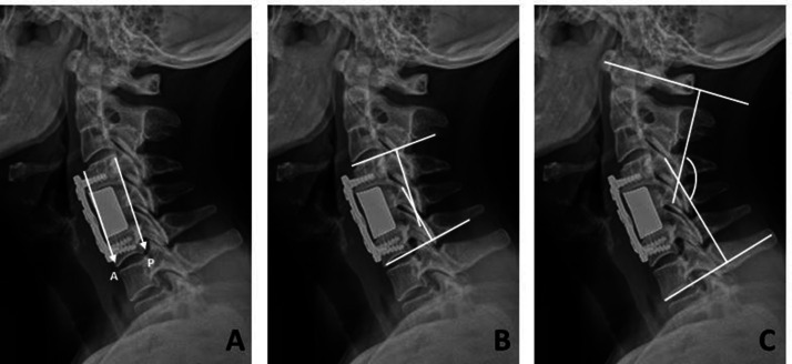
Clinical and radiologic follow-up was performed at 3, 6 months, and 1 year postoperatively and annually thereafter. Lateral radiographs of the cervical spine were used to measure cervical and segmental lordosis by means of the Cobb angle. Segmental sagittal alignment was defined as the angle between the cranial and caudal end plates of the vertebrae located above and below the affected segment and cervical sagittal alignment from C1 to C7. Both measures provided positive and negative values indicating lordotic or kyphotic angulation, respectively.
Fusion was defined as the absence of radiolucent lines within the perimeter of the implant evidenced on anteroposterior and lateral radiographic images of the cervical spine and an absence of movement on dynamic radiographs. Computed tomography (CT) scan reconstructions were used in case of doubtful fusion status.
Implant subsidence was evaluated by means of lateral radiographs of the cranial and caudal end plates of the segments located above and below the operated segments, respectively. Measurement of the distance between the superior end plate of the superior vertebral body and the inferior end plate of the inferior vertebral body was based on the anterior and posterior end plate height. Based on location, implant subsidence was classified according to 4 directions of implant sinkage: anterior, posterior, cranial, and caudal. Subsidence was considered to be mild if the loss of height was under 3 mm and severe if the loss was over 3 mm3,10,11 (Figure 1).
Figure 1.
(A) Implant subsidence was assessed on lateral radiographs at the cranial and the caudal end plates of the upper and the lower vertebrae in the affected segment. The distance between the upper end plate of the upper vertebral body and the lower end plate of the lower vertebral body was measured at the anterior and the posterior points of the end plate. Severe subsidence was defined as a loss of height of >3 mm. (B) Segmental sagittal alignment was defined as the angle between the cranial and the caudal end plates of the upper and the lower vertebrae in the affected segment. (C) Cervical lordosis was measured using Cobb angle from C1 to C7. A, anterior segment height; P, posterior segment height.
Statistical Analysis
The sample size was calculated based on a literature search for case series of treatment for degenerative cervical disc disease that studied sample sizes of between 30 and 70 participants.4,7,8,10–12 We determined that a sample size of 53 was sufficient for this study, using a precision of 0.8 and an alpha error of 0.05.
A database created using Microsoft Excel for Windows was used to enter data from field work carried out to gather patient information. Analysis was performed by an independent team of statisticians hired for the specific purposes of the study. Comparison was performed using contingency tables with the corresponding χ 2 test of independence and comparison of related and matched samples. The null hypothesis (“variables are independent”) was ruled out in cases where P values were below 0.05. All statistical analyses were performed using the SPSS software package.
Source of Funding
The authors did not receive grants or outside funding in support of their research or preparation of this manuscript. They did not receive payments or other benefits or a commitment or agreement to provide such benefits from a commercial entity. No commercial entity paid or directed, or agreed to pay or direct, any benefits to any research fund, foundation, educational institution, or other charitable or nonprofit organization with which the authors are affiliated or associated.
RESULTS
Of the 53 patients included in the study, 30 were men (56.6%) and 23 were women (43.4%), with a mean age of 50.96 years (range, 29–76). The mean duration of symptoms until surgery was 21.68 months. Nineteen patients (35.8%) presented symptoms for less than 1 year and in 90.5% of cases the time between symptom onset and surgery ranged from 4 months to 4 years. Mean length of postoperative follow-up at the time of writing was 4.85 years (range, 2–9).
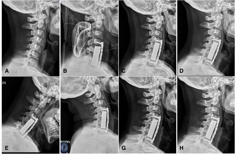
A total of 44 patients (83.0%) underwent 1-level surgery, and the most common vertebra in these surgical procedures was C6 (n = 27, 61.4%), followed by C5 (n = 15, 34.0%), and C4 in 4.5% (n = 2). Nine patients (16.9%) underwent 2-level surgery; in these interventions, the most common vertebrae operated were C5-C6 in 55.5% of cases (n = 5) followed by C4-C5 in 33.3% (n = 3), and then C6-C7 in 11.1% (n = 1) (Figure 2). In all patients, surgical decompression was followed by placement of a TTM-lordotic implant to reconstruct the anterior cervical column after corpectomy; however, 41 patients (77.3%) were outfitted with an additional semiconstrained anterior cervical plate for added construct stability (Table 1).
Figure 2.
(A) A 48-year-old man who underwent a 2-level corpectomy of C5 and C6 with fusion from C4 to C7. (B) Immediate postoperative lateral radiograph at 24 h. (C) Postoperative lateral radiograph at 3 months showing mild subsidence of the tantalum trabecular metal (TTM) implant into the C4 vertebral body. (D) Postoperative lateral radiograph at 6 months showing no further subsidence of the TTM implant. (E and F) Flexion and extension postoperative lateral radiograph at 12 months demonstrates stable positioning of the TTM implant. (G) Postoperative lateral radiograph at 2 years confirms stable and satisfactory placement of the TTM implant. (H) Postoperative lateral radiograph at 5 years with preserved alignment and placement.
Table 1.
Demographic characteristics, diagnosis, and the operated Level.
| Characteristic | Finding |
| Age, y, mean (range) | 50.96 (29–76) |
| Sex, % (n) | |
| Female | 43.39 (23) |
| Male | 56.61 (30) |
| Duration of symptoms, mo, mean (range) | 21.68 (4–48) |
| Diagnosis, % (n) | |
| Herniated disc | 41.51 (22) |
| Spondylosis | 58.49 (31) |
| Follow-up after surgery, y, mean (range) | 4.85 (2–9) |
| Number of levels fused, % (n) | |
| 1 | 83.01 (44) |
| C4 | 4.54 (2) |
| C5 | 34.09 (15) |
| C6 | 61.36 (27) |
| Number of levels fused, % (n) | |
| 2 | 19.98 (9) |
| C4-C5 | 33.33 (3) |
| C5-C6 | 55.55 (5) |
| C6-C7 | 11.11 (1) |
| Additional semiconstrained anterior cervical plate, % (n) | 77.35 (41) |
The primary diagnosis was herniated cervical disc in 22 patients (41.5%) and spondylosis in 31 (58.5%). Patients with nondegenerative diseases such as infection, tumors, or fracture/dislocation were excluded from the analysis. Of the patients studied, 64.1% (n = 34) had clinical and/or radiologic findings indicating myelopathy at the time of surgery, 30.2% (n = 16) presented pain in the cervical spine and brachialgia, and 5.6% (n = 1) had brachialgia alone.
X-ray images obtained during follow-up showed a fusion rate of 98.1% (52/53) by the end of follow-up. The only patient in whom fusion could not be demonstrated was a 73-year-old man with rheumatoid arthritis who underwent 2-level ACCF without anterior cervical plate and screws supplementation and who presented early stage subsidence and migration of the implant. He was reoperated at 7 months to replace the construct with a structural allograft and an anterior cervical plate. He required an additional corpectomy and the extension of the fusion to one level distally due to the complete destruction of the caudal vertebral body.
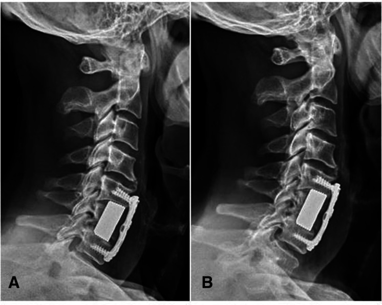
Mild or severe subsidence was observed in 44 cases (83.%). At the end of follow-up, mild subsidence (<3 mm) occurred in 37 cases (69.8%) (Figure 3), while there were 7 cases (13.2%) of severe subsidence (>3 mm). The greatest degree of subsidence developed in the first 3 months postoperatively (P = 0.003). The most common site of severe subsidence was the anterior region of the cranial endplate (4/7). Subsidence was more common among patients not outfitted with an anterior cervical plate (11/12, 91.6%) than in patients in whom a plate was used (33/41, 80.5%); though this association did not reach statistical significance (P = 0.587), the greater frequency of severe subsidence observed in cases in which no anterior plate was employed was statistically significant (P < 0.05) (Table 2).
Figure 3.
A 59-year-old woman who underwent 1-level corpectomy of C6. (A) Postoperative lateral radiograph at 3 months showing parallel contact between the vertebral end plate and the tantalum trabecular metal (TTM) implant (lordotic vertebral end plates with lordotic implant surfaces) maximizing the contact surface area between the implant and the vertebral end plate. Note the difficulties encountered in adapting the anterior cervical plate to the shape of the anterior wall of the vertebral body due to significant degenerative changes. (B) Postoperative lateral radiograph at 3 years showing mild subsidence of the TTM implant into the C5 vertebral body and confirming stable and satisfactory placement of the implant.
Table 2.
Subsidence occurrence in 53 patients treated with an anterior cervical corpectomy and fusion tantalum implant.
| Subsidence, % (n) | |||
| Additional Semiconstrained Anterior Cervical Plate | None | Mild | Severe |
| With (n = 41) | 19.51 (8) | 75.6 (31) | 4.87 (2) |
| Without (n = 12) | 8.33 (1) | 50 (6) | 41.66 (5) |
By measuring cervical lordosis, lordosis of the affected segment, and the anterior and posterior end plate height, we obtained values for sagittal changes. The height of the affected segment assessed by means of the anterior and posterior end plate height increased significantly in the immediate postoperative period following placement of a TTM-lordotic implant (P = 0.021). However, this height gradually decreased over follow-up. Though the final end plate height at the conclusion of follow-up was greater than the preoperative height, at 1-year and at 2-year follow-up, there were no significant differences found in the height of the affected segment when compared to the preoperative height (P = 0.42 and P = 0.25, respectively). No patients developed a significant increase in implant subsidence beyond the second year of follow-up.
Mean preoperative cervical lordosis (C1-C7) was 39.4° ± 12.5°, measuring 38.4° ± 13.8° at 48 hours postoperatively, 42.2° ± 11.3° at 6 months, 44.7° ± 11.9° at 1 year, and 44.5° ± 12.7° at the end of follow-up. As a result, we found an average increase in cervical lordosis of 5.1° ± 8.2° at the end of follow-up, though this difference did not reach statistical significance (P > 0.05).
Preoperatively, the Cobb angle of the affected segment, that is, between the cranial and caudal end plates of the vertebrae located above and below the segment targeted by the operation was 1.6° ± 5.8°, and this value was 5.9° ± 4.2° in the immediate postoperative period, and 3.6° ± 4.3° at the end of follow-up. The initial improvement in Cobb values for the affected segment during the immediate postoperative period was not maintained throughout follow-up, as an improvement of only 1.9° ± 6.1° of segmental lordosis was recorded at the end of follow-up (P > 0.05) (Table 3).
Table 3.
Radiologic follow-up: cervical lordosis of 53 patients treated with an anterior cervical corpectomy and fusion tantalum implant.
| Mean ± SD | ||
| Time | C1-C7 Cobb Angle | Segmental Cobb Angle |
| Preoperative | 39.46 ± 12.52 | 1.64 ± 5.84 |
| Postoperative | 38.39 ± 13.85 | 5.95 ± 4.23 |
| 6 mo | 42.18 ± 11.37 | 3.92 ± 4.55 |
| 1 y | 44.73 ± 11.93 | 3.95 ± 5.93 |
| Final follow-up | 44.52 ± 12.77 | 3.62 ± 5.38 |
| Improvement/P value | 5.06 ± 8.26/P > 0.05 | 1.98 ± 6.02/P > 0.05 |
An analysis of cervical and radicular pain before and after surgery showed a significant preoperative average VAS of 7.3 and 5.1, respectively, decreasing to 2.6 and 2.5 at the end of follow-up (P < 0.05). NDI scores fell significantly, from an average of 47.8 before surgery to 21.3 at the end of follow-up (P < 0.05). The improvement in NDI scores shows a statistically significant relationship with pain relief measured by VAS (P > 0.05), reflecting a substantial reduction in disability as a result of decreased cervical and radicular pain (Table 4).
Table 4.
Clinical outcomes of 53 patients treated with an anterior cervical corpectomy and fusion tantalum implant.
| % (n) | ||
| Outcome Measure | Preoperative | Postoperative |
| Cervical VAS | 7.3 (2.2) | 2.6 (2.9) |
| Radicular VAS | 5.1 (3.7) | 2.5 (2.4) |
| Neck Disability Index | 47.8 (21.5) | 21.3 (23.4) |
VAS, visual analog scale.
Three months after surgery, 98% of patients (n = 52) reported an improvement over their baseline situation before the operation (reduction of 50% or more in pain measured by VAS); the patient who required reoperation due to implant extrusion was the only case in which no improvement was reported. During follow-up, 9 patients (20.7%) reported progressive worsening over time, however, at no time during the follow-up was the initial level of pain that preceded surgery ever attained.
Regarding postoperative complications, 2 patients needed reoperation. One was a 74-year-old male patient with a medical history of high blood pressure and paroxysmal atrial fibrillation on oral anticoagulant therapy who underwent a subsequent intervention to drain a pre-esophageal hematoma at 24 hours postoperatively, though no active intraoperative vascular bleeding was detected; a 73-year-old male patient, with a medical history of rheumatoid arthritis, underwent reoperation 7 months after the initial surgery due to migration and extrusion of the implant (mentioned above). One patient presented an intraoperative dural tear, which was successfully treated by lumbar drainage and 7 days of restricted activity. There were no complications related to the anterior cervical plate. None of the patients developed a surgical site infection, esophageal laceration, epidural hematoma, or injury to the vertebral artery. Two patients complained of pain in the arm and neck. Both underwent conservative treatment and the symptoms resolved within 3 months. Four patients developed dysphagia, likely related to excessive retraction of the esophagus and trachea during decompression. The symptoms of all 4 resolved spontaneously within 6 months.
At the end of follow-up, 79.2% of patients (42/53) expressed satisfaction with the treatment received and stated that they would be willing to voluntarily undergo the same treatment again.
DISCUSSION
In this study, we determine the rate of fusion following ACCF with lordotic TTM implants based on radiographic and clinical findings, with special focus placed on the relevance and influence of implant subsidence on sagittal alignment and its clinical implications. The favorable clinical outcomes obtained using this procedure are the result of direct decompression of neural elements and immediate stabilization of the cervical spine anterior to the involved segment.13,14
With the arrival of the first artificial implants, reconstruction of the anterior cervical spine following ACCF using autologous bone grafts and allografts is being gradually abandoned due to the high rates of pseudarthrosis and other complications related to bone grafts, from subsidence to partial or total extrusion of the graft.
The aims of grafts used in corpectomy are to maintain an appropriate level of decompression, provide immediate postoperative stability to the segment, and to restore and maintain the resected segment height, alignment, and sagittal profile of the cervical spine. Furthermore, the procedure must promote bone fusion and act as an effective bridge between adjacent vertebral segments. Since the introduction of the first artificial implants in corpectomy, implants have been made using different types of materials (ie, titanium, tantalum, polyether ether ketone, polyamide/hydroxyapatite, carbon fiber) and designs such as metallic cylindrical mesh and expandable cylinders. The biomechanical properties of artificial corpectomy implants differ from those of bone grafts. Despite the advances in surgical technique and in the materials used for reconstruction, this procedure continues to have a high rate of complications related to subsidence and construct failure, especially in multilevel ACCF performed in patients of advanced age, in patients with low bone density, or when the construct goes through to C7 and T1.3–7,11,14,15
Rates of implant subsidence have been reported to range from 4% to 80% when cylindrical titanium mesh cage implants are used. Chen et al,14 in a study with 300 patients, found a subsidence rate of 79.7% (n = 239), including 19.0% (n = 57) with severe subsidence (>3 mm). In the study, severe subsidence correlated with the occurrence of complications, including neck pain, instrumentation failure, and a substantially lower rate of neurologic recovery. Ji et al12 reported an overall rate of subsistence >3 mm of 43.5%. The authors identified excessive distraction in intervertebral height and low bone mineral density to be significant risk factors for subsidence. Osteoporosis is associated with an increased risk of instrumentation failure during spine surgery. Current literature has evidenced the growing number of elderly patients with spondylotic myelopathy of the cervical spine who require multilevel corpectomy to achieve appropriate decompression of the spinal cord. Low bone mineral density has been identified as an independent risk factor for the appearance of subsidence. In an attempt to increase construct stability in ACCF with artificial implants for osteoporotic patients, it has been suggested to start antiosteoporosis treatment 6 months before surgery, to carry out additional posterior stabilization and instrumentation, and to use an anterior plate with cement-augmented screws.10,12,16,17
Previous research has reported the occurrence of subsidence of bone grafts within the vertebral body and also of a wide range of artificial corpectomy implants designed for reconstruction. Zhong et al11 studied 56 patients who underwent single-level ACCF with the use of a polyamide/hydroxyapatite implant and an anterior cervical plate. In total, 45 patients developed implant subsidence, with an incidence rate of 80% (45/56). Weber et al18 compared rates of subsidence and revision surgery associated with 4 widely used ACCF implants (fibular allograft, titanium mesh cage, titanium expandable cage, and carbon fiber cage). To a greater or lesser degree, all of the implants studied were associated with presence of subsidence. The literature currently defines subsidence of more than 3 mm as the cut point for classification of significant cases of subsidence.3,14 Subsidence is a frequent finding in the early postoperative period and though the clinical relevance of slight subsidence remains controversial, severe subsidence is correlated with poor neurologic recovery and poor clinical outcomes. Additionally, excessive subsidence in adjacent vertebral bodies may cause bulging of the yellow ligament, foraminal stenosis, instability, reconstruction failure, and recurrence of neurologic symptoms.3,10,14
Daubs, who studied 23 patients undergoing surgery with the use of titanium mesh cage implants, reported that a 30% (n = 7) rate of fixation failure with severe subsidence of the cage and distal extrusion of the anterior cervical plate. Revision surgery was performed in 6 of the 7 cases of failure; in all cases, the cage was removed and replaced with a fibular allograft. Due to the total destruction of the caudal vertebral body by the cage, all patients required an additional level corpectomy and distal extension of the fusion by one level. Additionally, all revision surgeries included a posterior approach with posterior instrumentation and fusion using bone graft from the iliac crest.5
In our study, severe subsidence in the TMM implant was detected in 7 patients (13.20%); the anterior aspect of the cranial end plate was the most common site of severe subsidence. This finding contrasts with the results of previous research4,5,14,19 using nonlordotic implants, where the posterior border of the inferior end plate was the most common site of subsidence. Jang et al4 reported subsidence of the titanium mesh cage in 93.3% of their 30-patient series. The authors observed significantly greater degrees of subsidence in the posterior part of the segment as a result of several factors: the posterior region of the vertebral end plate is weaker and more spongy; the posterior end plate is subjected to greater damage with the removal of posterior osteophytes or calcified ligaments; and finally, due to cervical lordosis and the location of the implant, the center of gravity of the segment is in the posterior region and is thus subjected to greater compression than the anterior part of the segment.
Our data show that the occurrence of severe implant subsidence in TTM implants on the anterior part of the cranial vertebral end plate was related to incongruence between the kyphotic degenerative cranial end plate and the lordotic surface of the implant. A lack of parallel positioning between the implant and the vertebral end plate reduces contact size, destabilizes load distribution due to the minimization of the contact area, and favors the occurrence of severe subsidence due to impaction (Figure 4). When contact between the vertebral end plate and the TTM implant occurs through parallel, nonconverging surfaces (eg, lordotic vertebral end plates with lordotic implant surfaces), the contact spaces between the implant and vertebral end plates are maximized, causing a more even load distribution, which can be important in preventing subsidence. Cawley et al3 in a study with 102 patients using carbon fiber cages with 4° of lordosis filled with autologous bone graft for reconstruction of the anterior cervical spine following ACCF, reported a 13% rate of severe subsidence, including 5 cases of posterior implant migration, all related to the mismatch of the implant contact surface due to the obliquity of the vertebral end plate anatomy. Use of implants whose contact surface area coincides with the anatomy and orientation of vertebral end plates, thus maximizing the area of contact between the implant and vertebral end plates, is likely to provide better load distribution effect, which may play an important role in preventing subsidence. In such a case, the low modulus of elasticity of tantalum, which resembles that of subchondral and cancellous bone, allows for better load transferal and minimizes stress shielding. The porosity of contact surfaces provided by trabecular metal results in a high coefficient of friction. Once placed, TTM implants are highly resistant to extrusion. However, the sharp teeth of certain implants, designed to increase initial stability and resistance to extrusion by penetrating the vertebral end plate, may actually erode the end plate, weakening it and favoring subsidence and failure.4,6,7,10,11,14
Figure 4.
A 41-year-old man who underwent 1-level corpectomy of C6. (A) Immediate postoperative lateral radiograph at 2 weeks showing incongruity between the vertebral end plate and the tantalum trabecular metal (TTM) implant (kyphotic degenerative cranial vertebral end plate and lordotic implant). (B and C) Postoperative lateral radiograph at 3 and 6 months showing progressive subsidence of the TTM implant into the C5 and C7 vertebral body. (D) Postoperative lateral radiograph at 1 year showing no further subsidence of the TTM implant. (E) Postoperative lateral radiograph at 3 years showing solid fusion (no radiolucency in the perimeter surrounding the TTM implant) with preserved alignment and placement.
Distortions to the normal anatomy of the cervical spine as a result of degenerative changes alter end plate orientation and introduce irregularity. Despite the advances in materials and techniques used for reconstruction, currently used corpectomy implants are limited in their ability to adapt contact surfaces (cranial and caudal) to such changes in orientation and irregularities in degenerative vertebral end plates, which minimize the contact surface at the bone-implant interface and increase the risk of subsidence due to an asymmetrical load distribution. With a greater variety in the size, shape, and orientation of implant contact surfaces, or by exploring the feasibility of modular implants (as in hip and knee implants), surgeons could improve the adaptability of the implants to the space created following corpectomy and reduce the likelihood of subsidence and implant failure.
Proper preparation of vertebral end plates during segmental decompression may also play an important role in implant subsidence. To avoid subsidence, authors recommend careful endplate curettage, preserving as much subchondral cortical bone as possible; placement of the implant in a slightly anterior position, as the posterior subchondral bone lacks the firmness of the bone on the anterior side; maximizing the contact surface between the vertebral end plate and the implant so that this contact is as parallel and congruent as possible; and paying appropriate attention to the appropriateness of the implant size so as to avoid intraoperative overdistraction, especially in the more elastic segments.12
Over nearly 2 decades, conventional TTM-lordotic implants have had demonstrated safety for use in anterior cervical discectomy and fusion, though empirical data on the use of these implants in the context of corpectomy remain scarce.20 TTM has sparked great interest in orthopedic surgery due to its porousness (75%–85%) and high structural similarity to cancellous bone. This porosity also favors osseointegration, formation of new bone, and neovascularization, and has been shown to be more osteoconductive than other commercial available biomaterials.21 Furthermore, TTM is biologically inert, meaning that it can be implanted in human tissue without substantial concerns about the immune response to the material.22 No bone graft was added in any of the patients in the present study. The osteoconductive properties of TTM eliminate the need for bone grafts to ensure solid bone fusion.
The use of an anterior cervical plate to increase the stability of the construct has been considered to be standard practice. Though the anterior cervical plate is technically demanding when applied as part of multilevel ACCF, it is not entirely free of morbidity and possible complications. In our study, subsidence, and particularly severe subsidence, was more frequent among patients in whom an anterior cervical plate had not been used than among those in whom the plate had been used. Placement of an anterior plate provides additional stability, increases the rate of bone fusion, and prevents implant dislocation and late kyphotic deformities.4,19,23,24 Furthermore, precontouring of the anterior cervical plate aids in restoring cervical lordosis. The authors use a semiconstrained screw-plate interface to supplement final arthrodesis stability. Semiconstrained plates enable surgeons to place compression screws while the cams of the plate impede extrusion. When turned, the cam compresses the screw head independently of its position against the other side of the hemispherical orifice. This offers several advantages: first, the screw is not constrained by predetermined angulation, and the surgeon may choose the most appropriate angle and orientation; second, the screw not locked to the plate allows some sagittal oscillation capacity. The latter phenomenon provides the plate with certain tolerance against load transmission due to corpectomy implant subsidence and minimizes the risk of implant failure. Semiconstricted plating systems provide an intermediate constriction between dynamic anterior cervical plates and static plates. Nonetheless, Chen et al14 did not find significant differences in the incidence of subsidence with the use of dynamic or static anterior cervical plates. To limit potential implant subsidence, it has been recommended to minimize plate length and strategically place screws adjacent to the implant in the dense vertebral end plate bone, so that this may act as a buttress.25
We used indirect evidence to radiographically demonstrate successful fusion, such as the absence of peri-implant radiolucence or a lack of movement as observed on dynamic x-ray images. In the event of questionable fusion, CT scan reconstruction was requested. MRI is a useful tool for assessing decompression of neural structures. The artifacts of TTM implants are limited to the structures immediately surrounding the implant. The spinal canal and neural foramina can be visible, thus allowing evaluation of decompression. However, MRI is not useful for determining fusion, as the artifact created makes it impossible to precisely interpret the interface between the implant and the vertebral body.
TTM implants are a valid option for the treatment of degenerative cervical disc disease with ACCF. TTM implants provide immediate stability to the anterior cervical spine with no complications in the donor site and do not require supplementary bone grafting. However, CT-based studies of TTM implants evidence larger quantities of metallic artifacts than of hollow implants due to the fact that TTM implants are solid blocks, and TTM implants are not free from risk of subsidence and other possible complications that may appear in the future due to their current widespread use. With a greater variety in the size, shape, and angle of implant contact surfaces, or by exploring the feasibility of modular implants, surgeons could improve the adaptability of the implants to the space created following corpectomy and reduce the likelihood of subsidence and implant failure. We recommend the use of cervical plates and screws independently of the screw-plate interface preferred by the surgeon. Precontoured, semiconstrained anterior cervical plates help to restore cervical lordosis, provide certain tolerance of corpectomy implant subsidence, and minimize the risk of implant failure.
This study presents certain limitations. First, our research is based on the work of 5 different surgeons, each with differing levels of experience with TTM. Second, the case series includes no group for comparison. Last, the analysis of the sample did not take into account factors that may influence the surgical outcome, such as advanced age, sex, smoking, and osteoporosis. As a result, studies carry out with longer follow-up periods and randomized prospective trials using different types of corpectomy implants are desirable.
CONCLUSION
ACCF with anterior cervical reconstruction using TTM-lordotic implants for treatment of cervical degenerative disease has high fusion rates and good clinical outcome. The osteoconductive properties of TTM provide immediate stabilization and eliminate the need for bone grafts to ensure solid bone fusion.
Before fusion occurs, asymptomatic implant settlement (<3 mm) into the vertebral body is inevitable. According to the findings of this study, the greatest degree of subsidence develops in the first 3 months postoperatively. No patients presented a significant increase in implant subsidence beyond the second year of follow-up. The most common site of severe subsidence was the anterior region of the cranial end plate. Lack of parallelism and reduced contact surface between the implant and the vertebral end plates are major risk factors for severe further subsidence (>3 mm), which may negatively affect the clinical outcomes. With a greater variety in the size, shape, and orientation of implant contact surfaces, or by exploring the feasibility of modular implants, surgeons could improve the adaptability of the implants to the space created following corpectomy and reduce the likelihood of severe subsidence and implant failure.
Additional anterior cervical plating helps to restore cervical lordosis, provide certain tolerance of corpectomy implant subsidence, and minimize the risk of implant failure.
Acknowledgements
Study presented at the 36th GEER-Spanish Spine Society meeting 2022.
References
- 1.Fei Q, Li J, Su N, et al. Comparison between anterior cervical discectomy with fusion and anterior cervical corpectomy with fusion for the treatment of cervical spondylotic myelopathy: a meta-analysis. Ther Clin Risk Manag. 2015;11:1707–1718. 10.2147/TCRM.S94290 [DOI] [PMC free article] [PubMed] [Google Scholar]
- 2.Thalgott JS, Xiongsheng C, Giuffre JM. Single stage anterior cervical reconstruction with titanium mesh cages, local bone graft, and anterior plating. Spine J. 2003;3(4):294–300. 10.1016/s1529-9430(02)00588-0 [DOI] [PubMed] [Google Scholar]
- 3.Cawley DT, Alzakri A, Fujishiro T, et al. Carbon-fibre cage reconstruction in anterior cervical corpectomy for multilevel cervical spondylosis: mid-term outcomes. J Spine Surg. 2019;5(2):251–258. 10.21037/jss.2019.03.08 [DOI] [PMC free article] [PubMed] [Google Scholar]
- 4.Jang JW, Lee JK, Lee JH, Hur H, Kim TW, Kim SH. Effect of posterior subsidence on cervical alignment after anterior cervical corpectomy and reconstruction using titanium mesh cages in degenerative cervical disease. J Clin Neurosci. 2014;21(10):1779–1785. 10.1016/j.jocn.2014.02.016 [DOI] [PubMed] [Google Scholar]
- 5.Daubs MD. Early failures following cervical corpectomy reconstruction with titanium mesh cages and anterior plating. Spine (Phila Pa 1976). 2005;30(12):1402–1406. 10.1097/01.brs.0000166526.78058.3c [DOI] [PubMed] [Google Scholar]
- 6.Hee HT, Majd ME, Holt RT, Whitecloud TS, Pienkowski D. Complications of multilevel cervical corpectomies and reconstruction with titanium cages and anterior plating. J Spinal Disord Tech. 2003;16(1):1–8. 10.1097/00024720-200302000-00001 [DOI] [PubMed] [Google Scholar]
- 7.Hartmann S, Kavakebi P, Wipplinger C, et al. Retrospective analysis of cervical corpectomies: implant-related complications of one- and two-level corpectomies in 45 patients. Neurosurg Rev. 2018;41(1):285–290. 10.1007/s10143-017-0854-8 [DOI] [PubMed] [Google Scholar]
- 8.Tomé-Bermejo F, Morales-Valencia JA, Moreno-Pérez J, et al. Degenerative cervical disc disease: long-term changes in sagittal alignment and their clinical implications after cervical interbody fusion cage subsidence: a prospective study with standalone lordotic tantalum cages. Clin Spine Surg. 2017;30(5):E648–E655. 10.1097/BSD.0000000000000293 [DOI] [PubMed] [Google Scholar]
- 9.Lequin MB, Verbaan D, Bouma GJ. Posterior lumbar interbody fusion with stand-alone trabecular metal cages for repeatedly recurrent lumbar disc herniation and back pain. J Neurosurg Spine. 2014;20(6):617–622. 10.3171/2014.2.SPINE13548 [DOI] [PubMed] [Google Scholar]
- 10.Fengbin Y, Jinhao M, Xinyuan L, Xinwei W, Yu C, Deyu C. Evaluation of a new type of titanium mesh cage versus the traditional titanium mesh cage for single-level, anterior cervical corpectomy and fusion. Eur Spine J. 2013;22(12):2891–2896. 10.1007/s00586-013-2976-1 [DOI] [PMC free article] [PubMed] [Google Scholar]
- 11.Zhong W, Liang X, Tang K, Luo X, Quan Z, Jiang D. Nanohydroxyapatite/polyamide 66 strut subsidence after one-level corpectomy: underlying mechanism and effect on cervical neurological function. Sci Rep. 2018;8(1):12098. 10.1038/s41598-018-30678-1 [DOI] [PMC free article] [PubMed] [Google Scholar]
- 12.Ji C, Yu S, Yan N, et al. Risk factors for subsidence of titanium mesh cage following single-level anterior cervical corpectomy and fusion. BMC Musculoskelet Disord. 2020;21(1):32. 10.1186/s12891-019-3036-8 [DOI] [PMC free article] [PubMed] [Google Scholar]
- 13.Majd ME, Vadhva M, Holt RT. Anterior cervical reconstruction using titanium cages with anterior plating. Spine (Phila Pa 1976). 1999;24(15):1604–1610. 10.1097/00007632-199908010-00016 [DOI] [PubMed] [Google Scholar]
- 14.Chen Y, Chen D, Guo Y, et al. Subsidence of titanium mesh cage: a study based on 300 cases. J Spinal Disord Tech. 2008;21(7):489–492. 10.1097/BSD.0b013e318158de22 [DOI] [PubMed] [Google Scholar]
- 15.Wang JC, Hart RA, Emery SE, Bohlman HH. Graft migration or displacement after multilevel cervical corpectomy and strut grafting. Spine (Phila Pa 1976). 2003;28(10):1016–1021. 10.1097/01.BRS.0000061998.62204.D7 [DOI] [PubMed] [Google Scholar]
- 16.Hartmann S, Thomé C, Tschugg A, Paesold J, Kavakebi P, Schmölz W. Cement-augmented screws in a cervical two-level corpectomy with anterior titanium mesh cage reconstruction: a biomechanical study. Eur Spine J. 2017;26(4):1047–1057. 10.1007/s00586-017-4951-8 [DOI] [PubMed] [Google Scholar]
- 17.Yan D, Wang Z, Deng S, Li J, Soo C. Anterior corpectomy and reconstruction with titanium mesh cage and dynamic cervical plate for cervical spondylotic myelopathy in elderly osteoporosis patients. Arch Orthop Trauma Surg. 2011;131(10):1369–1374. 10.1007/s00402-011-1317-2 [DOI] [PubMed] [Google Scholar]
- 18.Weber MH, Fortin M, Shen J, et al. Graft subsidence and revision rates following anterior cervical corpectomy. Clin Spine Surg. 2017;30(9):E1239–E1245. 10.1097/BSD.0000000000000428 [DOI] [PubMed] [Google Scholar]
- 19.Nakase H, Park YS, Kimura H, Sakaki T, Morimoto T. Complications and long-term follow-up results in titanium mesh cage reconstruction after cervical corpectomy. J Spinal Disord Tech. 2006;19(5):353–357. 10.1097/01.bsd.0000210113.09521.aa [DOI] [PubMed] [Google Scholar]
- 20.King V, Swart A, Winder MJ. Tantalum trabecular metal implants in anterior cervical corpectomy and fusion: 2-year prospective analysis. J Clin Neurosci. 2016;32:91–94. 10.1016/j.jocn.2016.03.026 [DOI] [PubMed] [Google Scholar]
- 21.Levine BR, Sporer S, Poggie RA, Della Valle CJ, Jacobs JJ. Experimental and clinical performance of porous tantalum in orthopedic surgery. Biomaterials. 2006;27(27):4671–4681. 10.1016/j.biomaterials.2006.04.041 [DOI] [PubMed] [Google Scholar]
- 22.Uribe JS, Sangala JR, Duckworth EAM, Vale FL. Comparison between anterior cervical discectomy fusion and cervical corpectomy fusion using titanium cages for reconstruction: analysis of outcome and long-term follow-up. Eur Spine J. 2009;18(5):654–662. 10.1007/s00586-009-0897-9 [DOI] [PMC free article] [PubMed] [Google Scholar]
- 23.Shaw KA, Griffith M, Mottern ET, Gloystein DM, Devine JG. Preventing construct subsidence following cervical corpectomy: the bump-stop technique. Asian Spine J. 2018;12(1):156–161. 10.4184/asj.2018.12.1.156 [DOI] [PMC free article] [PubMed] [Google Scholar]
- 24.Lee SH, Sung JK. Anterior cervical stabilization using a semi-constrained cervical plate and titanium mesh cage for single level corpectomy. J Clin Neurosci. 2008;15(11):1227–1234. 10.1016/j.jocn.2007.09.019 [DOI] [PubMed] [Google Scholar]
- 25.DiAngelo DJ, Foley KT, Vossel KA, Rampersaud YR, Jansen TH. Anterior cervical plating reverses load transfer through multilevel strut-grafts. Spine (Phila Pa 1976). 2000;25(7):783–795. 10.1097/00007632-200004010-00005 [DOI] [PubMed] [Google Scholar]


