Abstract
Abstract
Higher endotoxin in the circulation may indicate a compromised state of host immune response against coinfections in severe COVID-19 patients. We evaluated the inflammatory response of monocytes from COVID-19 patients after lipopolysaccharide (LPS) challenge. Whole blood samples of healthy controls, patients with mild COVID-19, and patients with severe COVID-19 were incubated with LPS for 2 h. Severe COVID-19 patients presented higher LPS and sCD14 levels in the plasma than healthy controls and mild COVID-19 patients. In non-stimulated in vitro condition, severe COVID-19 patients presented higher inflammatory cytokines and PGE-2 levels and CD14 + HLA-DRlow monocytes frequency than controls. Moreover, severe COVID-19 patients presented higher NF-κB p65 phosphorylation in CD14 + HLA-DRlow, as well as higher expression of TLR-4 and NF-κB p65 phosphorylation in CD14 + HLA-DRhigh compared to controls. The stimulation of LPS in whole blood of severe COVID-19 patients leads to lower cytokine production but higher PGE-2 levels compared to controls. Endotoxin challenge with both concentrations reduced the frequency of CD14 + HLA-DRlow in severe COVID-19 patients, but the increases in TLR-4 expression and NF-κB p65 phosphorylation were more pronounced in both CD14 + monocytes of healthy controls and mild COVID-19 patients compared to severe COVID-19 group. We conclude that acute SARS-CoV-2 infection is associated with diminished endotoxin response in monocytes.
Key messages
Severe COVID-19 patients had higher levels of LPS and systemic IL-6 and TNF-α.
Severe COVID-19 patients presented higher CD14+HLA-DRlow monocytes.
Increased TLR-4/NF-κB axis was identified in monocytes of severe COVID-19.
Blunted production of cytokines after whole blood LPS stimulation in severe COVID-19.
Lower TLR-4/NF-κB activation in monocytes after LPS stimulation in severe COVID-19.
Keywords: COVID-19, Inflammation, Monocytes, Lipopolysaccharide
Introduction
The coronavirus disease-2019 (COVID-19) is the disease caused by the severe acute respiratory syndrome coronavirus 2 (SARS-CoV-2) that induces a multiorgan damage and impairs several functional physiological units, such as the cardiorespiratory and neurological systems [1]. The clinical characteristics of COVID-19 are widely diverse, although most subjects are asymptomatic or exhibit only mild symptoms, a significant number of patients require hospitalization, including intensive care unit (ICU) admission [2]. Despite several progresses were recently made to understand the immunopathology of COVID-19, the decisive pathological processes that are responsible for patient morbidity and mortality remain unknown.
The main causes of death by COVID-19 include respiratory failure and the development of sepsis, a condition that has been observed in several deceased patients [3]. In this sense, the predominant theory is that an overexuberant immune response mediated by strong proinflammatory cytokinemia drives lung injury, multiorgan lesion, and a procoagulant state [4, 5]. On the other hand, an overproduction of inflammatory cytokines may lead to a state of immunologic collapse and sterile inflammation that predisposes the host to a secondary infection [3, 6]. The higher levels of pro- and anti-inflammatory cytokines, including but not only interleukin (IL)-6, IL-10, tumor necrosis factor-alpha (TNF-α), and interferons, contribute to the augment of the tissue damage in parallel with functional paralysis of both innate and adaptive immune cells [7, 8]. In such scenario, new bacterial or viral infections increases the risk of mortality and morbidity [9–11].
In fact, we and others recently reported higher levels of bacterial translocation markers in the peripheral blood of ICU COVID-19, which was associated with death [7, 12, 13]. Severe COVID-19 shares several immunopathological features with septic conditions, including T cell exhaustion, lymphocyte apoptosis, leukopenia, and increased immunosuppressive myeloid-derived suppressor cells (CD14 + HLA-DRlow) phenotype [13, 14] Moreover, endotoxin tolerance is one of the main mechanisms responsible for the appearance of secondary infections due to impairment of proinflammatory cytokine production after a secondary immunogenic stimulus, low antigen presentation, and high phagocytic ability [15, 16]. Here, we aimed to investigate the impact of lipopolysaccharide (LPS) whole blood assay on the cytokine production and the expression of toll-like receptor-4 (TLR-4) and the activation of the master inflammatory transcription NF-κB in CD14 + HLA-DRlow/+ monocytes of COVID-19 patients.
Methods
Ethics procedure and participants
This study was conducted in accordance with the ethical guidelines of the 1975 Declaration of Helsinki and was approved by the Moinhos de Vento Ethics Committee N 3,977,144 (Porto Alegre/Brazil). All the participants provided an informed consent. Unvacinated Patients who fulfilled the clinical criteria for mild and severe COVID-19 and have the diagnosis confirmed by SARS-CoV-2 nucleic acid testing of nasal and pharyngeal throat swab specimens using real-time RT-PCR assay were included in this study [7]. The clinical data of the patients included in the study are summarized in Supplementary Table I. Blood samples (5 mL) were obtained from controls and severe COVID-19 patients into K2EDTA tubes (Becton & Dickinson, USA) within 6 h from hospital admission. All healthy controls have no history of any significant systemic diseases or malignancy and were negative for SARS-CoV-2 acute infection (based on a routine diagnostic RT-PCR test).
Disease severity was classified according to the World Health Organization classification after completing the follow-up questionnaire. Clinical and socio-demographic data were collected from the patient’s electronic medical records upon admission to the unit.
Liquid chromatography-tandem mass spectrometry determination of LPS
LPS concentration was determined by quantitation of 3-hydroxytetradecanoic acid as described previously [7]. Briefly, 150 µL of plasma was hydrolyzed with 75 µL of NaCl 150 mM, 300 µL of HCl 8 mol for 4 h at 90 °C, and extracted with 4 mL of a hexane. Samples were dried under a nitrogen stream and resuspended in 50 μL of methanol immediately prior to the injection in the analytical system. A Nexera UFLC system coupled to a LCMS-8040 triple quadrupole mass spectrometer (Shimadzu, Kyoto, Japan) was used for the analysis. The electrospray parameters were set in the negative ion mode ([M-H]−) as follows: capillary voltage, 3000 V; desolvation line temperature, 250 °C; heating block temperature, 500 °C; drying gas, 18/L min; and nebulizing gas, 2 L/min. Collision-induced dissociation was obtained with 230 kPa argon pressure. Analyses were carried out with multiple reaction monitoring (MRM) by using the following fragmentation: m/z 243.1 → m/z 59.0. The chromatographic separation was conducted with an Acquity UPLC® C18 column (2.1 × 50 mm, 1.7-μm particle size) (Waters Corporation, Ireland). The analyses were performed in gradient elution mode with a flow rate of 0.4 mL/min and the gradient mobile phase system consisted of water (solvent A) and acetonitrile (solvent B) both fortified with 0.2 % acetic acid as follows: 0–1.5 min, 75–100% of B; 1.5–2.0 min, 100% of B; 2.0–2.1 min, 100–75 % of B; 2.1–5.5 min, 75 % B. The column oven was kept at 50 °C. The data were processed using LabSolutions software (Shimadzu, Kyoto, Japan).
Whole blood ex vivo assay
Blood samples collected into k3EDTA tubes were aliquoted in tubes to a volume of 1 mL and incubated (5%CO2, 37 °C) with two different LPS concentrations (1 and 10 ng/mL, Sigma-Aldrich, USA) or without immunogenic stimulus for 2 h [17]. At the end of the assay, the tubes were centrifuged, and the plasma was stored in microtubes and frozen at −80 °C until the day of cytokine analysis. The remaining blood was used to analyze the expression of TLR-4 and the activation of NF-κB in CD14 + HLA-DR−/+ monocytes.
sCD14, PGE2, and cytokine analyses
The supernatant concentrations of interleukin (IL)-6, IL-10, C–C motif ligand 2 (CCL2), and tumor necrosis factor-alpha (TNF-α) were analyzed by bead-based multiplex assays using Luminex Technology following the manufacturer’s recommendations (MILLIPLEX MAP Human Cytokine/Chemokine, Millipore, USA). The soluble form of CD14 was analyzed using Human CD14 ELISA Kit from Invitrogen (USA), with detection limits ranging 8.23–8000 ng/mL. Prostaglandin E2 (PGE2, Invitrogen, ThermoFisher, USA, detection limits of 39.1–10.000 pg/mL) and IL-1β (Intitrogen, USA, detection limits of 2.0–500 pg/mL) concentrations were analyzed in plasma and supernatant by enzyme‐linked immunosorbent assay (ELISA) in microplate reader (EzReader, EUA) following the manufacturer’s recommendations.
Flow cytometry
Whole blood samples (100 µL) were incubated with monoclonal surface antibodies (all anti-human) at 4 °C for 20 min in accordance with the following combination: (a) FITC-conjugated anti-CD14, Pe-conjugated anti-CD284; Percp-Cy5.5 anti-HLA-DR; (b) FITC-conjugated anti-CD14, Pe-conjugated anti-NF-κBp65 (Ser529); Percp-Cy5.5 anti-HLA-DR. Then, samples were incubated for 10 min with lysing buffer (BD Biosciences) and then centrifuged at 500 g for 5 min. The supernatant was discarded, and samples were resuspended in phosphate buffered saline (PBS — 1 mL) and then centrifuged at 500 g for 5 min. Finally, the samples were resuspended in 0.5 mL PBS and analyzed in flow cytometry. For intracellular analysis, tubes with whole blood stained with anti-CD4 or anti-CD14 antibodies were incubated with fixation and permeabilization buffers following the manufacturer’s recommendation (Ebioscience, EUA). After the period, samples were incubated with Phospo-NF-κBp65 (Ser529) Pe (Ebioscience, EUA) monoclonal during 30 min. Then, cells were washed and resuspended in Flow Cytometry Staining Buffer (Ebioscience, EUA). Cell phenotype was acquired using CELLQuest Pro Software (BD Bioscience) on a FACSCalibur flow cytometer (BD Bioscience).
Statistical analysis
Data were analyzed using SPSS 20.0 (SPSS Inc, IBM, Chicago, IL). After the normality test by Shapiro–Wilk, all values were presented as mean ± standard deviation (SD). Categorical variables were presented as relative frequency and analyzed by chi-square test. Baseline systemic immune response (i.e., without endotoxin stimulation) were analyzed by one-way analysis of variance followed by Bonferroni’s post hoc for multiple comparisons. To identify associations between LPS and symptoms, we used Mann–Whitney U test to compare SARS-CoV-2-infected patients with and without specific disease symptoms. Pearson’s coefficient test was performed to check correlations between LPS and the clinical variables of severe patients. Two-way repeated measurement analysis of variance, taking time and group as factors, was performed followed by Bonferroni’s post hoc for multiple comparisons. A p ≤ 0.05 was adopted in all analysis.
Results
Clinical and sociodemographic characteristics
Severe COVID-19 patients had higher time from symptoms onset (p < 0.001) and higher prevalence of hypertension, diabetes mellitus, chronic obstructive pulmonary disease, asthma, HIV, and dyslipidemia (p < 0.001 for all comparisons). Furthermore, patient’s electronic medical records indicate that 42.9% of severe COVID-19 patients required oxygen use during hospitalization, and 72% had secondary infections (Table 1). All healthy controls tested negative for SARS-CoV-2 before the blood collection.
Table 1.
Demographic and clinical participant’s characteristics
| Healthy controls (n = 5) | Mild COVID-19 (n = 8) | Severe COVID-19 (n = 7) | p | |
|---|---|---|---|---|
| Age (y) | 52.3 ± 18.5 | 47.6 ± 12.3 | 56.6 ± 17.2 | 0.08 |
| Sex (male) | 23.1 | 42.9 | 57.1 | 0.30 |
|
Racial or ethnic group • Caucasian • Non-Caucasian |
75 25 |
69.6 30.4 |
77.3 22.7 |
0.34 |
| Body mass (kg) | - | 79.1 ± 16.7 | 84.5 ± 11.7 | 0.42 |
| BMI (kg/m2) | - | 28.7 ± 3.9 | 29.6 ± 1.9 | 0.33 |
| Days from symptom onset | - | 3.7 ± 1.4 | 9.3 ± 2.4 | 0.001 |
| Secondary infections (%) | - | - | 72 | 0.001 |
|
Underlying medical condition (%) • Hypertension • Diabetes mellitus • COPD • Asthma • HIV • Dyslipidemia |
14.3 - 14.3 - - - |
28.6 28.6 - 9.1 14.3 14.3 |
0.002 0.001 0.001 0.001 0.001 0.001 |
|
| Oxygen use during hospitalization | - | - | 42.9 | 0.001 |
Boldface indicate statistical significance between-groups
Quantitative data presented as mean ± standard deviation. Between-group comparison through one-way ANOVA (p < 0.05)
Qualitative data presented as relative frequency. Between-group comparison through Qui-square test (p < 0.05)
BMI body mass index, COPD chronic obstructive pulmonary disease, HIV human immunodeficiency virus
Increased systemic lipopolysaccharide and cytokine levels and altered monocyte phenotype in the peripheral blood of severe COVID-19 patients
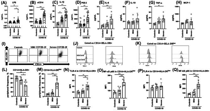
Figure 1 shows the concentrations of LPS, sCD14, IL-1β, and PGE-2 (healthy controls, n = 14; mild COVID-19, n = 25; severe COVID-19, n = 22) as well as the levels of cytokines (healthy controls, n = 5; mild COVID-19, n = 8; severe COVID-19, n = 7) in the blood of healthy controls and COVID-19 patients. Higher LPS levels (Fig. 1A; p = 0.05 vs. healthy controls and mild COVID-19 groups), sCD14 (Fig. 1B; p = 0.001 vs. healthy controls and mild COVID-19), IL-1β (Fig. 1C; p = 0.001 vs. healthy controls, p = 0.01 vs. mild COVID-19), PGE-2 (Fig. 1D; p < 0.001 vs. healthy control and mild COVID-19), IL-6 (Fig. 1E; p = 0.001 vs. healthy controls; p < 0.05 vs. mild COVID-19), and TNF-α (Fig. 1G; p = 0.01 vs. healthy controls) were identified in the blood of severe COVID-19 subjects. Mild COVID-19 patients presented higher IL-1β plasma concentrations than healthy controls (p < 0.01).
Fig. 1.
Analysis of systemic levels of LPS (A), soluble CD14 (B), cytokines concentrations (C, IL-1β; E, IL-6; F, IL-10; G, TNF-α; H, MCP-1), and prostaglandin E2 (D) in the plasma of healthy controls, mild COVID-19, and severe COVID-19 patients. Representative dot plot of CD14 + HLA-DR low/+ (I), the expression of TLR-4 and NF-κB p65 in CD14 + HLA-DR + (J), and the expression of TLR-4 and NF-κB p65 in CD14 + HLA-DRlow (K) monocytes are showed. Finally, whole blood was used to evaluate the peripheral frequency of CD14 + HLA-DR + (L) and CD14 + HLA-DRlow (M), the expression of TLR-4 (N) and NF-κB p65 (O) in CD14 + HLA-DR + , and the expression of TLR-4 (P) and NF-κB p65 (Q) in CD14 + HLA-DR.low in healthy controls, mild COVID-19, and severe COVID-19 patients. Between-group comparison performed through one-way ANOVA followed by Bonferroni’s post hoc. (p < 0.05). *p < 0.05; **p < 0.01; ***p < 0.001
In addition, higher frequency of CD14 + HLA-DRlow monocytes (Fig. 1M; p = 0.001 vs. healthy controls; p = 0.01 vs. mild COVID-19) presenting higher NF-κB p65 phosphorylation (Fig. 1Q; p = 0.001 vs. both healthy controls and mild COVID-19 patients) as well as lower CD14 + HLA-DR + monocytes (Fig. 1L; p = 0.01 vs. healthy controls; p < 0.05 vs. mild COVID-19 patients) expressing higher TLR-4 (Fig. 1N; p = 0.01 vs. both control and mild COVID-19 groups) and NF-κB p65 phosphorylation (Fig. 1O; p < 0.001 vs. both groups comparison) were identified in the peripheral blood of severe COVID-19 subjects.
Systemic LPS concentrations associate with COVID-19 symptoms and clinical cardiovascular parameters
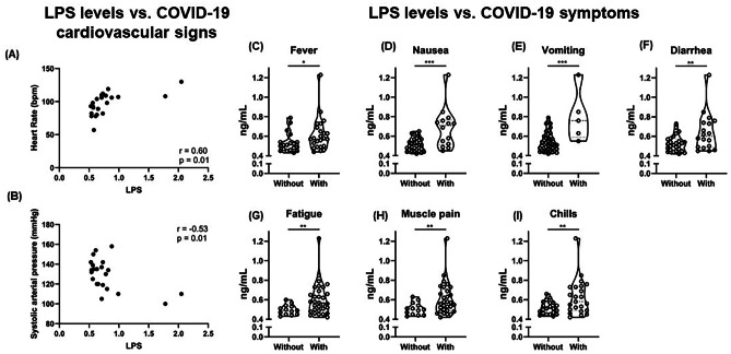
Figure 2 presents the systemic LPS concentrations in COVID-19 patients with or without clinical disease symptoms. Interestingly, LPS levels were higher in patients reporting fever (Fig. 2C; p = 0.05), nausea (Fig. 2D; p < 0.001), vomiting (Fig. 2E; p < 0.001), diarrhea (Fig. 2F; p < 0.01), fatigue (Fig. 2G; p < 0.01), muscle pain (Fig. 2H; p < 0.01), and chills (Fig. 2I; p < 0.01). Then, we correlated clinical cardiorespiratory parameters of severe COVID-19 patients with LPS and sCD14 levels. Systemic LPS inversely correlated with systolic blood pressure (Fig. 2B; r = − 0.53; p < 0.01) but positively correlated with heart rate (Fig. 2A; r = 0.60; p < 0.01).
Fig. 2.
The association of systemic microbial translocation markers with clinical symptoms of acute SARS-CoV-2 infection. LPS was correlated with heart rate (A) and systolic blood pressure (B) in severe SARS-CoV-2-infected patients by Pearson’s coefficient correlation test (p < 0.05). The levels of LPS were compared in mild and severe COVID-19 patients with and without fever (C), nausea (D), vomiting (E), diarrhea (F), fatigue (G), muscle pain (H), and chills (I) through Mann–Whitney U test and the significant results were presented (p < 0.05). *p < 0.05; **p < 0.01; ***p < 0.001
Whole blood LPS stimulation induces lower inflammatory response in severe COVID-19
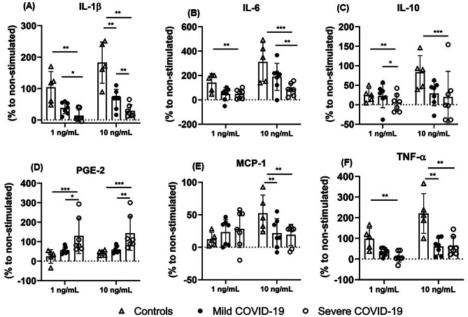
Next, we performed a whole blood ex vivo stimulation with LPS at two different concentrations (1 ng/mL or 10 ng/mL) for 2 h (5%CO2, 37 °C). After the incubation period, the plasma was collected and kept at − 80 °C to analyze cytokines production, while the remaining blood was used to verify the expression of TLR-4 and NF-κB p65 phosphorylation in CD14 + HLA-DRlow/+ monocytes by flow cytometry. After stimulation with LPS at 1 ng/mL, increased IL-1β (Fig. 3A; p = 0.01), IL-6 (Fig. 3B; p = 0.01), IL-10 (Fig. 3C; p = 0.01), and TNF-α (Fig. 3F; p = 0.01) production were found in healthy controls compared to severe COVID-19 patients, and higher IL-1β and IL-10 production by whole blood of mild compared to severe COVID-19 (p < 0.05) (Fig. 3). LPS (10 ng/mL)-stimulated whole blood cultures of healthy controls elicited a great increase in the production of IL-1β (p = 0.01 vs. mild COVID-19 and severe COVID-19), IL-6 (p = 0.001 vs. severe COVID-19), IL-10 (p = 0.001 vs. severe COVID-19), CCL2 (Fig. 3E; p = 0.01 vs. mild COVID-19; p = 0.01 vs. severe COVID-19), and TNF-α (p = 0.01 vs. mild COVID-19; p = 0.01 vs. severe COVID-19). Increased IL-1β and IL-6 production was also found after ex vivo LPS (10 ng/mL) stimulation of blood cells of mild COVID-19 patients compared to severe COVID-19 group (p = 0.01). Furthermore, LPS-stimulated whole blood of severe COVID-19 patients produced higher PGE-2 after 1 ng/mL (Fig. 3D; p < 0.001 vs. healthy controls; p < 0.05 vs. mild COVID-19) and 10 ng/mL (p < 0.001 vs. healthy controls; p < 0.01 vs. mild COVID-19) stimulation.
Fig. 3.
The production of IL-1β (A), IL-6 (B), IL-10 (C), PGE-2 (D), MCP-1 (E), and TNF-α (F) by ex vivo LPS-stimulated whole blood of healthy controls and mild and severe COVID-19 patients. Whole blood samples of controls, mild COVID-19, and severe COVID-19 patients were aliquoted in tubes to a volume of 1 mL and incubated (5%CO2, 37 °C) with two different LPS concentrations (1 and 10 ng/mL, Sigma-Aldrich, USA) or without immunogenic stimulus for 2 h. The supernatant concentrations of interleukin (IL)-6, IL-10, C–C motif ligand 2 (CCL2), and tumor necrosis factor-alpha (TNF-α) were analyzed by multiplex assays. The supernatant Prostaglandin E2 and IL-1β concentrations were analyzed by enzyme‐linked immunosorbent assay (ELISA). The production of cytokines was evaluated by the % of difference compared to the non-stimulated condition. **(p < 0.01) and ***(p < 0.001) denote statistical difference (p < 0.01)
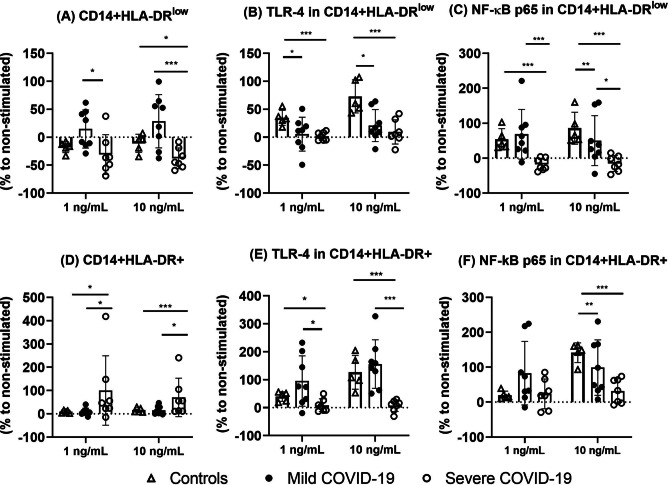
Severe COVID-19 patients presented lower proportions of CD14 + HLA-DRlow (Fig. 4A; 1 ng/mL, p < 0.05 vs. mild COVID-19; 10 ng/mL, p < 0.05 vs. healthy controls; 10 ng/mL, p = 0.001 vs. mild COVID-19) (Fig. 4) and increased frequencies of CD14 + HLA-DR + (Fig. 4D; 1 ng/mL, p < 0.05 vs. healthy controls; 1 ng/mL, p < 0.05 vs. mild COVID-19; 10 ng/mL, p < 0.001 vs. healthy controls; 10 ng/mL, p < 0.05 vs. mild COVID-19) after ex vivo LPS challenge. Furthermore, healthy controls presented increased TLR-4 in CD14 + HLA-DRlow after both 1 ng/mL (Fig. 4B; p < 0.05 vs. mild COVID-19; p = 0.001 vs. severe COVID-19) and 10 ng/mL LPS challenge (p = 0.05 vs. mild COVID-19; p = 0.001 vs. severe COVID-19). The NF-κB p65 phosphorylation was lower in CD14 + HLA-DRlow monocytes of severe COVID-19 patients after treatment with 1 ng/mL (Fig. 4C; p = 0.001 vs. healthy controls; p = 0.001 vs. mild COVID-19) and 10 ng/mL of LPS (p = 0.001 vs. healthy controls; p < 0.05 vs. mild COVID-19), although significant difference was also found in healthy controls vs. mild COVID-19 comparison (p = 0.01) after 10 ng/mL LPS stimulation. In CD14 + HLA-DR + monocytes of severe COVID-19, LPS stimulation induced lower TLR-4 expression (Fig. 4E; 1 ng/mL, p < 0.05 vs. healthy controls; 1 ng/mL, p < 0.05 vs. mild COVID-19; 10 ng/mL, p = 0.001 vs. healthy controls; 10 ng/mL, p < 0.005 vs. mild COVID-19). Finally, healthy controls presented higher NF-κB p65 phosphorylation in CD14 + HLA-DR + monocytes (Fig. 4F) compared to mild COVID-19 (p = 0.01) and severe COVID-19 (p = 0.001) patients.
Fig. 4.
The frequencies of CD14 + HLA-DR.low (A) monocytes expressing TLR-4 (B) and NF-κB (C) and CD14 + HLA-DR + (D) expressing TLR-4 (E) and NF-κB (F) in ex vivo LPS-stimulated whole blood of healthy controls and mild and severe COVID-19 patients. Whole blood samples of controls, mild COVID-19, and severe COVID-19 patients were aliquoted in tubes to a volume of 1 mL and incubated (5%CO2, 37 °C) with two different LPS concentrations (1 and 10 ng/mL, Sigma-Aldrich, USA) or without immunogenic stimulus for 2 h. The phenotype of monocytes was evaluated by flow cytometry. The frequencies of each cell subtype were evaluated by the % of difference compared to the non-stimulated condition. *(p < 0.05), **(p < 0.01), and ***(p < 0.001) denote statistical difference (p < 0.01)
Discussion
Here, we aimed to evaluate the acute inflammatory response and the modulation of monocyte phenotype after an ex vivo whole blood LPS challenge in patients with different degrees of COVID-19 severity. We hypothesized that acute SARS-CoV-2 infection impairs the ability of immune system to respond against a secondary pathogen, suggesting a temporary immunosuppression. We main found that severe COVID-19 patients presented: (a) Increased LPS and sCD14 concentrations in their peripheral blood associated with clinical COVID-19 symptoms and cardiovascular data; (b) Increased frequencies of CD14 + HLA-DRlow and lower CD14 + HLA-DR + monocytes in the peripheral blood; (c) Increased TLR-4 expression in CD14 + HLA-DR + and higher NF-κB p65 phosphorylation in both CD14 + HLA-DRlow and CD14 + HLA-DR + monocytes. Furthermore, after the stimulation of whole blood with different concentrations of the classic TLR-4 ligand LPS, we main found (a) lower production of inflammatory cytokines associated with increased PGE-2 production by stimulated whole blood of severe COVID-19 patients; (b) severe COVID-19 group presented decreased CD14 + HLA-DRlow and higher CD14 + HLA-DR + monocytes after 1 and 10 ng/mL LPS stimulation; (c) impaired TLR-4 expression and NF-κB p65 phosphorylation were found in both CD14 + HLA-DRlow and CD14 + HLA-DR + monocytes of mild and severe COVID-19 patients after 1 and 10 ng/mL LPS stimulation; and (d) severe COVID-19 patients presented the most pronounced impairment in monocyte modulation after LPS stimulation. Taken together, our results denote that acute SARS-CoV-2 infection is associated with diminished endotoxin response in monocytes of patients with severe disease. However, we like to reinforce that this study was conducted with a small sample size of mild and severe COVID-19 patients. Thus, future studies should be conducted with a larger number of participants to evaluate the role of LPS tolerance in COVID-19 pathogenesis.
The present results are in line with previous data published by our group [7] and others [18] demonstrating an association between increased markers of bacterial translocation and proinflammatory cytokines in severe COVID-19 subjects at hospital admission. Strong endotoxemia were also linked to tissue damage and multiorgan disease after SARS-CoV-2 once that increased LPS-binding protein (LBP) remained higher during hospitalization in patients with severe COVID-19 [12]. Here, we also observed increased PGE-2 levels in severe COVID-19 patients. Higher pro-resolving and inflammatory lipid mediators, such as eicosanoids and docosanoids, were found in the blood and bronchoalveolar lavage of severe COVID-19 patients which suggests a role of pro- and anti- inflammatory lipids to contribute to the progression of disease severity [19]. The production of PGE-2 during active SARS-CoV-2 infection occurs through the upregulation of cyclo-oxygenase-2 and reduced 15-hydroxyprostaglandin-dehydrogenase enzyme activity in infected lung epithelial cells [20]. Furthermore, higher level of PGE-2 was found in the blood of severe COVID-19 patients with high viral load [21] and reduces anti-viral immune response by targeting B-cell proliferation and differentiation [20]. Finally, systemic immune activation and chronic inflammation are linked to microbial translocation and endotoxemia after viral infection in patients with HIV infection [22, 23]. Collectively, these data indicate that viral infections, such as SARS-CoV-2, can induce an enhanced endotoxemia that may contribute to the systemic inflammation and hamper the immune response [24, 25].
Gut leakage and microbial product translocation were previously associated with cardiovascular involvement and inflammasome activation in hospitalized COVID-19 patients [12]. In fact, we identified a positive correlation between systemic LPS levels and heart rate in severe COVID-19 group. Thus, tachycardia observed during severe SARS-CoV-2 infection [26] may be related by the disruption of sympathetic nervous system and endothelial damage induced by hyperinflammation and endotoxemia. Furthermore, LPS levels inversely correlated with systolic arterial pressure which suggest that alterations in blood perfusion and conductance are impacted by endotoxins. In fact, endotoxemic shock patients present falls in blood pressure, and LPS infusion in rats reduces mean arterial blood pressure (MAP) by deregulation in molecular pathways involved in oxidative stress response and coagulation [27]. Finally, we identified higher levels of LPS in SARS-CoV-2-infected patients presenting gastrointestinal (i.e., nausea, vomiting, and diarrhea) or systemic inflammatory symptoms (i.e., fever, fatigue, muscle pain, and chills). In this sense, gastrointestinal symptoms may indicate intestinal damage and systemic microbial translocation during SARS-CoV-2 infection. Furthermore, since LPS levels were higher in severe COVID-19, the data presented here reinforces the implications of disruption in gut functions as a potential force to COVID-19 severity [7].
We also observed elevated frequencies of CD14 + HLA-DRlow monocytes, an immunosuppressive phenotype of myeloid cells, in the peripheral blood of severe COVID-19 patients. This result is in line with previous studies who also found increased proportions of HLA-DRlow immature monocytes in hospitalized and ICU COVID-19 patients [28, 29]. CD14 + HLA-DRlow monocytes are part of an immature cells that can mediate suppression of the immune response [30]. Interestingly, immunosuppressive monocytic phenotype remained unchanged over the hospitalization and is linked to a dysfunctional immune response. When transitioning to an immunosuppressive state, monocytes are highly sensitive and become inactive, leading to immunoparalysis [31]. Furthermore, immunosuppressive myeloid phenotype in severe COVID-19 patients was associated with the development of secondary infections, and 28- and 60-day mortality [28]. Thus, taken together, increased endotoxemia, inflammatory cytokines, and the accumulation of CD14 + HLA-DRlow monocytes in the peripheral blood of severe COVID-19 patients may be related to the immunosuppressive condition and higher secondary infection rates observed in these patients.
Low-grade endotoxemia is a common event in SARS-CoV-2-infected patients [32], and usually related to a continuous stimulation of TLR-4 in innate immune cells. Furthermore, the spike protein of SARS-CoV-2 synergically interacts with LPS, leading to a boosting of proinflammatory actions mediated by the phosphorylation of NF-κB in monocytic THP-1 cell line [33]. Similarly, it was found that the interaction between spike protein and LPS enhances in vivo pro-inflammatory cytokine production, suggesting that an early innate immune response occurs by the activation of transcription inflammatory genes after the co-presence of LPS and proteins related to SARS-CoV-2 [34–36]. The proportions of CD14 + HLA-DRlow and CD14 + HLA-DR + monocytes in severe COVID-19 patients altered after endotoxin stimulation (1 and 10 ng/mL), since has been observed a decreased frequency of immunosuppressive HLA-DRlow monocyte after LPS incubation. Possibly the endotoxin stimulation may increase the activation and antigen-presenting activity to adaptive immune cells by monocytes [37], although the functionality may be compromised as observed by low inflammatory cytokine secretion. On the other hand, we cannot exclude the hypothesis of higher monocyte apoptosis induced by LPS stimulation concomitant with SARS-CoV-2 infection. Spike protein activates apoptosis pathways and NLRP-3 inflammasome cascade linked to cell death [24, 33, 38], and CD14 + HLA-DRlow may be more prone to apoptosis during certain situations such as SARS-CoV-2 and endotoxin co-stimulation. Future studies should be conducted to address this question in COVID-19 condition. Here, we found an increased expression of TLR-4 on CD14 + HLA-DR + monocytes in conjunct with higher NF-κB p65 phosphorylation in both CD14 + HLA-DR + and CD14 + HLA-DRlow monocytes of severe COVID-19 patients. In fact, it was previously reported an overactivation of TLR-4 by the concomitant presence of systemic endotoxemia and SARS-CoV-2-related proteins observed in in vitro experiments conducted in previous reports [36, 39, 40]. Furthermore, previous in silico studies predicted cell surface TLR-4 is most likely to be involved in recognizing molecular patterns of SARS-CoV-2 to induce inflammatory response at early stages of infection and the inhibition of NF-κB was able to suppress IL-1β production induced by spike protein [41], confirming the crucial role of TLR-4/NF-κB axis in the hyperinflammatory response of COVID-19 pathogenesis.
Clinically, TLR-4 and signaling molecules are upregulated in leukocytes from COVID-19 patients in a condition that mimics bacterial sepsis. Furthermore, Shambat and colleagues [42] showed that a higher degree of SARS-CoV-2 viremia mediates hypercytokinemia and is positively associated with bacterial superinfections. Interestingly, the authors showed that TNF-α and other cytokines synergistically mediate impaired antibacterial effector functions and downregulate the HLA-DR expression of monocytes from COVID-19 patients [42]. We cannot exclude that others intracellular pathways linked to TLR4 may also be blunted during SARS-CoV-2 infection. Recent studies indicate that SARS-CoV-2 ORF9b protein suppress TRIF signaling and the secretion of IFN type I and type III [43, 44], which may be linked to the decreases in TLR sensitization during acute infection. Future studies should be conducted to evaluate additional roles of TLR-downstream pathways in LPS tolerance during acute SARS-CoV-2 infection.
Here, we employed an ex vivo LPS-whole blood stimulation to verify the ability of systemic immune cells of SARS-CoV-2-infected patients to respond to an endotoxin challenge. Interestingly, severe COVID-19 patients presented lower production of IL-1β, IL-6, IL-10, CCL2, and TNF-α after LPS stimulation at 1 and 10 ng/mL. Thus, our results suggest that SARS-CoV-2 infection hampers the cytokine production after a secondary stimulus, which may predispose the host to secondary infections. The impaired cytokine production after endotoxin challenge found here may be related to the higher PGE-2 production identified in the supernatant of whole blood stimulation. Previous in vitro studies identified low cytokine production and impaired cell activation by LPS-stimulated macrophages after PGE-2 treatment, reinforcing the immunomodulatory role of lipid mediators during inflammation [45].
PGE-2 is produced by macrophage during high endotoxemia and contributes to vasodilation, pain, fever, and multiple organ failure during conditions such as septic shock syndrome [46]. Furthermore, PGE-2 has a negative feedback loop through the enhancement in the expression of phosphatase 1 that impairs the activity of MAPK p38 and suppress cytokine release during disturbances of inflammatory response [47]. PGE-2 suppress the production of TNF-α by in vitro LPS-stimulated murine macrophages in a dose-dependent manner, confirming the regulatory action on acute inflammation [48]. In this sense, it seems that major immunologic abnormalities in COVID-19 are a profound defect in host immune response due to high inflammation condition induced by primary SARS-CoV-2 infection. There was no evidence of exaggerated TNF-α production in response to ex vivo LPS stimulation of mononuclear cells from COVID-19 patients compared to septic or critically illness non septic patients, despite increased plasma inflammatory cytokines [49]. Furthermore, mononuclear cells from healthy volunteers presented impaired TNF-α and IL-6 production after exposure to spike and nucleocapsid recombinant proteins from SARS-CoV-2 and then challenged with LPS, indicating endotoxin tolerance induced by SARS-CoV-2 proteins. Collectively, the data indicate that COVID-19 induces an immunocollapse that predisposes the patients to a secondary infection.
Since LPS triggers NF-κB activation and cytokine production through the engagement with TLR-4 on the cell surface of CD14 + monocytes, we evaluated the role of both innate inflammatory mediators after an ex vivo endotoxin challenge in whole blood from COVID-19 patients. We identified increased proportions of CD14 + HLA-DR + and lower CD14 + HLA-DRlow in the whole blood of severe COVID-19 patients compared to both mild COVID-19 and healthy control groups after ex vivo endotoxin stimulation. These results are in contrast with previous data that reported lower HLA-DR expression in monocytes after antigenic challenge [16, 42, 50]. The divergent data may be linked to the different conditions used for stimulation, since the reported impaired immune response to Candida albicans [50], Staphylococcus aureus, and Streptococcus pneumoniae [42] by monocytes from COVID-19 patients may indicate that other pattern recognition receptors (PRRs) are impaired during acute SARS-CoV-2. Future studies should be conducted to clarify the impact of COVID-19 on innate PRR response.
We observed for the first time that compared to healthy controls, both mild and severe COVID-19 patients presented impaired TLR-4 expression in CD14 + HLA-DRlow monocytes after ex vivo LPS stimulation and lower TLR-4 expression in CD14 + HLA-DR + monocytes from severe COVID-19 patients following LPS challenge. Collectively, these data may indicate that SARS-CoV-2 infection induces a lower endotoxin recognition by the decreased expression of TLR-4 despite the increases in activation status as demonstrated by higher HLA-DR expression in monocytes. Furthermore, in response to LPS, the NF-κB p65 phosphorylation was lower in CD14 + HLA-DRlow/+ monocytes of severe COVID-19 patients. LPS tolerance is a critical event observed in septic condition [51] and others critical ill situations induced by several negative regulators of TLR-4 signal transduction, such as IRAK-M, aryl hydrocarbon receptor, tryptophan catabolism, and other molecules that negatively regulate NF-κB activation, mainly BCL-3 and NF-κBp50 [52]. In this sense, SARS-CoV-2 antigens may contribute to the LPS tolerance during acute infection, since the continuous stimulation of TLR-4 and NF-κB by viral peptides may exhaust the innate pathways [33, 41]. The activation and translocation of NF-κB occur rapidly following exposure of monocytes to spike protein of coronavirus, suggesting that the recognition pattern by TLR is continuously activated during acute SARS-CoV infection [38]. Furthermore, SARS-CoV-2 nucleocapsid protein triggers the phosphorylation of NF-κB p65 in vitro and promotes M1 macrophage polarization which increases lung injury in mice model [53]. On the other hand, PGE-2 reduces the activation of NF-κB and PI3k-Akt signaling during endotoxin stimulation with strong implications in cytokine production and monocyte activation [54]. Furthermore, others possible mechanisms induced during LPS tolerance may contribute to the lower innate immune response observed in COVID-19 patients. The myeloid cells reprogramming during LPS tolerance acquire an anti-inflammatory phenotype due to the formation of NF-κB p50-p50 homodimeric form, which reduces the pro-inflammatory cytokine secretion concomitant to the increases in the immunoregulatory IL-10 secretion [55, 56]. However, we did not observe increased IL-10 production during LPS stimulation in whole blood of COVID-19 patients. As the inflammatory phenotype of acute SARS-CoV-2 infection is characterized by higher amounts of IL-10, we believe that a feedback loop may blunts any additional IL-10 production during co-infections or endotoxin stimulation. Thus, the continuously stimulation of TLR-4/NF-κB axis during acute SARS-CoV-2 infection may saturate the inflammatory pathways in a model to reduce the ability of innate response against a secondary endotoxin/LPS stimuli, which may indicate reduced immunosurveillance against a secondary infection.
Conclusion
In summary, our data provide evidence indicating that SARS-CoV-2 infection induces lower cytokine production and TLR-4/NF-κB activation in monocytes from severe COVID-19 after LPS stimulation. In addition, COVID-19 patients had systemic alterations in the peripheral blood, such as increased LPS and cytokine levels, higher frequency of immunosuppressive CD14 + HLA-DRlow, and higher expression of TLR-4 and NF-κB activation. Taken together, these results may indicate that the pro-inflammatory condition and microbial translocation markers observed in acute SARS-CoV-2 infection may induce endotoxin tolerance associated with the potential development of secondary infections.
Acknowledgements
We thank all subjects who agreed to participate in the study and donate blood.
Author contribution
GP Dorneles, PC Teixeira, A Peres, S Eller, SG Fonseca, and PRT Romão conceived and planned the experiments. GP Dorneles, PC Teixeira, S Eller, and TF Oliveira carried out the experiments. A Peres, LCR Júnior, SG da Fonseca, MC Monteiro, and EM Wedland contributed to the interpretation of the results. SG da Fonseca and PRT Romão took the lead in writing the manuscript. All authors provided critical feedback and helped shape the research, analysis, and manuscript.
Funding
We are grateful to the Coordenação de Aperfeiçoamento de Pessoal de Nível Superior (CAPES) – Finance Code 001 and Universidade Federal de Ciências da Saúde de Porto Alegre (Programa de Fomento a Pesquisa). This study was supported in part by the Hospital Moinhos de Vento through the Program for Supporting the Institutional Development of the Public Health System (PROADISUS), supported by the Ministry of Health of Brazil. GPD is supported by postdoctoral fellowship from CAPES. PRTR, AP, and MCM are grateful to CNPq for the PQ productivity scholarship.
Availability of data and materials
After request, first author can provide data.
Declarations
Ethics approval and consent to participate
Experimental protocols were approved by the Moinhos de Vento Ethics Committee N 3977144 (Porto Alegre/Brazil) and performed according to the declaration of Helsinki. All participants were informed before entering the study and informed consent was obtained from the subjects.
Consent for publication
All authors express their consent to the publication of this manuscript in this journal.
Competing interests
The authors declare no competing interests.
Footnotes
Gilson P Dorneles and Paula C Teixeira are considered first authors due to their equal collaboration.
Publisher's Note
Springer Nature remains neutral with regard to jurisdictional claims in published maps and institutional affiliations.
References
- 1.Chen G, Wu D, Guo W, Cao Y, Huang D, Wang H et al (2020) Clinical and immunologic features in severe and moderate forms of coronavirus disease 2019. 0–30
- 2.Pascarella G, Strumia A, Piliego C, Bruno F, Del Buono R, Costa F et al (2020) COVID-19 diagnosis and management: a comprehensive review. J Intern Med [DOI] [PMC free article] [PubMed]
- 3.Colantuoni A, Martini R, Caprari P, Ballestri M, Capecchi PL, Gnasso A et al (2020) COVID-19 sepsis and microcirculation dysfunction. Front Physiol 11. Available from: https://pubmed.ncbi.nlm.nih.gov/32676039/. Accessed 24 Jan 2022 [DOI] [PMC free article] [PubMed]
- 4.Tay MZ, Poh CM, Rénia L, MacAry PA, Ng LFP. The trinity of COVID-19: immunity, inflammation and intervention. Nat Rev Immunol. 2020;20(6):363–374. doi: 10.1038/s41577-020-0311-8. [DOI] [PMC free article] [PubMed] [Google Scholar]
- 5.Coperchini F, Chiovato L, Croce L, Magri F, Rotondi M (2020) The cytokine storm in COVID-19: an overview of the involvement of the chemokine/chemokine-receptor system. Cytokine and Growth Factor Reviews, vol. 53, Elsevier Ltd. pp. 25–32. Available from: https://pubmed.ncbi.nlm.nih.gov/32446778/. Accessed 17 May 2021 [DOI] [PMC free article] [PubMed]
- 6.Hotchkiss RS, Monneret G, Payen D (2013) Sepsis-induced immunosuppression: from cellular dysfunctions to immunotherapy. Nat Rev Immunol 13(12):862. Available from: https://www.ncbi.nlm.nih.gov/pmc/articles/PMC4077177/. Accessed 24 Jan 2022 [DOI] [PMC free article] [PubMed]
- 7.Teixeira PC, Dorneles GP, Santana Filho PC, da Silva IM, Schipper LL, Al Postiga I et al (2021) Increased LPS levels coexist with systemic inflammation and result in monocyte activation in severe COVID-19 patients. Int Immunopharmacol 108125. Available from: https://linkinghub.elsevier.com/retrieve/pii/S156757692100761X. Accessed 13 Sep 2021 [DOI] [PMC free article] [PubMed]
- 8.de Candia P, Prattichizzo F, Garavelli S, Matarese G (2020) T cells: warriors of SARS-CoV-2 infection. Trends Immunol 1–13. Available from: http://www.ncbi.nlm.nih.gov/pubmed/33277181. Accessed 25 Jan 2022 [DOI] [PMC free article] [PubMed]
- 9.Wang F, Nie J, Wang H, Zhao Q, Xiong Y, Deng L, et al. Characteristics of peripheral lymphocyte subset alteration in COVID-19 pneumonia. J Infect Dis. 2020;221(11):1762–1769. doi: 10.1093/infdis/jiaa150. [DOI] [PMC free article] [PubMed] [Google Scholar]
- 10.Musuuza JS, Watson L, Parmasad V, Putman-Buehler N, Christensen L, Safdar N (2021) Prevalence and outcomes of co-infection and superinfection with SARS-CoV-2 and other pathogens: a systematic review and meta-analysis. PLoS One 16(5). Available from: https://pubmed.ncbi.nlm.nih.gov/33956882/. Accessed 25 Jan 2022 [DOI] [PMC free article] [PubMed]
- 11.Garcia-Vidal C, Sanjuan G, Moreno-García E, Puerta-Alcalde P, Garcia-Pouton N, Chumbita M, et al. Incidence of co-infections and superinfections in hospitalized patients with COVID-19: a retrospective cohort study. Clin Microbiol Infect. 2021;27(1):83–88. doi: 10.1016/j.cmi.2020.07.041. [DOI] [PMC free article] [PubMed] [Google Scholar]
- 12.Hoel H, Heggelund L, Reikvam DH, Stiksrud B, Ueland T, Michelsen AE et al (2021) Elevated markers of gut leakage and inflammasome activation in COVID-19 patients with cardiac involvement. J Intern Med 289(4):523–31. Available from: https://pubmed.ncbi.nlm.nih.gov/32976665/. Accessed 5 May 2021 [DOI] [PMC free article] [PubMed]
- 13.Kahn R, Schmidt T, Golestani K, Mossberg A, Gullstrand B, Bengtsson AA et al (2020) Mismatch between circulating cytokines and spontaneous cytokine production by leukocytes in hyperinflammatory COVID-19. J Leukoc Biol [DOI] [PMC free article] [PubMed]
- 14.Payen D, Cravat M, Maadadi H, Didelot C, Prosic L, Dupuis C et al (2020) A longitudinal study of immune cells in severe COVID-19 patients. Front Immunol 11:2759. Available from: www.frontiersin.org. Accessed 18 May 2021 [DOI] [PMC free article] [PubMed]
- 15.Seeley JJ, Ghosh S (2017) Molecular mechanisms of innate memory and tolerance to LPS. J Leukoc Biol 101(1):107–19. Available from: https://pubmed.ncbi.nlm.nih.gov/27780875/. Accessed 25 Jan 2022 [DOI] [PubMed]
- 16.Avendaño-Ortiz J, Lozano-Rodríguez R, Martín-Quirós A, Maroun-Eid C, Terrón-Arcos V, Montalbán-Hernández K et al (2021) SARS-CoV-2 proteins induce endotoxin tolerance hallmarks: a demonstration in patients with COVID-19. J Immunol 207(1):162–74. Available from: https://pubmed.ncbi.nlm.nih.gov/34183364/. Accessed 25 Jan 2022 [DOI] [PubMed]
- 17.Dorneles GP, Reiter KC, dos Passos A, Marmett B, da Silva IM, dos Santos MAL et al (2021) Differential effects of whole blood heat treatment on the ex vivo inflammatory profile of untrained and trained men. Cytokine 142(September 2020) [DOI] [PubMed]
- 18.Sirivongrangson P, Kulvichit W, Payungporn S, Pisitkun T, Chindamporn A, Peerapornratana S et al (2020) Endotoxemia and circulating bacteriome in severe COVID-19 patients. Intensive Care Med Exp 8(1):1–15. Available from: https://icm-experimental.springeropen.com/articles/10.1186/s40635-020-00362-8. Accessed 25 Jan 2022 [DOI] [PMC free article] [PubMed]
- 19.Archambault AS, Zaid Y, Rakotoarivelo V, Turcotte C, Doré É, Dubuc I et al (2021) High levels of eicosanoids and docosanoids in the lungs of intubated COVID-19 patients. FASEB J 35(6). Available from: https://pubmed.ncbi.nlm.nih.gov/34033145/. Accessed 31 May 2022 [DOI] [PMC free article] [PubMed]
- 20.Ricke-Hoch M, Stelling E, Lasswitz L, Gunecsh AP, Kasten M, Zapatero-Belinchon FJ et al (2021) Impaired immune response mediated by prostaglandin E2 promotes severe COVID-19 disease. PLoS One 16(8). Available from: https://pubmed.ncbi.nlm.nih.gov/34347801/. Accessed 27 Oct 2021 [DOI] [PMC free article] [PubMed]
- 21.Romão PR, Teixeira PC, Schipper L, da Silva I, Santana Filho P, Júnior LCR, et al. Viral load is associated with mitochondrial dysfunction and altered monocyte phenotype in acute severe SARS-CoV-2 infection. Int Immunopharmacol. 2022;1(108):108697. doi: 10.1016/j.intimp.2022.108697. [DOI] [PMC free article] [PubMed] [Google Scholar]
- 22.De Castro FDOF, Silva JM, Dorneles GP, De Sousa Barros JB, Ribeiro CB, Noronha I et al (2019) Distinct inflammatory profiles in HIV-infected individuals under antiretroviral therapy using cannabis, cocaine or cannabis plus cocaine. AIDS 33(12):1831–42. Available from: https://pubmed.ncbi.nlm.nih.gov/31490211/. Accessed 5 May 2021 [DOI] [PubMed]
- 23.Ribeiro CB, de Castro F, de OF, Dorneles GP, de Sousa Barros JB, Silva JM, Tavares C et al (2021) The concomitant use of cannabis and cocaine coexists with increased LPS levels and systemic inflammation in male drug users. Cytokine 141(August 2020):1–7 [DOI] [PubMed]
- 24.Samsudin F, Raghuvamsi P, Petruk G, Puthia M, Petrlova J, MacAry P et al (2022) SARS-CoV-2 spike protein as a bacterial lipopolysaccharide delivery system in an overzealous inflammatory cascade. J Mol Cell Biol. Available from: https://pubmed.ncbi.nlm.nih.gov/36240490/. Accessed 18 Dec 2022 [DOI] [PMC free article] [PubMed]
- 25.Puthia M, Tanner L, Petruk G, Schmidtchen A (2022) Experimental model of pulmonary inflammation induced by SARS-CoV-2 spike protein and endotoxin. ACS Pharmacol Transl Sci (3):141–8. Available from: https://pubmed.ncbi.nlm.nih.gov/35774232/. Accessed 18 Dec 2022 [DOI] [PMC free article] [PubMed]
- 26.Niehues P, Wegner FK, Wolfes J, Willy K, Ellermann C, Vollenberg R et al (2022) Incidence and predictors of cardiac arrhythmias in patients with COVID-19 induced ARDS. J Cardiol. Available from: https://pubmed.ncbi.nlm.nih.gov/35589465/. Accessed 31 May 2022 [DOI] [PMC free article] [PubMed]
- 27.Blangy-Letheule A, Persello A, Michelland S, Cunin V, Souab F, Aillerie V et al (2021) The secretome deregulations in a rat model of endotoxemic shock. Oxid Med Cell Longev 2021. Available from: https://pubmed.ncbi.nlm.nih.gov/34349874/. Accessed 31 May 2022 [DOI] [PMC free article] [PubMed]
- 28.Marais C, Claude C, Semaan N, Charbel R, Barreault S, Travert B et al (2021) Myeloid phenotypes in severe COVID-19 predict secondary infection and mortality: a pilot study. Ann Intensive Care 11(1):1–10. Available from: https://annalsofintensivecare.springeropen.com/articles/10.1186/s13613-021-00896-4. Accessed 27 Jan 2022 [DOI] [PMC free article] [PubMed]
- 29.Payen D, Cravat M, Maadadi H, Didelot C, Prosic L, Dupuis C, et al. A longitudinal study of immune cells in severe COVID-19 patients. Front Immunol. 2020;23(11):2759. doi: 10.3389/fimmu.2020.580250. [DOI] [PMC free article] [PubMed] [Google Scholar]
- 30.Gabrilovich DI, Nagaraj S (2009) Myeloid-derived suppressor cells as regulators of the immune system. Nat Rev Immunol 9(3):162–74. Available from: https://www.nature.com/articles/nri2506. Accessed 27 Jan 2022 [DOI] [PMC free article] [PubMed]
- 31.Volk HD, Reinke P, Krausch D, Zuckermann H, Asadullah K, Muller JM et al (1996) Monocyte deactivation--rationale for a new therapeutic strategy in sepsis. Intensive Care Med 22(4). Available from: https://pubmed.ncbi.nlm.nih.gov/8923092/. Accessed 27 Jan 2022 [DOI] [PubMed]
- 32.Oliva A, Cammisotto V, Cangemi R, Ferro D, Miele MC, De Angelis M et al (2021) Low-grade endotoxemia and thrombosis in COVID-19. Clin Transl Gastroenterol 12(6):e00348. Available from: https://pubmed.ncbi.nlm.nih.gov/34092777/. Accessed 27 Jan 2022 [DOI] [PMC free article] [PubMed]
- 33.Petruk G, Puthia M, Petrlova J, Samsudin F, Strömdahl AC, Cerps S et al (2020) SARS-CoV-2 spike protein binds to bacterial lipopolysaccharide and boosts proinflammatory activity. J Mol Cell Biol 12(12):916–32. Available from: https://pubmed.ncbi.nlm.nih.gov/33295606/. Accessed 25 Jan 2022 [DOI] [PMC free article] [PubMed]
- 34.Tumpara S, Gründing AR, Sivaraman K, Wrenger S, Olejnicka B, Welte T et al (2021) Boosted pro-inflammatory activity in human PBMCs by lipopolysaccharide and SARS-CoV-2 spike protein is regulated by α-1 antitrypsin. Int J Mol Sci 22(15). Available from: https://pubmed.ncbi.nlm.nih.gov/34360706/. Accessed 27 Jan 2022 [DOI] [PMC free article] [PubMed]
- 35.Yalcinkaya M, Liu W, Islam MN, Kotini AG, Gusarova GA, Fidler TP et al (2021) Modulation of the NLRP3 inflammasome by Sars-CoV-2 Envelope protein. Sci Rep 11(1):24432. Available from: https://pubmed.ncbi.nlm.nih.gov/34952919/. Accessed 27 Jan 2022 [DOI] [PMC free article] [PubMed]
- 36.Al‐Ani B, ShamsEldeen AM, Kamar SS, Haidara MA, Al‐Hashem F, Alshahrani MY et al (2022) Lipopolysaccharide induces acute lung injury and alveolar hemorrhage in association with the cytokine storm, coagulopathy and AT1R/JAK/STAT augmentation in a rat model that mimics moderate and severe Covid-19 pathology. Clin Exp Pharmacol Physiol. Available from: https://pubmed.ncbi.nlm.nih.gov/35066912/. Accessed 27 Jan 2022 [DOI] [PubMed]
- 37.Knoll R, Schultze JL, Schulte-Schrepping J. Monocytes and macrophages in COVID-19. Front Immunol. 2021;21:2952. doi: 10.3389/fimmu.2021.720109. [DOI] [PMC free article] [PubMed] [Google Scholar]
- 38.Dosch SF, Mahajan SD, Collins AR (2009) SARS coronavirus spike protein-induced innate immune response occurs via activation of the NF-kappaB pathway in human monocyte macrophages in vitro. Virus Res 142(1–2):19–27. Available from: https://pubmed.ncbi.nlm.nih.gov/19185596/. Accessed 29 Jan 2022 [DOI] [PMC free article] [PubMed]
- 39.Gajanayaka N, Dong SXM, Ali H, Iqbal S, Mookerjee A, Lawton DA et al (2021) TLR-4 agonist induces IFN-γ production selectively in proinflammatory human M1 macrophages through the PI3K-mTOR- and JNK-MAPK-activated p70S6K pathway. J Immunol 207(9):2310–24. Available from: https://pubmed.ncbi.nlm.nih.gov/34551966/. Accessed 27 Jan 2022 [DOI] [PubMed]
- 40.Bickler SW, Cauvi DM, Fisch KM, Prieto JM, Sykes AG, Thangarajah H et al (2021) Extremes of age are associated with differences in the expression of selected pattern recognition receptor genes and ACE2, the receptor for SARS-CoV-2: implications for the epidemiology of COVID-19 disease. BMC Med Genomics 14(1). Available from: https://pubmed.ncbi.nlm.nih.gov/34030677/. Accessed 27 Jan 2022 [DOI] [PMC free article] [PubMed]
- 41.Zhao Y, Kuang M, Li J, Zhu L, Jia Z, Guo X et al (2021) SARS-CoV-2 spike protein interacts with and activates TLR41. Cell Res 31(7):818–20. Available from: https://www.nature.com/articles/s41422-021-00495-9. Accessed 27 Jan 2022 [DOI] [PMC free article] [PubMed]
- 42.Mairpady Shambat SI, Gómez-Mejia A, Schweizer TA, Huemer M, Chang CC, Acevedo C et al (2022) Hyperinflammatory environment drives dysfunctional myeloid cell effector response to bacterial challenge in COVID-19. Prince A (ed) PLOS Pathog 18(1):e1010176. Available from: https://journals.plos.org/plospathogens/article?id=10.1371/journal.ppat.1010176. Accessed 27 Jan 2022 [DOI] [PMC free article] [PubMed]
- 43.Han L, Zhuang MW, Deng J, Zheng Y, Zhang J, Nan ML et al (2021) SARS-CoV-2 ORF9b antagonizes type I and III interferons by targeting multiple components of the RIG-I/MDA-5–MAVS, TLR3–TRIF, and cGAS–STING signaling pathways. J Med Virol 93(9):5376–89. Available from: https://onlinelibrary.wiley.com/10.1002/jmv.27050. Accessed 28 Dec 2022 [DOI] [PMC free article] [PubMed]
- 44.Shemesh M, Aktepe TE, Deerain JM, McAuley JL, Audsley MD, David CT et al (2021) SARS-CoV-2 suppresses IFNβ production mediated by NSP1, 5, 6, 15, ORF6 and ORF7b but does not suppress the effects of added interferon. PLOS Pathog 17(8):e1009800. Available from: https://journals.plos.org/plospathogens/article?id=10.1371/journal.ppat.1009800. Accessed 18 Dec 2022 [DOI] [PMC free article] [PubMed]
- 45.Saleh LS, Vanderheyden C, Frederickson A, Bryant SJ (2020) Prostaglandin E2 and its receptor EP2 modulate macrophage activation and fusion in vitro. ACS Biomater Sci Eng 6(5):2668–81. Available from: https://pubmed.ncbi.nlm.nih.gov/33463295/. Accessed 31 May 2022 [DOI] [PubMed]
- 46.Eliopoulos AG, Dumitru CD, Wang CC, Cho J, Tsichlis PN (2002) Induction of COX-2 by LPS in macrophages is regulated by Tpl2-dependent CREB activation signals. EMBO J 21(18):4831–40. Available from: https://onlinelibrary.wiley.com/doi/full/10.1093/emboj/cdf478. Accessed 31 May 2022 [DOI] [PMC free article] [PubMed]
- 47.Tang T, Scambler TE, Smallie T, Cunliffe HE, Ross EA, Rosner DR et al (2017) Macrophage responses to lipopolysaccharide are modulated by a feedback loop involving prostaglandin E2, dual specificity phosphatase 1 and tristetraprolin. Sci Reports 7(1):1–13. Available from: https://www.nature.com/articles/s41598-017-04100-1. Accessed 31 May 2022 [DOI] [PMC free article] [PubMed]
- 48.Kunkel SL, Wiggins RC, Chensue SW, Larrick J (1986) Regulation of macrophage tumor necrosis factor production by prostaglandin E2. Biochem Biophys Res Commun 137(1):404–10. Available from: https://pubmed.ncbi.nlm.nih.gov/3459461/. Accessed 18 Dec 2022 [DOI] [PubMed]
- 49.Remy KE, Mazer M, Striker DA, Ellebedy AH, Walton AH, Unsinger J et al (2020) Severe immunosuppression and not a cytokine storm characterizes COVID-19 infections. JCI insight 5(17). Available from: https://pubmed.ncbi.nlm.nih.gov/32687484/. Accessed 27 Jan 2022 [DOI] [PMC free article] [PubMed]
- 50.Moser D, Biere K, Han B, Hoerl M, Schelling G, Choukér A et al (2021) COVID-19 impairs immune response to Candida albicans. Front Immunol 12. Available from: https://pubmed.ncbi.nlm.nih.gov/33717195/. Accessed 28 Jan 2022 [DOI] [PMC free article] [PubMed]
- 51.Collins PE, Carmody RJ (2015) The regulation of endotoxin tolerance and its impact on macrophage activation. Crit Rev Immunol 35(4):293–324. Available from: https://www.dl.begellhouse.com/journals/2ff21abf44b19838,446e9fae25faaa7b,4680a0db19162f48.html. Accessed 28 Jan 2022 [DOI] [PubMed]
- 52.Carmody RJ, Ruan Q, Palmer S, Hilliard B, Chen YH (2007) Negative regulation of toll-like receptor signaling by NF-κB p50 ubiquitination blockade. Science 317(5838):675–8. Available from: https://www.science.org/doi/abs/10.1126/science.1142953. Accessed 28 Jan 2022 [DOI] [PubMed]
- 53.Xia J, Tang W, Wang J, Lai D, Xu Q, Huang R et al (2021) SARS-CoV-2 N protein induces acute lung injury in mice via NF-ĸB activation. Front Immunol 12. Available from: https://pubmed.ncbi.nlm.nih.gov/34950152/. Accessed 28 Jan 2022 [DOI] [PMC free article] [PubMed]
- 54.Bryson TD, Ross J, Peterson E, Harding P. Prostaglandin E2 and an EP4 receptor agonist inhibit LPS-induced monocyte chemotactic protein 5 production and secretion in mouse cardiac fibroblasts via Akt and NF-κB signaling. Prostaglandins Other Lipid Mediat. 2019;1(144):106349. doi: 10.1016/j.prostaglandins.2019.106349. [DOI] [PMC free article] [PubMed] [Google Scholar]
- 55.Nürnberger F, Leisengang S, Ott D, Murgott J, Gerstberger R, Rummel C et al (2021) Manifestation of lipopolysaccharide-induced tolerance in neuro-glial primary cultures of the rat afferent somatosensory system. Inflamm Res 70(4):429–44. Available from: https://pubmed.ncbi.nlm.nih.gov/33582876/. Accessed 18 Dec 2022 [DOI] [PMC free article] [PubMed]
- 56.López-Collazo E, del Fresno C (2013) Pathophysiology of endotoxin tolerance: mechanisms and clinical consequences. Crit Care 17(6):1–11. Available from: https://ccforum.biomedcentral.com/articles/10.1186/cc13110. Accessed 18 Dec 2022 [DOI] [PMC free article] [PubMed]
Associated Data
This section collects any data citations, data availability statements, or supplementary materials included in this article.
Data Availability Statement
After request, first author can provide data.


