Abstract
Background and Objectives
Ocrelizumab improved clinical and MRI measures of disease activity and progression in three phase 3 multiple sclerosis (MS) studies. Post hoc analyses demonstrated a correlation between the ocrelizumab serum concentration and the degree of blood B-cell depletion, and body weight was identified as the most influential covariate on ocrelizumab pharmacokinetics. The magnitude of ocrelizumab treatment benefit on disability progression was greater in lighter vs heavier patients. These observations suggest that higher ocrelizumab serum levels provide more complete B-cell depletion and a greater delay in disability progression. The current post hoc analyses assessed population exposure–efficacy/safety relationships of ocrelizumab in patients with relapsing and primary progressive MS.
Methods
Patients in OPERA I/II and ORATORIO were grouped in exposure quartiles based on their observed individual serum ocrelizumab level over the treatment period. Exposure–response relationships were analyzed for clinical efficacy (24-week confirmed disability progression (CDP), annualized relapse rate [ARR], and MRI outcomes) and adverse events.
Results
Ocrelizumab reduced new MRI lesion counts to nearly undetectable levels in patients with relapsing or primary progressive MS across all exposure subgroups, and reduced ARR in patients with relapsing MS to very low levels (0.13–0.18). A consistent trend of higher ocrelizumab exposure leading to lower rates of CDP was seen (0%–25% [lowest] to 75%–100% [highest] quartile hazard ratios and 95% confidence intervals; relapsing MS: 0.70 [0.41–1.19], 0.85 [0.52–1.39], 0.47 [0.25–0.87], and 0.34 [0.17–0.70] vs interferon β-1a; primary progressive MS: 0.88 [0.59–1.30], 0.86 [0.60–1.25], 0.77 [0.52–1.14], and 0.55 [0.36–0.83] vs placebo). Infusion-related reactions, serious adverse events, and serious infections were similar across exposure subgroups.
Discussion
The almost complete reduction of ARR and MRI activity already evident in the lowest quartile, and across all ocrelizumab-exposure groups, suggests a ceiling effect. A consistent trend of higher ocrelizumab exposure leading to greater reduction in risk of CDP was observed, particularly in the relapsing MS trials, and was not associated with a higher rate of adverse events. Higher ocrelizumab exposure may provide improved control of disability progression by reducing disease activity below that detectable by ARR and MRI, and/or by attenuating other B-cell–related pathologies responsible for tissue damage.
Classification of Evidence
This analysis provides Class III evidence that higher ocrelizumab serum levels are related to greater reduction in risk of disability progression in patients with multiple sclerosis. The study is rated Class III because of the initial treatment randomization disclosure that occurred after inclusion in the open-label extension.
Trial Registration Information
ClinicalTrials.gov Identifier: NCT01247324 (OPERA I), NCT01412333 (OPERA II), and NCT01194570 (ORATORIO).
Ocrelizumab, at an IV dose of 600 mg every 6 months, was the first CD20+ B-cell–selective humanized monoclonal antibody1 approved for the treatment of patients with relapsing multiple sclerosis (RMS) or primary progressive multiple sclerosis (PPMS).
Ocrelizumab had significant benefit on 12- and 24-week confirmed disability progression (12 W-CDP and 24 W-CDP), annualized relapse rate (ARR), and MRI measures vs comparator in pivotal phase 3 studies in patients with RMS2 or PPMS,3 with sustained efficacy in the respective open-label extension (OLE) periods.4,5 Further analysis of data from these studies6 and others7,8 has demonstrated that most disability progression in patients with RMS receiving disease-modifying therapies (DMTs) occurs independent of relapse activity, and is attributable to an underlying and continuous progressive disease course. Although ocrelizumab demonstrated substantial reductions of disability progression in patients with both RMS and PPMS, the need for providing a more pronounced effect on disability progression, or even stopping disability progression altogether, remains the greatest challenge to the field.
In pharmacokinetic and pharmacodynamic analyses of the OPERA I, OPERA II, and ORATORIO studies, a correlation between the individual ocrelizumab serum concentration over the treatment period for each patient and the level of B-cell depletion in blood was observed,9 where higher ocrelizumab exposure led to lower blood B-cell counts in patients with RMS and PPMS. The population pharmacokinetic analysis identified body weight as the most influential covariate on ocrelizumab serum levels. In the prespecified subgroup analyses of ocrelizumab efficacy in the pooled OPERA trials, body mass index (BMI) showed a significant treatment-by-subgroup interaction for both 12 W- and 24 W-CDP endpoints, indicating that the magnitude of ocrelizumab treatment benefit on disability progression was greater in lighter vs heavier patients (BMI ≤25 kg/m2 vs >25 kg/m2).10 A plausible explanation for this observation could be that higher ocrelizumab exposure in lighter patients leads to more complete B-cell depletion and to a more pronounced delay in disability progression. In this study, we investigated the relationship between higher ocrelizumab exposure and clinical, imaging, and safety outcomes, with a special focus on disability progression.
Methods
Standard Protocol Approvals, Registrations, and Patient Consents
The relevant institutional review boards/ethics committees approved the trial protocols (NCT01247324, NCT01412333, and NCT01194570). All patients provided written informed consent.
Trial Design and Patients
The conduct and clinical results from the pivotal phase 3 studies, and the pharmacokinetic–pharmacodynamic analyses, have been reported previously.2-5 In the phase 3 studies, patients received the approved dose of ocrelizumab (600 mg administered IV preceded by 100 mg methylprednisolone [or equivalent] IV every 6 months) or comparator. Patients with RMS were randomized 1:1 to ocrelizumab or subcutaneous interferon (IFN) β-1a 44 μg 3 times weekly for 96 weeks, whereas those with PPMS were randomized 2:1 to either ocrelizumab or placebo every 24 weeks until the last enrolled patient completed at least 120 weeks of study treatment and the planned total number of CDP events had been reached (median 7 doses).
Measures of Exposure
Ocrelizumab serum concentrations were measured with a validated ELISA with a lower limit of quantitation of 250 ng/mL, as described previously.9 Briefly, blood samples for pharmacokinetic assessments were drawn in OPERA: predose at weeks 1, 24, 48, and 72; 30 minutes postinfusion at week 72; and on days 84 and 96; and in ORATORIO: preinfusion on days 1 and 15, and at weeks 24, 48, 72, and 96; 30 minutes postinfusion on days 1 and 15 and at week 72; and at weeks 12, 84, and 120. The serum concentration data were analyzed using the population pharmacokinetic method. Concentration vs time curves over the 6-monthly dosing intervals were modeled from the individual estimated pharmacokinetic parameters, and subsequently Cmean, i.e., the average concentration over the treatment period, derived for each patient. Ocrelizumab exposure (Cmean) was defined as the average ocrelizumab serum concentration in an individual patient during their treatment with ocrelizumab. The exposure quartiles split all patients into 4 equal groups based on where an individual's exposure ranks in the range of ocrelizumab serum concentrations observed in the study patient population. For patients who received all planned doses, this corresponded to the average concentration over the entire treatment period of 96 weeks in patients with RMS in OPERA I and OPERA II, whereas treatment duration varied in patients with PPMS because of the event-driven design of the ORATORIO study (median [range] number of infusions: OPERA I/II, 4 [1–4]; ORATORIO, 7 [1–10]).
For investigation of the potential association between a patient's ocrelizumab serum level and clinical outcomes, ocrelizumab-treated patients were divided into subgroups based on the quartiles of the serum ocrelizumab exposure range. In patients with RMS, the ranges for ocrelizumab exposure Quartiles 1–4 were min–<15.4, ≥15.4–<18.7, ≥18.7–<22.2, and ≥22.2–max µg/mL, respectively. The corresponding ranges in patients with PPMS were min–<15.8, ≥15.8–<18.9, ≥18.9–<23.2, and ≥23.2–max µg/mL.9 To estimate treatment effects between ocrelizumab and control within comparable patient groups, ocrelizumab and IFN β-1a–treated patients were divided into subgroups based on BMI as a strong predictor of ocrelizumab exposure.
Endpoints and Populations Analyzed
Analyses of endpoints in patients with RMS and PPMS are based on the pooled population of OPERA I and OPERA II, and ORATORIO, respectively. Unless otherwise stated, analyses of endpoints are based on the complete double-blind controlled treatment phases (96 weeks for OPERA I and OPERA II [pooled data], >120 weeks for ORATORIO). For B-cell–related endpoints, the OLE was included for the ocrelizumab-treated patients from the pooled population of OPERA I and OPERA II, and the extended-controlled period and OLE for the ORATORIO population.4,5
Exposure–response analyses of ARR are presented only for patients with RMS because ARR was not specified as an endpoint in ORATORIO; the low number of protocol-defined relapses in ORATORIO also precluded any meaningful analysis. Analyses of 12 W-CDP, 24 W-CDP, MRI-based measures, and safety outcomes, including serious adverse events (SAEs), infusion-related reactions, and serious infections, are presented for both RMS and PPMS populations. MRI measures assessed were T1 gadolinium-enhancing lesions, new and/or enlarging T2 lesions, and percent change in whole brain volume from baseline; for brain volume change, MRI rebaselining at week 8 was also included to exclude MRI activity that occurs during the first 8 weeks of treatment before the potential treatment benefit of ocrelizumab is realized.
In addition to the exposure–response analyses of disability progression defined by the Expanded Disability Status Scale (EDSS) score (an increase from baseline of at least 1 point [or 0.5 points if the baseline EDSS score was >5.5]), analyses by baseline BMI quartile and blood B-cell depletion (3 groups based on the median blood CD19+ B-cell count over the treatment period of each patient, i.e., 0, 1–5, and >5 B cells/µL, or grouped by < and ≥ the median of the postbaseline median CD19+ B-cell count of each patient; blood CD19+ B-cell counts were assessed by flow cytometry), along with an exposure–response analysis of composite CDP (cCDP; first confirmed occurrence of an increase in the EDSS score and/or increase in Timed 25-Foot Walk [T25FW] or Nine-Hole Peg Test [9HPT] of ≥20%), were conducted.
Statistical Analyses
All patients with available ocrelizumab serum concentrations were included in the pharmacokinetic analysis.9 Analyses of BMI subgroups were based on the intention-to-treat population and randomized treatment assignment. Time-to-progression analyses of exposure–outcome dependencies were based on a Cox regression model with a binary exposure main effect (low and high) based on a median exposure cut. Analyses of treatment effect within exposure quartiles were based on a Cox regression model, with treatment as the main effect, where treatment groups consisted of all control patients (IFN β-1a or placebo), and ocrelizumab-treated patients divided into 4 groups based on ocrelizumab exposure quartiles. For univariate subgroup analyses, ocrelizumab-treated patients were divided into 2 groups based on a median exposure cut; in addition to the treatment effect, the respective baseline covariate and its interaction with treatment were included in the Cox regression model. For the multivariate subgroup analyses, all baseline covariates and their interactions with treatment were fitted in 1 Cox regression model. For the exposure response analyses, the average serum concentration for a patient over their treatment period (Cmean) was treated as a time-invariant measure, because a patient's characteristics determining their pharmacokinetics are not expected to change over the observation period. For analyses of blood B-cell counts, the median CD19+ B-cell count over all postbaseline dosing visits was calculated for every patient (measured before the ocrelizumab infusion at weeks 24, 48, 72, 96, and for ORATORIO week 120) and ocrelizumab-treated patients were allocated to 3 groups of 0, 1–5, and >5 B cells/µL (based on the sensitivity of the assay, repeated B-cell counts of 0 and >5 cells/µL were considered to represent certainty on blood B-cell levels, with 1–5 cells/µL being between the thresholds of certainty). Patients with an initial disability progression during the controlled treatment period who discontinued treatment early without a subsequent EDSS assessment were censored for OPERA or imputed as having a CDP event for ORATORIO. Analyses of relapse and MRI lesion rates were based on Poisson regression models with follow-up time or number of MRI scans as an offset variable. Analyses of brain volume changes were performed using Mixed Model Repeated Measures regression. These analyses were post hoc; hence, the studies were likely underpowered to detect a statistically significant difference. However, consistent results for outcomes across exposure quartiles and B-cell levels lend support to the conclusions derived from the data; hence, the word “trend” is used whenever there is a numeric difference without reaching statistical significance, and a consistent pattern in outcomes is observed across the exposure quartiles and B-cell levels.
Data Availability
For eligible studies, qualified researchers may request access to individual patient-level clinical data through a data request platform. At the time of writing, this request platform is Vivli, vivli.org/ourmember/roche/. For up-to-date details of Roche's Global Policy on the Sharing of Clinical Information and how to request access to related clinical study documents, see go.roche.com/data_sharing.
Results
Baseline Demographic and Disease Characteristics
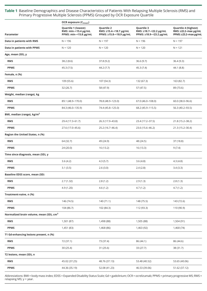
In patients with RMS (Table 1), ocrelizumab exposure-grouped baseline demographic and disease characteristics were comparable for most covariates. Higher proportions of patients were male, had a higher weight/BMI, or were from the United States in lower exposure quartiles (Quartiles 1 and 2) vs higher quartiles (Quartiles 3 and 4). The presence of T1 gadolinium-enhancing lesions was lower in patients within the lower vs the higher exposure quartiles; however, no effect was noted between baseline enhancing lesions and progression outcomes. In patients with PPMS (Table 1), the pattern of exposure-grouped baseline demographic and disease characteristics was generally consistent with those observed in patients with RMS.
Table 1.
Baseline Demographics and Disease Characteristics of Patients With Relapsing Multiple Sclerosis (RMS) and Primary Progressive Multiple Sclerosis (PPMS) Grouped by OCR Exposure Quartile
Annualized Relapse Rate Grouped by Ocrelizumab Exposure Quartile in Patients With RMS
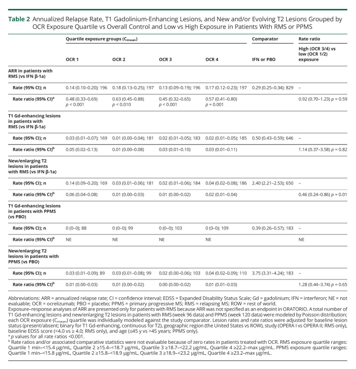
In ocrelizumab-treated patients with RMS, adjusted ARRs through week 96 were low and comparable across exposure quartiles (Quartile 1, 0.14 [95% confidence interval (CI): 0.10–0.20]; Quartile 2, 0.18 [95% CI: 0.13–0.25]; Quartile 3, 0.13 [95% CI: 0.09–0.19]; Quartile 4, 0.17 [95% CI: 0.12–0.23]); the ARR in patients receiving IFN β-1a was 0.29 (95% CI: 0.25–0.34). The treatment effect of ocrelizumab relative to the IFN β-1a arm on ARR (rate ratios [RR]) in individual exposure quartiles is reported in Table 2. Overall, no association between exposure and ARR was observed.
Table 2.
Annualized Relapse Rate, T1 Gadolinium-Enhancing Lesions, and New and/or Evolving T2 Lesions Grouped by OCR Exposure Quartile vs Overall Control and Low vs High Exposure in Patients With RMS or PPMS
Disability Progression in Patients With RMS
Confirmed Disability Progression Measured With the EDSS
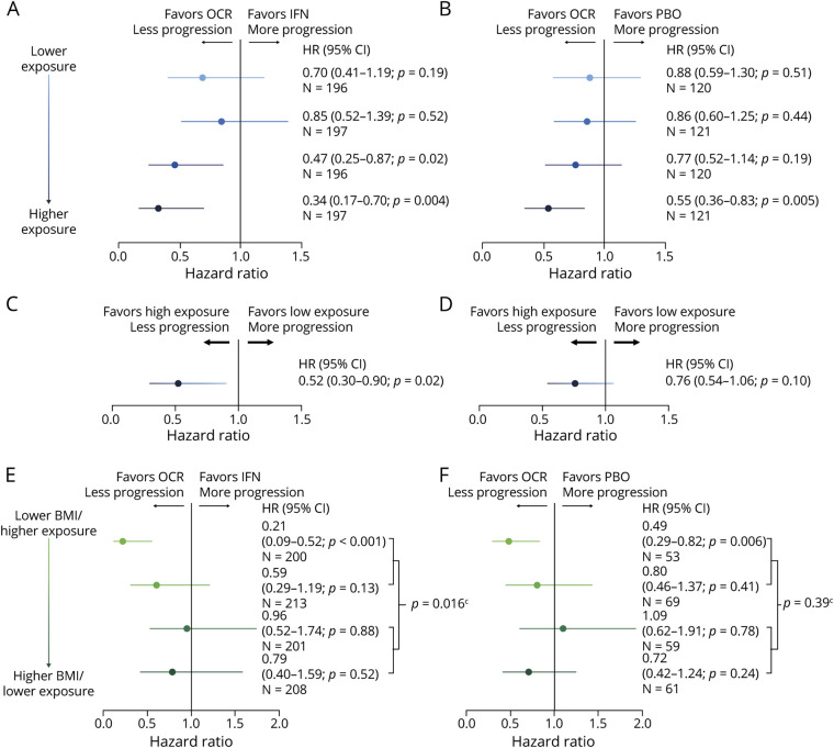
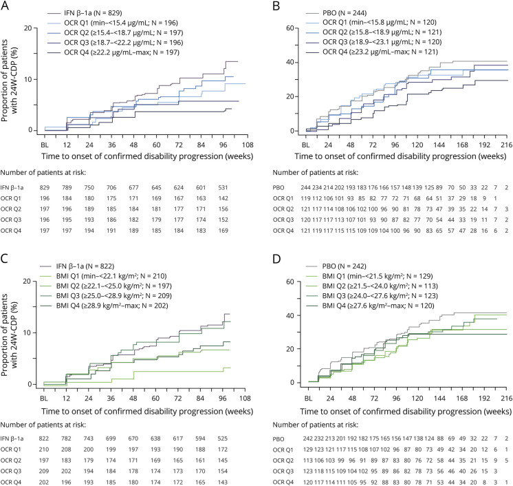
An exposure dependency of time to onset of 24 W-CDP was seen within ocrelizumab-treated patients with RMS, when comparing high-exposure (combined Quartiles 3 and 4 [3/4]) with low-exposure (combined Quartiles 1 and 2 [1/2]) patients (hazard ratio [HR] 0.52 [95% CI: 0.30, 0.90; p = 0.0178]; Figure 1). Exposure dependency of 24 W-CDP was observed in the individual quartile exposure-grouped Kaplan–Meier analysis (Figure 2) and the associated HRs in ocrelizumab-treated patients compared with all IFN β-1a–treated patients (Figure 1). Point estimates of 24 W-CDP HRs vs IFN β-1a were lower in the higher exposure quartiles (Quartile 3, 0.47 [95% CI: 0.25–0.87; p = 0.02]; Quartile 4, 0.34 [95% CI: 0.17–0.70; p = 0.004]) compared with the lower exposure quartiles (Quartile 1, 0.70 [95% CI: 0.41–1.19; p = 0.19]; Quartile 2, 0.85 [95% CI: 0.52–1.39; p = 0.52]).
Figure 1. Hazard Ratios of 24 W-CDP Grouped by (A and B) OCR Exposure Quartiles vs Overall Control Arm; (C and D) High (Quartiles 3/4) vs Low (Quartiles 1/2) OCR Exposure; (E and F) OCR vs Control Arm Within BMI Quartiles; and (E and F) Interaction Effects of OCR vs Control Arm Between High (Quartiles 3/4) and Low (Quartiles 1/2) BMI in Patients With RMSa (A, C, E) and PPMSb (B, D, F).
Hazard ratios were estimated by a stratified Cox regression model with treatment group as a covariate. Treated patients with missing Cmean values were excluded. a,bStratified by region (the United States vs ROW), abaseline EDSS (<4.0 vs ≥4.0), and bage (≤45 vs >45 years). cInteraction effects of OCR vs control between high and low BMI in RMS (HR, 95% CI: 2.39, 1.18–4.83) and PPMS (HR, 95% CI: 1.27, 0.74–2.16). 24 W-CDP = 24-week confirmed disease progression; BMI = body mass index; CI = confidence interval; Cmean = average OCR serum concentration in an individual patient over their treatment period; EDSS = Expanded Disability Status Scale; HR = hazard ratio; IFN = interferon; OCR = ocrelizumab; PBO = placebo; PPMS = primary progressive MS; Q = quartile; RMS = relapsing MS; ROW = rest of world.
Figure 2. Kaplan–Meier Analyses of 24 W-CDP Grouped by (A and B) OCR Exposure Quartiles vs Overall Control Arm or (C and D) BMI Quartiles vs Overall Control Arm in Patients With RMSa (A and C) or PPMSb (B and D).
a,bStratified by region (the United States vs ROW), abaseline EDSS (<4.0 vs ≥4.0), and bage (≤45 vs >45 years). Treated patients with missing Cmean values were excluded. 24 W-CDP = 24-week confirmed disease progression; BMI = body mass index; IFN = interferon; OCR = ocrelizumab; PBO = placebo; PPMS = primary progressive MS; RMS = relapsing MS; ROW = rest of world.
When comparing time to onset of 24 W-CDP between treatment arms (IFN β-1a vs ocrelizumab) within BMI quartiles, numerically, the greatest reductions in risk of disability progression were seen in the lower BMI quartiles (Figure 1). An important advantage of the BMI analysis is that it permits the control and treatment groups to be compared based on the same baseline patient characteristic as the treatment group, allowing for a randomized treatment comparison in the same patient population. Consistent with the observed exposure dependency, a significant treatment-by-baseline BMI (low ≤median vs high >median) interaction was also observed between patients for 24 W-CDP, where lighter patients received more benefit than heavier patients (Figure 1).
In ocrelizumab-treated patients with RMS, there was a trend of higher median B-cell levels during the double-blind period (DBP) being associated with greater rates of 24 W-CDP during the combined DBP and OLE (median circulating B-cell levels vs 0 cells/µL, HR [95% CI]: 1–5/µL, 1.36 [0.82–2.25] p = 0.23; >5/µL, 1.43 [0.79–2.62] p = 0.28) as well as during the OLE only (median circulating B-cell levels vs 0 cells/µL, HR [95% CI]: 1–5/µL, 1.31 [0.76–2.24] p = 0.36; >5/µL, 1.63 [0.88–3.03] p = 0.079; eFigure 1, links.lww.com/NXI/A801). In the DBP, when splitting ocrelizumab-treated patients with RMS at the median of the individual patient median B-cell levels (1 cells/µL; <median n = 305, ≥median n = 509) and comparing with IFN β-1a–treated patients (n = 829), patients with more B-cell depletion showed a greater reduction in risk of 24 W-CDP (HR [95% CI]: CD19 < median, 0.38 [95% CI: 0.21–0.68; p = 0.0011] vs IFN β-1a; CD19 ≥ median, 0.73 [95% CI: 0.51–1.05; p = 0.0881] vs IFN β-1a).
Composite Disability Progression
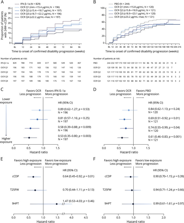
The ocrelizumab exposure dependency in 24 W-CDP (assessed by EDSS) was also observed in the composite measure of disability progression (cCDP) when comparing within ocrelizumab-treated patients (high vs low exposure; HR 0.64 [95% CI: 0.45–0.92; p = 0.0135]) and by ocrelizumab exposure quartile vs all IFN β-1a–treated patients (Figure 3). Among the subcomponents of 24 W-cCDP, an exposure trend was observed for T25FW (high vs low exposure; HR 0.70 [95% CI: 0.44–1.11; p = 0.1281]). There was no association between exposure and outcome observed for the 9HPT subcomponent.
Figure 3. Kaplan–Meier Analyses (A and B) and Hazard Ratios (C and D) of 24 W-cCDP Grouped by OCR Exposure Quartiles vs Overall Control, and Hazard Ratios of 24 W-cCDP, T25FW, and 9HPT (E and F) by High (Quartiles 3/4) vs Low (Quartiles 1/2) OCR Exposure in Patients With RMSa (A, C, E) or PPMSb (B, D, F).
Hazard ratios were estimated by a stratified Cox regression model with treatment group as a covariate. a,bStratified by region (the United States vs ROW), abaseline EDSS (<4.0 vs ≥4.0), and bage (≤45 vs >45 years). Treated patients with missing Cmean values were excluded. 9HPT = Nine-Hole Peg Test; 24 W-cCDP = 24-week composite confirmed disease progression; Cmean = average OCR serum concentration in an individual patient over their treatment period; EDSS = Expanded Disability Status Scale; IFN = interferon; OCR = ocrelizumab; PBO = placebo; PPMS = primary progressive MS; Q = quartile; RMS = relapsing MS; ROW = rest of world; T25FW = Timed 25-Foot Walk.
Disability Progression in Patients With PPMS
Confirmed Disability Progression Measured With the EDSS
In patients with PPMS, exposure dependency analysis of 24 W-CDP was directionally consistent with RMS but did not reach statistical significance (high vs low exposure: HR 0.76 [95% CI: 0.54–1.06; p = 0.099]); HRs of 24 W-CDP and associated Kaplan–Meier analyses (grouped by quartile and high vs low exposure, and BMI quartile and high vs low BMI) and overall 24 W-cCDP (grouped by quartile and high vs low exposure) are presented in Figures 1 and 2, respectively. Unlike RMS, in patients with PPMS there was no association of grouped median B-cell levels during the DBP with rates of 24 W-CDP during the combined DBP and OLE (median circulating B-cell levels vs 0 cells/µL, HR [95% CI]: 1–5/µL, 0.95 [0.71–1.28] p = 0.77; >5/µL, 1.14 [0.74–1.74] p = 0.72) or the OLE only (median circulating B-cell levels vs 0 cells/µL, HR [95% CI]: 1–5/µL, 0.90 [0.63–1.27] p = 0.59; >5/µL, 0.65 [0.34–1.23] p = 0.26; eFigure 1, links.lww.com/NXI/A801). When splitting ocrelizumab-treated patients with PPMS at the median of the individual patient median B-cell levels and comparing with placebo-treated patients, there was no association of median B-cell levels with rates of 24 W-CDP (HR [95% CI]: CD19 <median, 0.81 [95% CI: 0.59, 1.11; p = 0.20 vs placebo]; CD19 ≥median, 0.70 [95% CI: 0.51, 0.96; p = 0.03 vs placebo]).
Composite Disability Progression
Exposure dependency analysis of 24 W-cCDP was directionally consistent with RMS but did not reach statistical significance (high vs low exposure: HR 0.90 [95% CI: 0.70, 1.15; p = 0.39]). An exposure dependency trend on 24 W-cCDP was also observed across quartiles in patients with PPMS (Figure 3). The exposure dependency trend was also observed in time to 20% increase in T25FW (Quartile 1 HR: 0.88 [95% CI: 0.63–1.21; p = 0.43]; Quartile 2 HR: 0.66 [95% CI: 0.47–0.92; p = 0.014]; Quartile 3 HR: 0.79 [95% CI: 0.57–1.09; p = 0.15]; Quartile 4 HR: 0.64 [95% CI: 0.46–0.89; p = 0.008]). No association between exposure and outcome was observed for time to 20% increase in 9HPT (data not shown).
In general, for the above measures, consistent results were also observed for 12-week confirmed data in patients with either RMS or PPMS (eTable 1, links.lww.com/NXI/A801).
Influence of Baseline Patient Characteristics
In RMS, all subgroups, with the exceptions of region the United States and EDSS ≥4, showed numerically greater reduction in risk of disability progression in the high-exposure group compared with the low-exposure group in both the univariate and multivariate analyses (eFigure 2A and eTable 2, links.lww.com/NXI/A801). In the univariate subgroup analyses in PPMS, comparing the time to 24 W-CDP of ocrelizumab-treated patients with low and high exposures with all placebo patients, all subgroups with the exceptions of region the United States showed numerically greater or approximately identical reduction in risk of disability progression in the high-exposure group compared with the low-exposure group. Multivariate analysis results were consistent with the univariate analyses except for patients with a duration since symptom onset of ≤5 years, where the treatment effect in high-exposure patients with PPMS was markedly reduced (eFigure 2B and eTable 2, links.lww.com/NXI/A801). The interpretation of the multivariate analysis is limited by the small sample size.
Magnetic Resonance Imaging
T1 Gadolinium-Enhancing Lesions and New and/or Enlarging T2 Lesions
Owing to the near-zero rate of T1 gadolinium-enhancing lesions with ocrelizumab treatment in patients with RMS or PPMS, exposure dependency could not be reliably assessed. For new and/or evolving T2 lesions, exposure dependency was evident for new and/or evolving T2 lesion rates in patients with RMS (high vs low exposure, RR [95% CI]: 0.46 [0.24–0.86] p = 0.0124) but not PPMS. Of note, the observed dependency in RMS was entirely driven by an increased lesion rate in the lowest exposure quartile.
Brain Atrophy
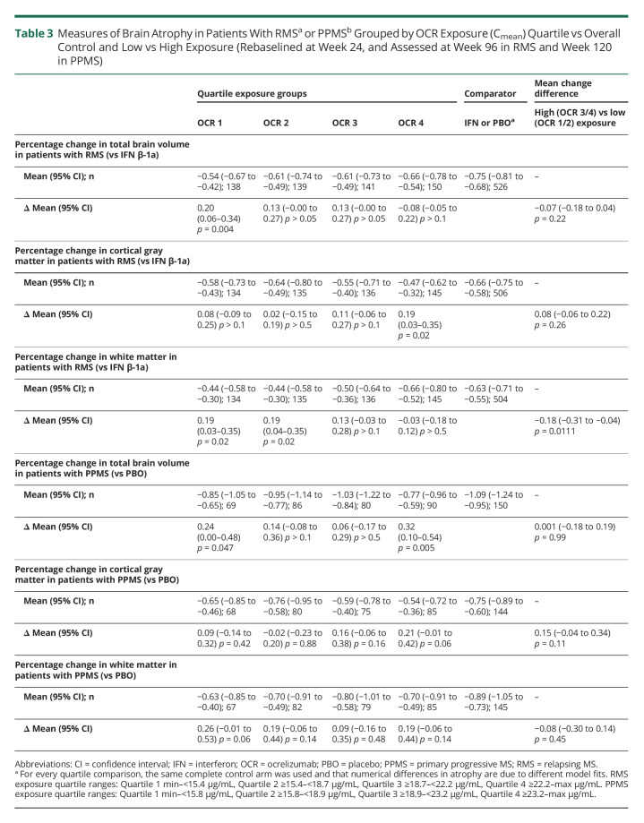
An exposure-dependent trend for whole brain volume change and an association with white matter volume change from rebaseline at week 24 to week 96, vs IFN β-1a, was seen in patients with RMS, with greater volume reduction at higher ocrelizumab exposure; no such associations were seen in patients with PPMS (Table 3). An exposure-dependent association with gray matter volume change from rebaseline at week 24 to week 96, vs comparator, was seen in patients with RMS and PPMS; however, less gray matter volume reduction was seen with higher ocrelizumab exposure (Table 3).
Table 3.
Measures of Brain Atrophy in Patients With RMSa or PPMSb Grouped by OCR Exposure (Cmean) Quartile vs Overall Control and Low vs High Exposure (Rebaselined at Week 24, and Assessed at Week 96 in RMS and Week 120 in PPMS)
For the above measures, consistent results were also observed when the study baseline (week 0) was used for volumetric assessments in patients with either RMS or PPMS (eTable 3, links.lww.com/NXI/A801).
Safety Endpoints
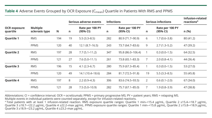
No association between higher exposure and the incidence of SAEs, infections, serious infections, or infusion-related reactions was observed in either RMS or PPMS (Table 4).
Table 4.
Adverse Events Grouped by OCR Exposure (Cmean) Quartile in Patients With RMS and PPMS
Discussion
Ocrelizumab, the first anti-CD20+ B-cell–selective monoclonal antibody approved for the treatment of RMS and PPMS, at a dose of 600 mg IV every 6 months, had a significant benefit on disability progression in the pivotal phase 3 studies2,3 and sustained efficacy and safety with continuous therapy up to 6.5–7 years in their OLEs.4,5,11 In a prespecified subgroup-efficacy analysis of the pooled OPERA I/II studies, a significant treatment-by-subgroup interaction was observed between patients by baseline BMI for both 12 W- and 24 W-CDP, where benefits were evident across both subgroups vs IFN β-1a, but lighter patients received more benefit than heavier patients.10 In this study, we report that higher ocrelizumab exposure, which leads to greater B-cell depletion in the blood,9 was also associated with a reduced rate of disability progression. Disability may progress in MS because of both relapsing disease activity with incomplete recovery (relapse-associated worsening), and progression independent of relapse activity (PIRA).6 We found that for most measures reflecting relapsing disease biology, including clinical (ARR) and MRI (new lesion) outcomes, no association with ocrelizumab exposure could be observed. This is potentially due to treatment-related reduction of these inflammatory pathology-related measures to such low levels that the effect size is no longer exposure-dependent (ceiling effect). An exception was seen for new and/or evolving T2 lesion rates in patients with RMS, possibly driven by an incomplete suppression of inflammation in the lowest exposure quartile, although the clinical relevance of differences between such low residual lesion rates at the boundaries of measurement sensitivity is uncertain.
In contrast to the inflammatory mechanisms involved in RMS disease activity, pathophysiologic processes underlying PIRA are believed to include slowly evolving chronic-active plaques, cortical demyelination and neuronopathy possibly related to B-cell rich inflammation in the meninges, diffuse white matter microgliosis and associated myelin injury, and age-related degeneration.12-16 Furthermore, an increasing body of evidence suggests that progression observed in patients with RMS treated with highly effective DMTs reflects the same or at least very similar pathology with progression observed in secondary progressive MS and PPMS.12-18 At present, it is not known to what degree each of these pathologies contributes to progression in different patients and/or different stages of disease, and which components of this pathology may respond to higher ocrelizumab exposure specifically, although ocrelizumab favorably affects chronic-active evolving lesions.19,20 The mostly statistically nonsignificant observations on different measures of brain volume loss are intriguing but could be dependent on methodologic constraints or differential effects on the gray and white matter compartments. An inverse exposure–response relationship was evident for white matter volume loss, perhaps through the strong reduction of lesion pathology and inflammatory processes by ocrelizumab that decreases the volume of white matter (pseudoatrophy).21 On measures of disease progression (EDSS-based CDP, cCDP including T25FW measure of gait, and 9HPT of hand motor function) where the observed effects of ocrelizumab are statistically and clinically relevant but incomplete, we observed a consistent trend toward higher efficacy with higher drug exposure across the 3 studies in univariate and multivariate analyses. Owing to the strong effects of ocrelizumab on relapse prevention in RMS, the majority of disability progression was relapse-independent,6 and when relapse-independent CDP (PIRA) was investigated separately here, similar associations were found (data not shown). In PPMS, the lack of statistical significance for the comparison in CDP and cCDP between high vs low exposure by median cut could be due to lower power (smaller sample size) or differences in the contributions of individual pathologies associated with progression. However, the highest exposure quartile was still associated with significant differences for both CDP and cCDP in PPMS.
As previously shown, the pharmacokinetics of ocrelizumab (measured in serum) can be accurately described by a 2-compartment pharmacokinetic model with time-dependent clearance, typical for an immunoglobulin G1 monoclonal antibody,22 and with body weight as the main covariate.9 Greater B-cell depletion, and less B-cell repletion between doses, was observed in patients with higher ocrelizumab exposure.9 It has previously been reported that repletion after anti-CD20 therapy is body surface area dependent,23 and specifically after ocrelizumab therapy repletion was shown to be BMI dependent.24 Other factors influencing the level of B-cell depletion with anti-CD20 therapies include the Fc-gamma receptor genotype, as described with rituximab.25 Preclinical data have also noted an effect of the route of administration on lymphoid B-cell depletion.26-28 IV vs subcutaneous dosing of anti-CD20 monoclonal antibodies show different pharmacokinetics (e.g., peak and mean blood concentration), which may manifest in differing clinical dynamic profiles. Only 2% of the body's total lymphocyte pool resides in peripheral blood,29 and B-cell depletion in lymphoid organs is less complete than in blood. Furthermore, the relationship between blood and extravascular B-cell repletion kinetics is unclear, although preclinical data indicate repletion in the bone marrow and lymphoid tissue before the blood.30
In the present analysis, a consistent pharmacodynamic effect was observed in patients with RMS, where an incomplete B-cell depletion or higher B-cell counts in blood before the next infusion showed a trend toward elevated risk of disease progression over the DBP and OLE, and higher median B-cell levels during the 96-week DBP showed a trend for more disability progression in the OLE. This suggests that maintaining lower B-cell levels over a longer time may lead to better disability outcomes. In PPMS, these associations were not observed; however, this post-hoc analysis was not powered to detect an effect of B-cell lowering on progression in PPMS.
It is important that there was no association between ocrelizumab exposure and analyzed adverse events (SAEs, serious infections, and infusion-related reactions) in patients with RMS or PPMS.
The analyses presented here are subject to the limitations of any post hoc investigation, including sample and effect sizes. As noted, Cmean was treated as a time-invariant measure because a patient's characteristics (e.g., body weight and sex) determining their pharmacokinetics are not expected to change during the course of the given observation period. The pharmacodynamics of ocrelizumab-mediated B-cell depletion are different compared with, e.g., drugs displaying a direct effect based on reversible receptor occupancy, which fluctuates in parallel to the pharmacokinetic profile. Owing to the mechanism of action of ocrelizumab, i.e., destruction of B cells,2,3,9,30 B cells are depleted with immediate onset upon contact with ocrelizumab, but return only slowly, after regeneration from stem or progenitor cells in bone marrow.31 The median time to B-cell repletion in blood was 72 weeks based on a lower limit of normal of 80 cells/µL, after dosing cessation.9 B-cell depletion kinetics in other body compartments and particularly the CNS is less clear. However, this may affect clinical efficacy and could be linked to cumulative effects over time on B cells in blood and tissues because B-cell depletion becomes more pronounced in tissues with repeated ocrelizumab administrations. This could be a potential explanation for the exposure-dependent effects on long-term outcomes such as disability progression demonstrated in this study.
The observed differences in the demographic characteristics of sex and weight/BMI, when grouped by exposure quartile, were expected because these characteristics were identified as covariates in the population pharmacokinetic analysis,9 and BMI was shown to affect disability outcome in a prespecified subgroup analysis for efficacy.10 Furthermore, although an association between ocrelizumab exposure quartile and efficacy on progression measures was observed, no causal dependence can be definitively concluded.
Treatment with ocrelizumab 600 mg is known to lead to rapid and near-complete depletion of B cells in blood, which is maintained over time with subsequent doses,9 accompanied with a rapid suppression of acute disease activity (suppression of MRI activity within 4 weeks and relapse activity within 8 weeks).32 It is, however, unclear to what extent efficacy and pathology track with levels of B cells in blood, and whether greater exposure and/or deeper depletion of B cells in tissue in the periphery and CNS is required to further modify disease and disability progression. The results from these post hoc exposure–response analyses suggest that a higher dose of ocrelizumab could lead to an improved efficacy on disability progression. However, the multivariate analysis did not identify whether this would benefit all or only subgroups of patients with MS. The absence of an increase of SAEs with higher ocrelizumab exposure encourages further exploration of the potential benefits of a higher dose in controlled prospective clinical studies. To this end, 2 double-blind, parallel-group, randomized phase 3b studies (one in RMS [MUSETTE; NCT04544436] and the other in PPMS [GAVOTTE; NCT04548999]) investigating the efficacy and safety of higher doses of ocrelizumab (1,200 mg for patients <75 kg or 1,800 mg for patients ≥75 kg, every 24 weeks) are ongoing and will inform whether higher ocrelizumab doses can further decrease the risk of disability progression.
Observational data from the OLE period of the phase 2 ocrelizumab trial indicated that both clinical relapses and new focal inflammatory lesions measured by MRI remain low even after some B-cell repletion occurs.33 Other data also suggested that a long-term favorable modulation of circulating B cells, and other immune parameters, follows treatment with anti-CD20 monoclonal antibodies.34-36 Some clinicians have interpreted these data to indicate that treatment intervals can be safely extended beyond the recommended 6-month schedule without any decrement in efficacy. The current data, indicating that higher serum levels of ocrelizumab were associated with improved efficacy against progression in both RMS and PPMS, strongly suggest that an every-6-month dosing schedule will provide better protection against disability progression than less frequent dosing.
In conclusion, higher ocrelizumab exposure and lower median B-cell levels in the blood were associated with a consistent and sustained benefit on rates of disability progression, across several outcome measures in RMS and, to some extent, PPMS. Higher ocrelizumab exposure and greater B-cell depletion may be important for the control of disability progression.
Acknowledgment
The authors thank all patients, their families, and the investigators who participated in the trials contained within these analyses. The authors are grateful to Gisèle von Büren (F. Hoffmann-La Roche Ltd) for additional critical review of this manuscript and technical advice. Initial assistance and discussion regarding data and analyses was provided by Andrew Stead of Articulate Science, UK, and funded by F. Hoffmann-La Roche Ltd. The authors had full editorial control of the manuscript and provided their final approval of all content.
Glossary
- 12W
12-week
- 24W
24-week
- 9HPT
Nine-Hole Peg Test
- ARR
annualized relapse rate
- BMI
body mass index
- cCDP
composite confirmed disability progression
- CDP
confirmed disability progression
- CI
confidence interval
- DBP
double-blind period
- DMT
disease-modifying therapy
- EDSS
Expanded Disability Status Scale
- HR
hazard ratio
- IFN
interferon
- MS
multiple sclerosis
- OLE
open-label extension
- PIRA
progression independent of relapse activity
- PPMS
primary progressive multiple sclerosis
- RMS
relapsing multiple sclerosis
- RR
rate ratio
- SAE
serious adverse event
- T25FW
Timed 25-Foot Walk
Appendix. Authors

Footnotes
Class of Evidence: NPub.org/coe
Study Funding
This work was supported by financial support from F. Hoffmann-La Roche Ltd, Basel, Switzerland, for the study and publication of the manuscript.
Disclosure
S.L. Hauser serves on the Board of Directors for Neurona and on scientific advisory boards for Accure, Alector, and Annexon; has previously consulted for BD, Moderna, and NGM Bio; and has received travel reimbursement and writing assistance from F. Hoffmann-La Roche Ltd and Novartis AG for CD20-related meetings and presentations. A. Bar-Or has received consulting fees from Accure, Atara Biotherapeutics, Biogen Idec., BMS/Celgene/Receptos, GlaxoSmithKline, Gossamer, Janssen/Actelion, MedImmune, Merck/EMD Serono, Novartis, Roche/Genentech, and Sanofi-Genzyme. He has carried out contracted research for Genentech and Biogen. He receives a salary from The University of Pennsylvania, Perelman School of Medicine. M.S. Weber receives research support from the Deutsche Forschungsgemeinschaft (DFG; WE 3547/5-1), from Novartis, Teva, Biogen Idec., Roche, Merck, and the ProFutura Program of the Universitätsmedizin Göttingen. He is serving as an editor for PLoS One. He has received travel funding and/or speaker honoraria from Biogen Idec., Merck-Serono, Novartis, Roche, Teva, Bayer, and Genzyme. H. Kletzl is an employee of and shareholder in F. Hoffmann-La Roche Ltd. A. Günther is an employee of F. Hoffmann-La Roche Ltd. M. Manfrini is an employee of and shareholder in F. Hoffmann-La Roche Ltd. F. Model was an employee of F. Hoffmann-La Roche Ltd during completion of the work related to this manuscript. He is currently an employee of Denali Therapeutics, South San Francisco, California, USA. F. Mercier is an employee of F. Hoffmann-La Roche Ltd. C. Petry is an employee of and shareholder in F. Hoffmann-La Roche Ltd. Q. Wang is an employee of F. Hoffmann-La Roche Ltd. H. Koendgen was an employee of and shareholder in F. Hoffmann-La Roche Ltd during completion of the work related to this manuscript. He is currently an employee of UCB Farchim SA, Bulle, Switzerland. L. Kappos has received research support in the last 3 years for his institution (University Hospital Basel) via steering committee, advisory board, and consultancy fees from Actelion, Allergan, Almirall, Bayer HealthCare, Baxalta, Biogen, Celgene Receptos, CSL Behring, Desitin, Eisai, Excemed, Genzyme, Japan Tobacco, Merck, Novartis, Pfizer, Roche, Sanofi, Santhera, and Teva; licence fees for Neurostatus-UHB products; and grants from Bayer, Biogen, Novartis, the Swiss MS Society, the Swiss National Research Foundation, Innosuisse, and the European Union and Roche Research Foundation to the Research of the MS Center in Basel. Go to Neurology.org/NN for full disclosures.
References
- 1.Klein C, Lammens A, Schäfer W, et al. Epitope interactions of monoclonal antibodies targeting CD20 and their relationship to functional properties. mAbs. 2013;5(1):22-33. doi: 10.4161/mabs.22771. [DOI] [PMC free article] [PubMed] [Google Scholar]
- 2.Hauser SL, Bar-Or A, Comi G, et al. Ocrelizumab versus interferon beta-1a in relapsing multiple sclerosis. N Engl J Med. 2017;376(3):221-234. doi: 10.1056/NEJMoa1601277. [DOI] [PubMed] [Google Scholar]
- 3.Montalban X, Hauser SL, Kappos L, et al. Ocrelizumab versus placebo in primary progressive multiple sclerosis. N Engl J Med. 2017;376(3):209-220. doi: 10.1056/NEJMoa1606468. [DOI] [PubMed] [Google Scholar]
- 4.Hauser SL, Kappos L, Arnold DL, et al. Five years of ocrelizumab in relapsing multiple sclerosis: OPERA studies open-label extension. Neurology. 2020;95(13):e1854-e1867. doi: 10.1212/WNL.0000000000010376. [DOI] [PMC free article] [PubMed] [Google Scholar]
- 5.Wolinsky JS, Arnold DL, Brochet B, et al. Long-term follow-up from the ORATORIO trial of ocrelizumab for primary progressive multiple sclerosis: a post-hoc analysis from the ongoing open-label extension of the randomised, placebo-controlled, phase 3 trial. Lancet Neurol. 2020;19(12):998-1009. doi: 10.1016/S1474-4422(20)30342-2. [DOI] [PubMed] [Google Scholar]
- 6.Kappos L, Wolinsky JS, Giovannoni G, et al. Contribution of relapse-independent progression vs relapse-associated worsening to overall confirmed disability accumulation in typical relapsing multiple sclerosis in a pooled analysis of 2 randomized clinical trials. JAMA Neurol. 2020;77(9):1132-1140. doi: 10.1001/jamaneurol.2020.1568. [DOI] [PMC free article] [PubMed] [Google Scholar]
- 7.Kappos L, Butzkueven H, Wiendl H, et al. Greater sensitivity to multiple sclerosis disability worsening and progression events using a roving versus a fixed reference value in a prospective cohort study. Mult Scler. 2018;24(7):963-973. doi: 10.1177/1352458517709619. [DOI] [PMC free article] [PubMed] [Google Scholar]
- 8.University of California San Francisco MS-EPIC Team; Cree BAC, Hollenbach JA, Bove R, et al. Silent progression in disease activity-free relapsing multiple sclerosis. Ann Neurol. 2019;85(5):653-666. doi: 10.1002/ana.25463. [DOI] [PMC free article] [PubMed] [Google Scholar]
- 9.Gibiansky E, Petry C, Mercier F, et al. Ocrelizumab in relapsing and primary progressive multiple sclerosis: pharmacokinetic and pharmacodynamic analyses of OPERA I, OPERA II and ORATORIO. Br J Clin Pharmacol. 2021;87(6):2511-2520. doi: 10.1111/bcp.14658. [DOI] [PMC free article] [PubMed] [Google Scholar]
- 10.Turner B, Cree BAC, Kappos L, et al. Ocrelizumab efficacy in subgroups of patients with relapsing multiple sclerosis. J Neurol. 2019;266(5):1182-1193. doi: 10.1007/s00415-019-09248-6. [DOI] [PMC free article] [PubMed] [Google Scholar]
- 11.Hauser SL, Kappos L, Montalban X, et al. Safety of ocrelizumab in patients with relapsing and primary progressive multiple sclerosis. Neurology. 2021;97(16):e1546-e1559. doi: 10.1212/WNL.0000000000012700. [DOI] [PMC free article] [PubMed] [Google Scholar]
- 12.Filippi M, Bar-Or A, Piehl F, et al. Multiple sclerosis. Nat Rev Dis Primers. 2018;4(1):43. doi: 10.1038/s41572-018-0041-4. [DOI] [PubMed] [Google Scholar]
- 13.Lassmann H. Multiple sclerosis pathology. Cold Spring Harb Perspect Med. 2018;8(3):a028936. doi: 10.1101/cshperspect.a028936. [DOI] [PMC free article] [PubMed] [Google Scholar]
- 14.Correale J, Gaitán MI, Ysrraelit MC, Fiol MP. Progressive multiple sclerosis: from pathogenic mechanisms to treatment. Brain. 2017;140(3):527-546. doi: 10.1093/brain/aww258. [DOI] [PubMed] [Google Scholar]
- 15.Magliozzi R, Howell O, Vora A, et al. Meningeal B-cell follicles in secondary progressive multiple sclerosis associate with early onset of disease and severe cortical pathology. Brain. 2007;130(4):1089-1104. doi: 10.1093/brain/awm038. [DOI] [PubMed] [Google Scholar]
- 16.Mahad DH, Trapp BD, Lassmann H. Pathological mechanisms in progressive multiple sclerosis. Lancet Neurol. 2015;14(2):183-193. doi: 10.1016/S1474-4422(14)70256-X. [DOI] [PubMed] [Google Scholar]
- 17.Lassmann H. Targets of therapy in progressive MS. Mult Scler. 2017;23(12):1593-1599. doi: 10.1177/1352458517729455. [DOI] [PubMed] [Google Scholar]
- 18.Ciotti JR, Cross AH. Disease-modifying treatment in progressive multiple sclerosis. Curr Treat Options Neurol. 2018;20(5):12. doi: 10.1007/s11940-018-0496-3. [DOI] [PubMed] [Google Scholar]
- 19.Elliott C, Belachew S, Wolinsky JS, et al. Chronic white matter lesion activity predicts clinical progression in primary progressive multiple sclerosis. Brain. 2019;142(9):2787-2799. doi: 10.1093/brain/awz212. [DOI] [PMC free article] [PubMed] [Google Scholar]
- 20.Elliott C, Wolinsky JS, Hauser SL, et al. Slowly expanding/evolving lesions as a magnetic resonance imaging marker of chronic active multiple sclerosis lesions. Mult Scler. 2019;25(14):1915-1925. doi: 10.1177/1352458518814117. [DOI] [PMC free article] [PubMed] [Google Scholar]
- 21.Zivadinov R, Jakimovski D, Gandhi S, et al. Clinical relevance of brain atrophy assessment in multiple sclerosis. Implications for its use in a clinical routine. Expert Rev Neurother. 2016;16(7):777-793. doi: 10.1080/14737175.2016.1181543. [DOI] [PubMed] [Google Scholar]
- 22.Mould DR, Sweeney KRD. The pharmacokinetics and pharmacodynamics of monoclonal antibodies–mechanistic modeling applied to drug development. Curr Opin Drug Discov Devel. 2007;10(1):84-96. [PubMed] [Google Scholar]
- 23.Ellwardt E, Ellwardt L, Bittner S, Zipp F. Monitoring B-cell repopulation after depletion therapy in neurologic patients. Neurol Neuroimmunol Neuroinflamm. 2018;5(4):e463. doi: 10.1212/NXI.0000000000000463. [DOI] [PMC free article] [PubMed] [Google Scholar]
- 24.Signoriello E, Bonavita S, Di Pietro A, et al. BMI influences CD20 kinetics in multiple sclerosis patients treated with ocrelizumab. Mult Scler Relat Disord. 2020;43:102186. doi: 10.1016/j.msard.2020.102186. [DOI] [PubMed] [Google Scholar]
- 25.Anolik JH, Campbell D, Felgar RE, et al. The relationship of FcγRIIIa genotype to degree of B-cell depletion by rituximab in the treatment of systemic lupus erythematosus. Arthritis Rheum. 2003;48(2):455-459. doi: 10.1002/art.10764. [DOI] [PubMed] [Google Scholar]
- 26.Vugmeyster Y, Howell K, Bakshi A, Flores C, Hwang O, McKeever K. B-cell subsets in blood and lymphoid organs in Macaca fascicularis. Cytometry A. 2004;61A(1):69-75. doi: 10.1002/cyto.a.20039. [DOI] [PubMed] [Google Scholar]
- 27.Gelzleichter T. Reduction and reconstitution of B-cells in peripheral blood and lymphoid tissues in cynomolgus monkeys following administration of ocrelizumab. Presented at the 2014 Joint ACTRIMS-ECTRIMS Meeting. ECTRIMS Online Library. Abstract P954.
- 28.Brown PC. Ocrelizumab Pharmacology Review. FDA.gov; 2016. Accessed March 1, 2021. accessdata.fda.gov/drugsatfda_docs/nda/2017/761053Orig1s000PharmR.pdf. [Google Scholar]
- 29.Dock J, Hultin L, Hultin P, et al. Human immune compartment comparisons: optimization of proliferative assays for blood and gut T lymphocytes. J Immunol Methods. 2017;445:77-87. doi: 10.1016/j.jim.2017.03.014. [DOI] [PMC free article] [PubMed] [Google Scholar]
- 30.Häusler D, Häusser-Kinzel S, Feldmann L, et al. Functional characterization of reappearing B cells after anti-CD20 treatment of CNS autoimmune disease. Proc Natl Acad Sci USA. 2018;115(39):9773-9778. doi: 10.1073/pnas.1810470115. [DOI] [PMC free article] [PubMed] [Google Scholar]
- 31.Nissimov N, Hajiyeva Z, Torke S, et al. B cells reappear less mature and more activated after their anti-CD20-mediated depletion in multiple sclerosis. Proc Natl Acad Sci USA. 2020;117(41):25690-25699. doi: 10.1073/pnas.2012249117. [DOI] [PMC free article] [PubMed] [Google Scholar]
- 32.Barkhof F, Kappos L, Wolinsky JS, et al. Onset of clinical and MRI efficacy of ocrelizumab in relapsing multiple sclerosis. Neurology. 2019;93(19):e1778-e1786. doi: 10.1212/WNL.0000000000008189. [DOI] [PMC free article] [PubMed] [Google Scholar]
- 33.Hauser S, Li D, Calabresi P, et al. Week 144 results of a phase II, randomised, multicenter trial assessing the safety and efficacy of ocrelizumab in patients with relapsing-remitting multiple sclerosis (RRMS). Neurology. 2013;80(suppl 7):S31.004. [Google Scholar]
- 34.Ellrichmann G, Bolz J, Peschke M, et al. Peripheral CD19+ B-cell counts and infusion intervals as a surrogate for long-term B-cell depleting therapy in multiple sclerosis and neuromyelitis optica/neuromyelitis optica spectrum disorders. J Neurol. 2019;266(1):57-67. doi: 10.1007/s00415-018-9092-4. [DOI] [PMC free article] [PubMed] [Google Scholar]
- 35.Palanichamy A, Jahn S, Nickles D, et al. Rituximab efficiently depletes increased CD20-expressing T cells in multiple sclerosis patients. J Immunol. 2014;193(2):580-586. doi: 10.4049/jimmunol.1400118. [DOI] [PMC free article] [PubMed] [Google Scholar]
- 36.Gingele S, Jacobus TL, Konen FK, et al. Ocrelizumab depletes CD20+ T cells in multiple sclerosis patients. Cells. 2018;8(1):12. doi: 10.3390/cells8010012. [DOI] [PMC free article] [PubMed] [Google Scholar]
Associated Data
This section collects any data citations, data availability statements, or supplementary materials included in this article.
Data Availability Statement
For eligible studies, qualified researchers may request access to individual patient-level clinical data through a data request platform. At the time of writing, this request platform is Vivli, vivli.org/ourmember/roche/. For up-to-date details of Roche's Global Policy on the Sharing of Clinical Information and how to request access to related clinical study documents, see go.roche.com/data_sharing.


