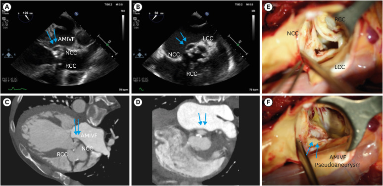
A 63-year-old man presented with dyspnea. Transthoracic echocardiography (TTE) revealed severe degenerative aortic stenosis with decreased left ventricular contractility. Transesophageal echocardiography (TEE) and cardiac computed tomography (CT) showed an outpouching structure at aortomitral intervalvular fibrosa (Figure 1A-D, Supplementary Video 1), compatible with pseudoaneurysm suggesting sequelae of previous infective endocarditis. As he did not suffer from fever or elevated inflammatory markers, surgical aortic valve replacement (SAVR) was performed. On surgery, when extracting the leaflets, subannular pouching was seen without evidence of active inflammation or infection (Figure 1E and F).
Figure 1.
Pre-operative TEE, CT images and Intra-op findings.
(A-D) Severe degenerative aortic stenosis with regurgitation and left ventricular dilatation. Outpouching structure at AMIVF with systolic expansion (blue arrows) suggesting pseudoaneurysm. (E) Thickened aortic valve leaflets were noted, especially on NCC and LCC. (F) When extracting the degenerative aortic valve leaflets, subannular pouching was seen between NCC and LCC sides without evidence of active inflammation or infection (blue arrows).
AMIVF = aortomitral intervalvular fibrosa; CT = computed tomography; LCC = left coronary cusp; NCC = noncoronary cusp; TEE = transesophageal echocardiography.
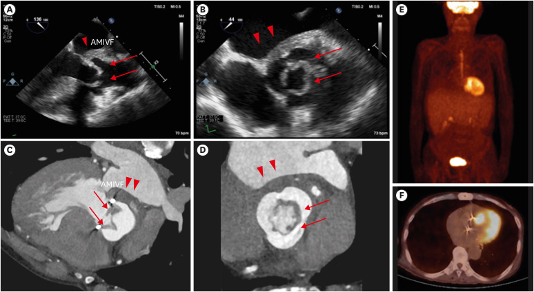
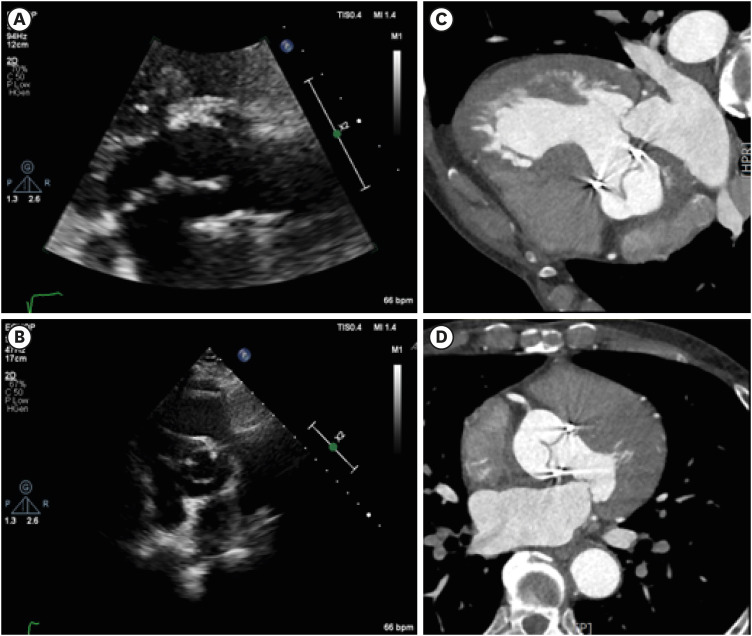
One-year follow-up TTE showed markedly increased aortic valve pressure gradient. TEE and cardiac CT revealed symmetric nodular thickening of bioprosthetic leaflets with opening limitation (Figure 2A-D). Based on the diagnosis of subclinical bioprosthetic valve thrombosis, anticoagulation therapy was started. During the hospitalization, the patient had persistent fever with bicytopenia and hepatosplenomegaly, but repetitive blood cultures were negative. There was no definite fever focus in 18F-fluorodeoxyglucose (18F-FDG) positron emission tomography (PET)-CT (Figure 2E). Since the patient worked on a farm in Dangjin, where Q fever is relatively prevalent, an indirect immunofluorescence assay for Coxiella burnetii was conducted and turned out positive. Additional coagulopathy tests showed positive for lupus anticoagulant, anti-cardiolipin immunoglobulin M (IgM), and anti-beta2 GPI IgM both at baseline and 12-week follow-up. Finally, the patient was diagnosed with Q fever endocarditis combined with antiphospholipid syndrome and treated with doxycycline with hydroxychloroquine and maintained anticoagulation. Recent TTE and cardiac CT follow-up showed complete resolution of bioprosthetic thrombus (Figure 3).
Figure 2.
One-year follow-up TEE, CT and 18F-FDG PET-CT images.
(A-D) Symmetric nodular thickening of bioprosthetic aortic valve leaflets with opening limitation (red arrows), suggestive of valve thrombus, combined with thickened left atrial wall on AMIVF side (red arrows heads); (E, F) There was no definite abnormal hypermetabolic lesion at the periprosthetic valve in 18F-FDG PET-CT, and there was only mild uptake on hepatosplenomegaly.
18F-FDG = 18F-fluorodeoxyglucose; AMIVF = aortomitral intervalvular fibrosa; CT = computed tomography; LCC = left coronary cusp; NCC = noncoronary cusp; PET = positron emission tomography.
Figure 3.
Follow-up TTE and CT images after anticoagulation and antibiotics.
(A-D) Symmetric nodular thickening of bioprosthetic AV leaflets were disappeared, suggesting complete resolution of prosthetic AV thrombus.
AV = aortic valve; CT = computed tomography; TTE = transthoracic echocardiography.
Detailed history taking and multimodal imaging are crucial for early diagnosis and treatment for a rare condition of Q fever endocarditis combined with antiphospholipid syndrome.
We obtained written informed consent from the patient.
Footnotes
Funding: The authors received no financial support for the research, authorship, and/or publication of this article.
Conflict of Interest: The authors have no financial conflicts of interest.
Data Sharing Statement: The data generated in this study is available from the corresponding author upon reasonable request.
Author Contributions: Conceptualization: Choi JY; Supervision: Na JO, Choi JY; Visualization: Yong HS, Choi JY; Writing- original draft: Lee J; Writing- review & editing: Lee J, Choi YJ, Yong HS, Baek MJ, Na JO, Choi JY.
SUPPLEMENTARY MATERIAL
Three-dimensional TEE of pseudoaneurysm.
Associated Data
This section collects any data citations, data availability statements, or supplementary materials included in this article.
Supplementary Materials
Three-dimensional TEE of pseudoaneurysm.


