Abstract
Background
Artificial intelligence (AI), when applied to computer vision using a convolutional neural network (CNN), is a promising tool in “difficult-to-diagnose” conditions such as malignant biliary strictures and cholangiocarcinoma (CCA). The aim of this systematic review is to summarize and review the available data on the diagnostic utility of endoscopic AI-based imaging for malignant biliary strictures and CCA.
Methods
In this systematic review, PubMed, Scopus and Web of Science databases were reviewed for studies published from January 2000 to June 2022. Extracted data included type of endoscopic imaging modality, AI classifiers, and performance measures.
Results
The search yielded 5 studies involving 1465 patients. Of the 5 included studies, 4 (n=934; 3,775,819 images) used CNN in combination with cholangioscopy, while one study (n=531; 13,210 images) used CNN with endoscopic ultrasound (EUS). The average image processing speed of CNN with cholangioscopy was 7-15 msec per frame while that of CNN with EUS was 200-300 msec per frame. The highest performance metrics were observed with CNN-cholangioscopy (accuracy 94.9%, sensitivity 94.7%, and specificity 92.1%). CNN-EUS was associated with the greatest clinical performance application, providing station recognition and bile duct segmentation; thus reducing procedure length and providing real-time feedback to the endoscopist.
Conclusions
Our results suggest that there is increasing evidence to support a role for AI in the diagnosis of malignant biliary strictures and CCA. CNN-based machine leaning of cholangioscopy images appears to be the most promising, while CNN-EUS has the best clinical performance application.
Keywords: Artificial intelligence, endoscopic ultrasound, cholangioscopy, malignant biliary strictures, cholangiocarcinoma
Introduction
Cholangiocarcinoma (CCA) is a malignant bile duct cancer arising from epithelial cells of the intrahepatic, perihilar or distal bile ducts [1-5]. The etiology of CCA includes primary sclerosing cholangitis (PSC), hepatobiliary flukes, Caroli’s syndrome and congenital hepatic fibrosis [1,2,6]. CCA is highly lethal because most patients are diagnosed at an advanced stage [5,7]. The incidence and mortality rate of CCA are increasing worldwide, and it accounts for approximately 20% of all hepatobiliary cancer-related deaths [3,4,7]. The only effective cure for CCA is the surgical resection of localized lesions. However, the prognosis of CCA remains extremely poor, with 5-year survival rates after surgery rarely exceeding 35% [6,8].
The diagnosis of malignant biliary strictures and CCA is challenging. When a patient with a biliary stricture is approached, endoscopic retrograde cholangiopancreatography (ERCP) is usually used initially. ERCP-based diagnosis of biliary stricture through use of either brush cytology or intraductal biopsies is limited by their poor sensitivity (43% and 48%, respectively) [9]. Hence a significant proportion of strictures remain indeterminate, which has led to the development of cholangioscopy-based techniques.
Cholangioscopy provides endoscopic direct visualization of the biliary system and the possibility of targeted biopsies under direct vision. In a meta-analysis of 21 studies, single-operator cholangioscopy with targeted biopsies was the most accurate diagnostic imaging modality for cholangiocarcinoma in patients with PSC-induced biliary strictures, despite having a modest sensitivity of 65% (95% confidence interval [CI] 35-87%) [1]. A recent multicenter trial demonstrated that cholangioscopy improved the sensitivity of visual identification of malignant biliary strictures from 65-95%, with a concomitant specificity of visual impression of 89% [10]. However, more than 25% of patients presumed to have malignant strictures during cholangioscopy show benign pathology after major surgical intervention [11]. Interpretation of the visual findings during cholangioscopy remains challenging, even for experienced endoscopists [12].
Endoscopic ultrasound (EUS) has become a valuable tool in the evaluation of the pancreaticobiliary system. Multiple studies have reported on the use of EUS-fine needle aspiration (FNA) for the diagnosis of malignant extrahepatic biliary strictures and CCA (i.e., distal bile duct due to accessibility). In a meta-analysis of 6 studies, the overall pooled sensitivity of EUS-FNA for the diagnosis of CCA was 66% (95%CI 57-74%) [13]. Although EUS-FNA is useful in CCA detection, there have also been concerns over the risk of tumor seeding or needle track seeding [14]. Therefore, endoscopic visualization via EUS without FNA may be a safer approach to the diagnosis of CCA. The lack of a sensitive and specific early diagnostic marker, coupled with the scarcity of alternative curative treatments to surgical resection, produces a dismal prognosis in patients with malignant biliary strictures and CCA, who have an estimated life expectancy of 6-12 months.
Artificial intelligence (AI) is a branch of computer science that uses computational methods to simulate human intelligence [15]. AI based on deep learning (DL), a type of machine learning that enables end-to-end learning of very complex functions from raw data, has triggered tremendous global interest in recent years. The convolutional neural network (CNN) is a type of DL algorithm that hardcodes translational invariance, a key feature of image data. DL with CNN has been widely adopted in image recognition, and the use of AI has been increasing gradually in medical diagnosis and prognosis [16,17]. Currently, large amounts of imaging data, coupled with data on clinical outcomes, have led to the emergence of AI within endoscopy as a new field of hepatobiliary research [12]. AI methods in medical imaging include the traditional flowchart of radiomics analysis and DL algorithms (Fig. 1) [7]. The traditional flowchart includes segmentation of regions of interest (ROI), feature extraction, feature selection and modeling. It relies on radiomics features extracted from the ROI and conventional machine learning algorithms. DL algorithms also fall under radiomics, but do not require region annotation. The process includes some hidden layers, where extraction of radiomics features, selection and ultimate modeling are performed simultaneously during training [7,16,17].
Figure 1.

Traditional flow chart and deep learning algorithms
In recent studies, the impact of AI tools on the evaluation of endoscopic bile duct images has recently been assessed to develop and validate CNN-based algorithms for the automatic detection and differentiation of malignant biliary strictures and CCA [18-22]. To the best of our knowledge, the literature lacks a systematic review and meta-analysis of the available evidence that has examined the diagnostic performance of endoscopic AI-based imaging in the diagnosis of malignant bile duct strictures and CCA. The aim of this systematic review is to summarize and review the available data on the diagnostic utility of endoscopic AI-based imaging for malignant biliary strictures and CCA. We also aim to propose future challenges and directions for endoscopic AI-based imaging in the diagnosis of malignant biliary strictures and CCA.
Materials and methods
Literature search
A systematic literature review was performed according to the Preferred Reporting Items for Systematic Reviews and Meta-Analyses (PRISMA) guidelines [23]. We searched PubMed, Scopus and Web of Science databases to identify all potentially relevant studies published from January 2000 to June 2022. Additional published proceedings were also abstracted from major hepatology and gastrointestinal meetings up to June 2022. Scientific meetings included Digestive Disease Week and United European Gastroenterology Week, along with other sponsored meetings by the American College of Gastroenterology, the American Association for the Study of Liver Diseases, and the European Association for the Study of the Liver. All relevant articles were included, irrespectively of language, year of publication, type of publication or publication status. The search queries were carefully built with the guidance of a professional librarian, using search terms related to endoscopic AI-based imaging and malignant biliary strictures or CCA. The specific search string was as follows: ((Malignant biliary strictures OR Cholangiocarcinoma OR CCA OR Bile duct cancer OR Cancer of biliary duct OR carcinoma of bile duct) AND (Medical imag *Endoscopic imag OR Ultrasound OR cholangioscopy) AND (Computer aided OR Artificial intelligence OR Deep learning OR Machine learning) AND (Image preprocessing OR Segment *OR Feature ex- traction OR Feature selection OR Region of interest OR Classification OR Recogni *OR Detect *OR Predict *) AND (Performance OR Accuracy OR Precision OR Recall OR F-score OR Metric *)). Two reviewers independently screened the titles and abstracts of all the articles according to predefined inclusion and exclusion criteria. Any differences were resolved by mutual agreement and in consultation with the third reviewer. We searched for additional references by cross-checking the bibliographies of retrieved full-text papers. All biomedical studies that evaluated endoscopic AI-based imaging models assisting in malignant biliary strictures or CCA diagnosis were included. Duplicates were discarded using the EndNote reference management software. Following the elimination of duplicates, a careful screening of titles and abstracts was performed to identify papers relevant to our research topic. We extracted the following data from each study: 1) application; 2) name of first author; 3) year of publication; 4) clinical aim; 5) pathology; 6) type of data; 7) data; 8) AI classifier; 9) benchmark measure; and 10) results.
Selection criteria
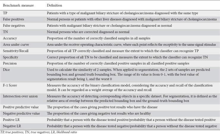
Only studies involving endoscopic AI-based imaging in the identification of malignant biliary strictures or CCA, with availability of data for the construction of 2×2 contingency tables, were included. The numbers of true positives, true negatives, false positives, and false negatives were retrieved. We removed studies with insufficient data and those with a sample size of <10. We determined the utility of visual EUS and cholangioscopic AI-based findings in the detection of malignant bile duct strictures and CCA. AI-based performance benchmarks of interest included: accuracy, sensitivity, specificity/recall, area under curve, precision, Dice, intersection over union, and F-1 score (Table 1).
Table 1.
Artificial intelligence performance evaluator metrics
Index test
The index test in our analysis was the use of any endoscopic AI-based imaging modality with studies reporting evidence of malignant biliary strictures or CCA.
Assessment of methodological quality
Quality assessment of diagnostic accuracy studies (QUADAS-2) was used to assess quality in this study [24]. QUADAS-2 is an evidence-based tool for assessment of quality in systematic reviews of diagnostic accuracy studies. It is structured so that 4 key domains are rated for risk of bias, and concerns regarding applicability to the research question were used to evaluate the studies. Each key domain has a set of signaling questions to assess bias and applicability. Disagreement among raters was resolved by consensus with the other authors. We used tabular and graphical displays in Review Manager 5 (RevMan 5.4) to summarize the QUADAS-2 assessments.
Results
Characteristics of included studies
An initial literature search generated 131 articles. We screened 93 articles after duplicates were removed. The titles of these were reviewed in accordance with the predefined inclusion criteria, yielding 18 potentially relevant articles reviewed in depth. Among these, 5 studies (n=1465) that met the inclusion criteria were included in the systematic review and meta-analysis [18-22]. A PRISMA flow chart of the search results is shown in Fig. 2.
Figure 2.
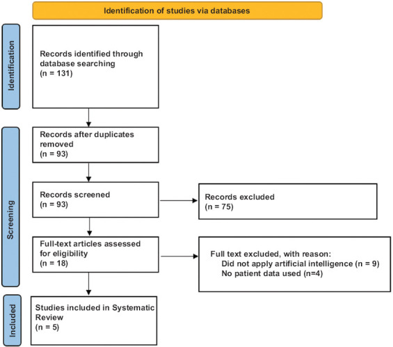
PRISMA flow diagram for studies identified for the systematic review
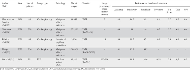
Of the 5 included studies, 4 (n=934; 3,775,819 images) used CNN in combination with cholangioscopy, while 1 study (n=531; 13,210 images) used CNN with EUS. The average image processing speed of CNN with cholangioscopy was 7-15 msec per frame, while that of CNN with EUS was 200-300 msec per frame. The characteristics of the included studies and their performance metrics are shown in Table 2.
Table 2.
Characteristics of included studies, summary of artificial intelligence endoscopic imaging modalities and performance benchmark measures for detection of malignant biliary strictures and cholangiocarcinoma
Quality assessment of included studies
The quality of the eligible studies was assessed by QUADAS-2 criteria and is reported in Fig. 3. There was a low risk of bias regarding the selection of patients, index test and reference standards; however, the 4 studies involving cholangioscopy did not clearly account for risk of bias in the flow and timing of the study. There were patient selection applicability concerns in the study by Reibero et al and index test applicability concerns in the study by Yao et al, notably due to variable index definitions [20,21].
Figure 3.
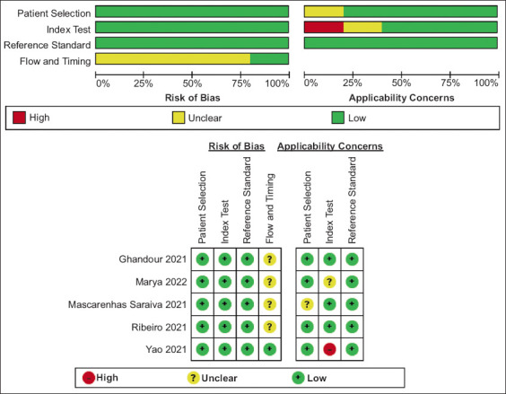
QUADAS-2 quality assessment of included studies. Risk of bias and applicability concerns graph: review authors’ judgements about each domain presented as percentages across included studies
Clinical utility
A Fagan plot was employed to determine the meaningfulness or clinical utility [25]. The Fagan nomogram is a graphical tool for estimating how much the result of a diagnostic test changes the probability that a patient has a disease. The Fagan nomogram for diagnosis of malignant biliary strictures/CCA using CNN in endoscopic imaging is shown in Fig. 4. With a pretest probability (20%) of malignant biliary stricture or CCA, if a patient tests positive, the post-test probability that the patient truly has malignant biliary stricture/CCA would be approximately 69%. Alternatively, if the patient tests negative, the post-test probability that the patient has malignant biliary stricture or CCA would be approximately 3%.
Figure 4.
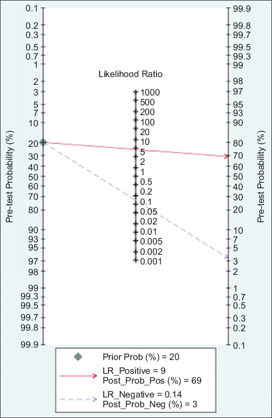
Fagan nomogram of endoscopic artificial intelligence-based imaging in the diagnosis of malignant biliary strictures and cholangiocarcinoma
LR, likelihood ratio
Application of clinical performance
Among the studies included in this systematic review and meta-analysis, CNN with EUS imaging had the best clinical performance application, while CNN with cholangioscopy for diagnosis of malignant biliary stricture/CCA needs to be further verified. The EUS bile duct scanning segmentation system significantly improved the accuracy of endoscopic station recognition and bile duct segmentation and may shorten the learning time for the diagnosis of CCA [21]. In addition, it could ensure stable and smooth operation on a private computer, completely affordable to practicing gastroenterologists in private practice. Above all, the system could run automatically, which would provide real-time guidance for endoscopists and reduce unnecessary work. Therefore, this proposed system was of great clinical impact. However, compared to EUS, cholangioscopy with CNN had a faster image processing speed (200 msec vs. 7 sec per frame) and therefore may be associated with a shorter overall procedure time [19,21].
Discussion
Diagnosing malignant biliary strictures and CCA remains challenging despite the availability of several endoscopic modalities. Currently, there are no clear international guidelines on the optimal diagnostic modality for malignant biliary strictures or CCA. Cytologic or tissue diagnosis, obtained during ERCP by brushing, biopsies or both, is limited by their poor sensitivity [9]. Cholangioscopy and EUS provide direct visualization of strictures and allow for targeted biopsies and FNA, respectively, which may help diagnose or rule out malignancy in indeterminate strictures. In previous systematic reviews and meta-analyses, we demonstrated that the pooled sensitivity and specificity of EUS-FNA to detect CCA as the etiology of biliary strictures were 66% and 100%, respectively, while the pooled sensitivity and specificity for diagnosis of cholangioscopy-guided biopsies in the diagnosis of CCA were 66.2% and 97.0%, respectively [1,13]. In the current systematic review, the highest performance metrics were observed with CNN-cholangioscopy (accuracy 94.9, sensitivity 94.7%, and specificity 92.1%). Thus, the introduction of AI algorithms such as CNN-cholangioscopy may significantly enhance the diagnostic armamentarium in patients with suspected malignant biliary strictures or CCA. In addition, given the high accuracy of AI-based endoscopic imaging for the diagnosis of malignant biliary strictures/CCA, patients with highly suspicious lesions by EUS or cholangioscopic images suitable for surgery may be able to proceed to surgical resection even if tissue biopsy results are negative for malignancy.
One of the major potential benefits of using AI-based endoscopic imaging for the diagnosis of malignant biliary strictures/CCA, without further tissue sampling such as biopsies or FNA, is that AI-based endoscopic imaging alone, without further invasive testing, is likely to result in fewer procedure-associated adverse events. For example, Kalaitzakis et al reported post-procedural cholangitis in 11% of patients after cholangioscopy with targeted biopsies, and there have also been concerns over the risk of tumor seeding or needle track seeding with EUS-FNA [26]. In a study from the Mayo Clinic, of 191 patients with locally unresectable hilar CCA, the incidence of peritoneal metastasis was 8% in those who did not undergo biopsy, compared with 83% in those with a diagnostic transperitoneal FNA (P=0.009) [14]. According to this report, the Mayo Clinic transplantation protocol excludes patients who have undergone biopsy of the primary tumor for neoadjuvant therapy and liver transplantation. The concern is that the EUS needle traverses the peritoneum and omental fat that will not be resected at the time of liver transplantation. Nevertheless, it is important to clarify that the role of AI is to assist with tissue diagnosis and improve targeted biopsies. At this time, tissue diagnosis is required to confirm the diagnosis of cholangiocarcinoma.
Although the potential of AI imaging in CCA diagnosis is promising, to observe practical benefits in real-world systems, it is critical to delineate some challenges. Notably, there is a clinically significant improvement in sensitivity when CNN algorithms are used with endoscopic imaging, with a minimal drop in its specificity. Data quality, data inconsistency and instability, and limitations of large size and diversity in support of new studies are some of the major concerns. DL algorithms require large datasets for validation, not readily available. Furthermore, the risk of overfitting should not be ignored. Overfitting is a risk in the development of AI systems that undermines the applicability of an algorithm in real-life settings. The inclusion of a large pool of frames extracted from full-length videos (with distinct resolution and viewing angles) has contributed to the mitigation of the possibility of overfitting [16,27]. In this systematic review, CNN with EUS imaging had better clinical performance application compared to DL using CNN with cholangioscopy, although comparative and externally validated studies are needed. Furthermore, the EUS literature is limited, with very few studies available at this time. The research community will need to create and populate public repositories to make resources publicly available for external validation of published AI imaging algorithms. More studies are needed on the clinical applicability of AI-based endoscopic imaging in the diagnosis of malignant biliary strictures/CCA. While AI may produce powerful predictions, this abstraction can lead to hesitation in deploying them. Moreover, the problem of liability emerges if AI is entrusted with medical activities. To close the gap between clinical practice and AI, future research may concentrate, not only on the technological aspects of the design of AI for clinical applications, but also on the development of ethical and legal systems for the implementation, validation and control of AI in clinical care. AI methods should operate in parallel with and under the supervision of clinicians until their accuracy and margin of error are considered appropriate and reasonable, respectively. It is important that researchers not focus only on the performance of algorithms, but rather on increasing their trustworthiness. There is a need for more studies to show that AI algorithms will help save diagnosis time. Such an AI system will need to be connected to doctor workstations and should be easy to use. Finally, clinical trials to show that AI systems will improve clinical outcomes such as mortality, as well as cost-effectiveness analyses of the implementation of AI-based endoscopic imaging for the diagnosis of malignant biliary strictures/CCA in routine clinical practice are paramount.
In summary, although no current screening strategies are recommended, in part because of the difficulty in distinguishing CCA from chronic inflammation in dominant benign strictures, the present life expectancy associated with CCA is unacceptable and necessitates a streamlined, universally agreed upon diagnostic approach. Our results suggest that there is increasing evidence to support a role of AI in the diagnosis of malignant biliary strictures and CCA. CNN-based machine learning of cholangioscopy and EUS images appears to be the most promising application for the visual diagnosis of malignant biliary strictures and CCA, though it is important to acknowledge that only a limited amount of data exist at this time. As with any computer vision machine learning modality, addressing “overfitting” and bias are important. Comparative and externally validated studies to establish the role of AI systems in patient-centered clinical outcomes are warranted. Furthermore, data describing the cost-effectiveness of using AI-based endoscopic imaging for the visual diagnosis of malignant biliary strictures, and CCA as a first-line diagnostic tool, are still needed before the approach can be widely accepted as a standard of care.
Summary Box
What is already known:
Cholangiocarcinoma (CCA) remains challenging to diagnose despite the availability of a variety of endoscopic modalities
Artificial intelligence (AI) may assist clinicians with the detection and differentiation of malignant biliary strictures and CCA
What the new finding is:
Convolutional neural network-based machine learning in cholangioscopy and endoscopic ultrasound imaging appears to be a promising AI-associated application for the diagnosis of malignant biliary strictures and CCA
Biography
Harvard Medical School, Boston, MA, USA; Yale University School of Medicine, New Haven, CT, USA; University of Oxford, United Kingdom; Houston Methodist Hospital, TX, USA; University of Utah School of Medicine, Salt Lake City, USA; Johns Hopkins University, Baltimore, MD, USA; Digestive Health Institute, Orlando Health, FL, USA
Footnotes
Conflict of Interest: None
Grant Support: Supported by NIH CTSA Grant Number TL1 TR001864 (B. Njei)
References
- 1.Njei B, McCarty TR, Varadarajulu S, Navaneethan U. Systematic review with meta-analysis:endoscopic retrograde cholangiopancreatography-based modalities for the diagnosis of cholangiocarcinoma in primary sclerosing cholangitis. Aliment Pharmacol Ther. 2016;44:1139–1151. doi: 10.1111/apt.13817. [DOI] [PubMed] [Google Scholar]
- 2.Njei B, McCarty TR, Varadarajulu S, Navaneethan U. Cost utility of ERCP-based modalities for the diagnosis of cholangiocarcinoma in primary sclerosing cholangitis. Gastrointest Endosc. 2017;85:773–781. doi: 10.1016/j.gie.2016.08.020. [DOI] [PubMed] [Google Scholar]
- 3.Njei B. Intrahepatic cholangiocarcinoma:are we doing the right thing? Gastrointest Cancer Res. 2014;7:1–3. [PMC free article] [PubMed] [Google Scholar]
- 4.Njei B. Changing pattern of epidemiology in intrahepatic cholangiocarcinoma. Hepatology. 2014;60:1107–1108. doi: 10.1002/hep.26958. [DOI] [PubMed] [Google Scholar]
- 5.Njei B, Konjeti VR, Sanchez H. The curious case of a Klatskin tumor. Conn Med. 2013;77:591–594. [PubMed] [Google Scholar]
- 6.Brindley PJ, Bachini M, Ilyas SI, et al. Cholangiocarcinoma. Nat Rev Dis Primers. 2021;7:65. doi: 10.1038/s41572-021-00300-2. [DOI] [PMC free article] [PubMed] [Google Scholar]
- 7.Yang CM, Shu J. Cholangiocarcinoma evaluation via imaging and artificial intelligence. Oncology. 2021;99:72–83. doi: 10.1159/000507449. [DOI] [PubMed] [Google Scholar]
- 8.Banales JM, Marin JJG, Lamarca A, et al. Cholangiocarcinoma 2020:the next horizon in mechanisms and management. Nat Rev Gastroenterol Hepatol. 2020;17:557–588. doi: 10.1038/s41575-020-0310-z. [DOI] [PMC free article] [PubMed] [Google Scholar]
- 9.Navaneethan U, Njei B, Lourdusamy V, Konjeti R, Vargo JJ, Parsi MA. Comparative effectiveness of biliary brush cytology and intraductal biopsy for detection of malignant biliary strictures:a systematic review and meta-analysis. Gastrointest Endosc. 2015;81:168–176. doi: 10.1016/j.gie.2014.09.017. [DOI] [PMC free article] [PubMed] [Google Scholar]
- 10.Gerges C, Beyna T, Tang RSY, et al. Digital single-operator peroral cholangioscopy-guided biopsy sampling versus ERCP-guided brushing for indeterminate biliary strictures:a prospective, randomized, multicenter trial (with video) Gastrointest Endosc. 2020;91:1105–1113. doi: 10.1016/j.gie.2019.11.025. [DOI] [PubMed] [Google Scholar]
- 11.Jang S, Stevens T, Kou L, Vargo JJ, Parsi MA. Efficacy of digital single-operator cholangioscopy and factors affecting its accuracy in the evaluation of indeterminate biliary stricture. Gastrointest Endosc. 2020;91:385–393.e1. doi: 10.1016/j.gie.2019.09.015. [DOI] [PubMed] [Google Scholar]
- 12.Goyal H, Mann R, Gandhi Z, et al. Application of artificial intelligence in pancreaticobiliary diseases. Ther Adv Gastrointest Endosc. 2021;14:2631774521993059. doi: 10.1177/2631774521993059. [DOI] [PMC free article] [PubMed] [Google Scholar]
- 13.Navaneethan U, Njei B, Venkatesh PG, Lourdusamy V, Sanaka MR. Endoscopic ultrasound in the diagnosis of cholangiocarcinoma as the etiology of biliary strictures:a systematic review and meta-analysis. Gastroenterol Rep (Oxf) 2015;3:209–215. doi: 10.1093/gastro/gou057. [DOI] [PMC free article] [PubMed] [Google Scholar]
- 14.Heimbach JK, Sanchez W, Rosen CB, Gores GJ. Trans-peritoneal fine needle aspiration biopsy of hilar cholangiocarcinoma is associated with disease dissemination. HPB (Oxford) 2011;13:356–360. doi: 10.1111/j.1477-2574.2011.00298.x. [DOI] [PMC free article] [PubMed] [Google Scholar]
- 15.Yu KH, Beam AL, Kohane IS. Artificial intelligence in healthcare. Nat Biomed Eng. 2018;2:719–731. doi: 10.1038/s41551-018-0305-z. [DOI] [PubMed] [Google Scholar]
- 16.Esteva A, Chou K, Yeung S, et al. Deep learning-enabled medical computer vision. NPJ Digit Med. 2021;4:5. doi: 10.1038/s41746-020-00376-2. [DOI] [PMC free article] [PubMed] [Google Scholar]
- 17.Ting DSW, Pasquale LR, Peng L, et al. Artificial intelligence and deep learning in ophthalmology. Br J Ophthalmol. 2019;103:167–175. doi: 10.1136/bjophthalmol-2018-313173. [DOI] [PMC free article] [PubMed] [Google Scholar]
- 18.Ghandour B, Hsieh HW, Akshintala V, et al. S1 Machine learning for classification of indeterminate biliary strictures during cholangioscopy. Am J Gastroenterol. 2021;116:S1. [Google Scholar]
- 19.Mascarenhas Saraiva M, Ribeiro T, Ferreira JPS, et al. Artificial intelligence for automatic diagnosis of biliary strictures malignancy status in single-operator cholangioscopy:a pilot study. Gastrointest Endosc. 2021;95:339–348. doi: 10.1016/j.gie.2021.08.027. [DOI] [PubMed] [Google Scholar]
- 20.Ribeiro T, Saraiva MM, Afonso J, et al. Automatic identification of papillary projections in indeterminate biliary strictures using digital single-operator cholangioscopy. Clin Transl Gastroenterol. 2021;12:e00418. doi: 10.14309/ctg.0000000000000418. [DOI] [PMC free article] [PubMed] [Google Scholar]
- 21.Yao L, Zhang J, Liu J, et al. A deep learning-based system for bile duct annotation and station recognition in linear endoscopic ultrasound. EBioMedicine. 2021;65:103238. doi: 10.1016/j.ebiom.2021.103238. [DOI] [PMC free article] [PubMed] [Google Scholar]
- 22.Marya NB, Powers PD, Petersen BT, et al. Identification of patients with malignant biliary strictures using a cholangioscopy-based deep learning artificial intelligence (with video) Gastrointest Endosc. 2023;97:268–278.e1. doi: 10.1016/j.gie.2022.08.021. [DOI] [PubMed] [Google Scholar]
- 23.Page MJ, McKenzie JE, Bossuyt PM, et al. The PRISMA 2020 statement:An updated guideline for reporting systematic reviews. Int J Surg. 2021;88:105906. doi: 10.1016/j.ijsu.2021.105906. [DOI] [PubMed] [Google Scholar]
- 24.Whiting P, Rutjes AW, Reitsma JB, Bossuyt PM, Kleijnen J. The development of QUADAS:a tool for the quality assessment of studies of diagnostic accuracy included in systematic reviews. BMC Med Res Methodol. 2003;3:25. doi: 10.1186/1471-2288-3-25. [DOI] [PMC free article] [PubMed] [Google Scholar]
- 25.Fagan TJ. Letter:Nomogram for Bayes theorem. N Engl J Med. 1975;293:257. doi: 10.1056/NEJM197507312930513. [DOI] [PubMed] [Google Scholar]
- 26.Kalaitzakis E, Sturgess R, Kaltsidis H, et al. Diagnostic utility of single-user peroral cholangioscopy in sclerosing cholangitis. Scand J Gastroenterol. 2014;49:1237–1244. doi: 10.3109/00365521.2014.936032. [DOI] [PubMed] [Google Scholar]
- 27.Ding Z, Shi H, Zhang H. Gastroenterologist-level identification of small-bowel diseases and normal variants by capsule endoscopy using a deep-learning model. Gastroenterology. 2019;157:1044–1054.e5. doi: 10.1053/j.gastro.2019.06.025. [DOI] [PubMed] [Google Scholar]


