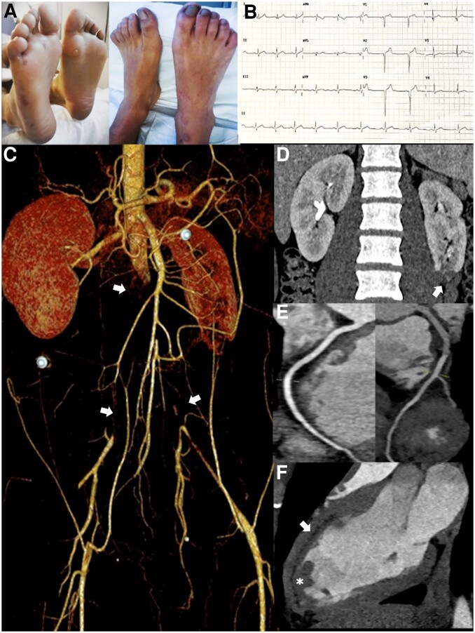
A 23-year-old man was admitted with a 3-week history of abdominal pain and intermittent claudication. On examination, he had cyanosis of the low extremities, pulselessness, livedo reticularis, and purpuric petechia of the soles (Panel A). The blood test showed severe thrombocytopenia (12 × 103/µL), a prolonged partial thromboplastin time ratio and elevated D-dimer levels, as well as persistent positive antibodies for lupus anticoagulant, anti-beta2-GP1 IgG, and anticardiolipin IgM/IgG. High-sensitivity cardiac troponin T measurement was negative.
The electrocardiogram showed a QS complex in the precordial leads V1–V3 (Panel B). Transthoracic cardiac echocardiography revealed a normal ejection fraction, akinesia of the mid-apical segments of the anteroseptal walls, in addition to an apical thrombus in the left ventricle (see Supplementary material online, Videos S1 and S2).
Computed tomography angiography (CTA) showed massive arterial thrombosis with total occlusion of the infrarenal abdominal aorta, bilateral common iliac arteries, bilateral popliteal arteries, with distal recanalization through collateral vessels (Panel C, arrows and Supplementary material online, Video S3). Furthermore, a focal parenchymal defect was documented in the lower pole of the left renal cortex indicative of probable infarction (Panel D, arrow).
Coronary CTA was ruled-out obstructive coronary artery disease and confirmed apical thrombus (Panels E-F, asterisk). The first-pass CTA phase showed a subendocardial hypoenhancement in the anteroseptal wall of the left ventricle, suggesting a myocardial infarction (Panel F, arrow).
We present an illustrative case of a patient who meets the classification criteria for probable catastrophic antiphospholipid syndrome, a rare and life-threatening disease, and highlight the utility of multimodal imaging in establishing the diagnosis and cardiovascular extension of thrombotic storm.
Supplementary Material
Contributor Information
Paula Monteagudo, Department of Internal Medicine, University Hospital Arnau de Vilanova, Ave. Rovira Roure 80, Lleida 25198, Spain; Institut de Reserca Biomedica de Lleida (IRBLLEIDA), Ave. Rovira Roure 80, Lleida 25198, Spain.
Marta Zielonka, Department of Cardiology, University Hospital Arnau de Vilanova, Ave. Rovira Roure 80, Lleida 25198, Spain; Institut de Reserca Biomedica de Lleida (IRBLLEIDA), Ave. Rovira Roure 80, Lleida 25198, Spain.
Albert Tugues Peiro, Department of Hematology, University Hospital Arnau de Vilanova, Ave. Rovira Roure 80, Lleida 25198, Spain; Institut de Reserca Biomedica de Lleida (IRBLLEIDA), Ave. Rovira Roure 80, Lleida 25198, Spain.
Eva Vicente Pascual, Department of Hematology, University Hospital Arnau de Vilanova, Ave. Rovira Roure 80, Lleida 25198, Spain; Institut de Reserca Biomedica de Lleida (IRBLLEIDA), Ave. Rovira Roure 80, Lleida 25198, Spain.
Kristian Rivera, Department of Cardiology, University Hospital Arnau de Vilanova, Ave. Rovira Roure 80, Lleida 25198, Spain; Institut de Reserca Biomedica de Lleida (IRBLLEIDA), Ave. Rovira Roure 80, Lleida 25198, Spain.
Supplementary material
Supplementary material is available at European Heart Journal – Case Reports online.
Consent: The authors confirm that written consent has been obtained from the patient for the submission and publication of this cardiovascular flashlight, including images and associated text, in accordance with the COPE guidelines.
Funding: None declared.
Data availability
The data underlying this manuscript will be shared on reasonable request to the corresponding author.
Associated Data
This section collects any data citations, data availability statements, or supplementary materials included in this article.
Supplementary Materials
Data Availability Statement
The data underlying this manuscript will be shared on reasonable request to the corresponding author.


