

Abstract
Mycoplasma species (spp.) are bacteria that are difficult to detect. Currently, the polymerase chain reaction (PCR) is considered the most effective diagnostic tool to detect these microorganisms in both human and veterinary medicine. There are 13 known species of human Mycoplasma and 15 species of canine Mycoplasma. Owing to the difficulties in identifying the individual species of Mycoplasma, there is a lack of information regarding which species are saprophytic and which are pathogenic. The prevalence of the individual species is also unknown. In addition, in both humans and dogs, the results of some studies on the impact of Mycoplasma are conflicting. The presence of Mycoplasma spp. on the epithelium of reproductive tract is often associated with infertility, although they are also detected in healthy individuals. The occurrence of Mycoplasma spp. is more common in dogs (even 89%) than in humans (1.3%–4%). This is probably because the pH of a dog’s genital is more conducive to the growth of Mycoplasma spp. than that of humans. Phylogenetically, human and canine Mycoplasma are related, and majority of them belong to the same taxonomic group. Furthermore, 40% of canine Mycoplasma spp. are placed in common clusters with those of human. This suggests that species from the same cluster can play a similar role in the canine and human reproductive tracts. This review summarizes the current state of knowledge about the impact of Mycoplasma on canine and human male fertility as well as the prospects of further development in this field.
Keywords: Mycoplasma, sperm morphology, sperm motility
INTRODUCTION
In human medicine, infertility is defined as a failure to conceive after 12 months of regular intercourse without contraception,1 and it affects 8%–12% of couples.2 Infectious organisms in the reproductive tract may affect male fertility. Although some researchers suggested a correlation between Mycoplasma and infertility in humans and dogs, this phenomenon has not been proved in other studies.3 It is suspected that these bacteria may be commensals, although it is difficult to estimate their role. This article summarizes the current state of knowledge about the impact of Mycoplasma species (spp.) on fertility in dogs and men.
Mycoplasma spp. are the smallest self-replicating organisms, belonging to the Mycoplasmataceae family, and are detectable in humans, animals, as well as in plants.4 There is a theory that Mycoplasma spp. evolved from Gram-positive bacteria, and phylogenetically they are close to Clostridia.4 Morphologically, Mycoplasma spp. stand out because of the total lack of a cell wall, and because they are included in the Mollicutes class (from Latin: mollis means soft, cutis means skin). The Mycoplasma cell contains only the organelles that are essential for growth and replication.4 Taxonomically, Mycoplasma spp. are divided into the following groups: anaeroplasma, asteroleplasma, hominis, pneumoniae, and spiroplasma.5 The majority of both canine (Ca) and human (Ho) genital Mycoplasma belong to the hominis group, which shows that they are relatively closely related. In the hominis group, among others, there are three clusters: hominis, bovis, and synoviae, in which both human and canine Mycoplasma are placed. The hominis cluster includes Mycoplasma (M.) arginini (Ca), M. gateae (Ca), M. spumans (Ca), M. buccale (Ho), M. faucium (Ho), M. hominis (Ho), and M. orale (Ho); the bovis cluster includes M. bovigenitalium (Ca), M. maculosum (Ca), M. opalescens (Ca), M. fermentans (Ho), M. primatum (Ho), and M. spermatofilum (Ho); and the synoviae cluster includes M. cynos (Ca), M. edwardii (Ca), M. felis (Ca), and M. canis (Ca).6 Therefore, on the basis of Mycoplasmataceae taxonomy, it has been estimated that 40% of canine species are in the same cluster as human (not published).
All phylogenetic are shown in Figure 1. The 16S ribosomal DNA sequences of Mycoplasma species were retrieved from GenBank (NCBI), as shown in Table 1. Alignment of the sequences was constructed using GeneDoc using Blosum62 matrix (gap open cost: 8, gap extend cost: 4). Aligned sequences were trimmed to the longest overlapping region and sequences of M. primatum, M. haemocanis, and M. arginini were rejected due to small overlapping region, and rest of the sequences were aligned again using aforementioned parameters. An evolutionary tree was constructed with Molecular Evolutionary Genetic Analysis (MEGA) software using the maximum likelihood method and Tamura-Nei model with bootstrap consensus inferred from 10 000 replicates.
Figure 1.
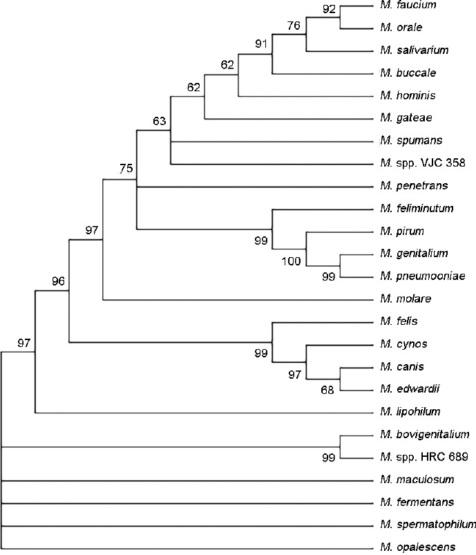
The evolutionary tree of 16S ribosomal DNA sequences of canine and human species of Mycoplasma. Numbers above the branches show the percentage of probability of the result. M.: Mycoplasma.
Table 1.
List of species of Mycoplasma and their numbers in the GenBank used to create a phylogenetic tree
| Species of Mycoplasma | GenBank number |
|---|---|
| Mycoplasma facium | NR_024983.1 |
| Mycoplasma orale | NR_043199.1 |
| Mycoplasma salivarium | NR_041745.1 |
| Mycoplasma hominis | NR_041881.1 |
| Mycoplasma gateae | NR_029180.1 |
| Mycoplasma spumans | NR_24980.1 |
| Mycoplasma spp. VJC 358 | AY246564.1 |
| Mycoplasma penetrans | RCH401000003.1 |
| Mycoplasma feliminutum | NR_029181.1 |
| Mycoplasma pirum | NR_029165.1 |
| Mycoplasma genitalium | NR_026155.1 |
| Mycoplasma pneumooniae | NR_041751.1 |
| Mycoplasma molare | NR_041931.1 |
| Mycoplasma felis | U09787.1 |
| Mycoplasma cynos | NR_025181.1 |
| Mycoplasma canis | AB680678.1 |
| Mycoplasma lipohilum | AB680693.1 |
| Mycoplasma bovigenitalium | AB680692.1 |
| Mycoplasma spp. HRC 689 | AF527624.1 |
| Mycoplasma maculosum | AB680679.1 |
| Mycoplasma fermentans | NR_044666.2 |
| Mycoplasma spermatophilum | NR_025069.1 |
| Mycoplasma opalescens | NR_025067.1 |
This affinity between species of human and canine Mycoplasma suggests that they could influence semen quality similarly. Accordingly, the dog can probably be treated as a model organism for research on mycoplasmosis of the genital tract. In addition, it is possible for canine Mycoplasma to colonize the human body. Klein et al.7 have isolated M. canis from human tissue after a dog bite.
The primary habitats of human and canine Mycoplasma are the mucous surfaces of the respiratory and urogenital tracts, eyes, digestive system, mammary glands, and joints.4 In addition, there is a report about their occurrence in pathological canine brain tissues.8 As well as other mollicutes, Mycoplasma spp. can be present intracellularly in the host’s cells. In both humans and animals, Mycoplasma is taken up by leukocytes and macrophages, but the mechanism of entry into the cells is still unclear. However, it has been described that this invasion may affect cell function.4 Díaz-García et al.9 demonstrated that M. hominis can also infect spermatozoa.9
Mycoplasma adheres to the surface of the epithelium in the reproductive tract, and this process is strong enough to prevent their elimination in their secretions or urine.4 It is also known that M. genitalium has a major surface adhesion complex known as the nucleoid-associated protein (NAP) on its surface, and because of this, it can adhere to surfaces and remains motile.10 Furthermore, no specific toxins or virulence factors of M. genitalium have been described, and it is suspected that the lipoproteins exposed on their surface can stimulate local inflammatory response in the reproductive tract.11 There is limited knowledge about the virulence factors of canine Mycoplasma spp. However, some species can cause hemolysis during culturing; therefore, it has been suggested that some of them can synthesize hemolytic enzymes.12 Genital Mycoplasma in humans and possibly in veterinary patients are natural inhabitants of the male urethra, and therefore, they can be present in spermatozoa during ejaculation.13 There are 13 known species of human Mycoplasma which occur in the genital tract including M. buccale, M. faucium, M. fermentans, M. genitalium, M. hominis, M. lipophilum, M. orale, M. penetrans, M. pirum, M. pneumoniae, M. primatum, M. salivarium, and M. spermatophilum,14 but the more common are M. genitalium and M. hominis.15
In the canine reproductive tract, M. arginini, M. bovigenitalium, M. canis, M. cynos, M. edwardii, M. feliminutum, M. felis, M. gateae, M. haemocanis, M. maculosum, M. molare, M. opalescens, Mycoplasma spp. HRC 689, Mycoplasma spp. VJC 358, and M. spumans can be detected, and the more common are M. canis, M. spumans, and M. maculosum.16 Both canine and human Mycoplasma are shown in Figure 2.
Figure 2.

Scientific classification of human and canine genital Mycoplasmas, based on: ncbi.nlm.nih.gov (strains) and patricbrc.org/view/Taxonomy/ (taxonomy). M.: Mycoplasma.
It has been estimated that their prevalence in the human reproductive tract in countries with high levels of development is 1.3%, while it is almost 4% in countries with lower levels of development.17 In veterinary medicine, the occurrence of Mycoplasma spp. in animals is more common. It has been estimated that among dogs, up to 89% can be Mycoplasma positive.18 There are possible reasons that Mycoplasma spp. is more common in dogs than in humans. On the one hand, dogs have more different sexual partners than humans, and in addition, people are using safeguards against contracting venereal diseases. On the other hand, Mycoplasma spp. may be present in the prepuce of some dogs before the first mating. The pH value of the canine reproductive tract may be potentially more suitable for the growth of this microorganism. The best pH conducive for Mycoplasma growth is between 7.8 and 8.19 In canine females, the pH value in the vagina is 7.4–8.320 and 6.3–6.7 in prepuce of males,21 as opposed to humans who have lower values of 5.71 in men’s prepuce22 and 3.8–4.5 in women’s vagina.23 The pH values of canine semen are as follows; first fraction: 6.37, second fraction: 6.37, and the third one is 7.2;24 and human semen pH values are between 7.2 and 8.25 The most important factor seems to be pH in the place of arising the Mycoplasma. In the tunica mucosa of the human reproductive tract, the pH is inappropriate for growth and development of these bacteria. This phenomenon can be a reason that Mycoplasma-positive results are more common in the dog than in the human reproductive tract. In a few publications, the presence of Mycoplasma was in semen, not the prepuce.26,27 Ultimately, the hypothesis is that in the canine reproductive tract, the environmental conditions are better for Mycoplasma spp. can be given. However, more research is needed to confirm this theory. Moreover, the prevalence of Mycoplasma in the respiratory tract is higher in dogs than that in man; in humans, it ranges from 2% to 35%,27 while in dogs, it ranges from 86% to 90%.28 Nevertheless, a study performed in mice showed that those infected by Mycoplasma intranasally were more resistant to Mycoplasma infections of the reproductive tract than the noninfected.29 Probably, a similar phenomenon can be observed in dogs and humans; however, further studies are required to confirm this suggestion.
Similar to Mycoplasma spp. are Ureaplasma (U.) spp. which reside in the urogenital tract. These bacteria, by evolution, have also lost their cell wall. In humans, there are two known species: U. urealyticum and U. parvum. Like Mycoplasma spp., Ureaplasma spp. are also considered to be a cause of infertility, but it has also been suggested that they could be a part of the normal genital flora.30 Since Mycoplasma and Ureaplasma are related and very similar, some researchers have named them together as “Mycoplasmas”, and their effect on the semen is examined together in studies.
SPECIFYING THE MYCOPLASMA
In the past, the main method of detecting Mycoplasma spp. was by culturing them, but owing to the high requirements of these bacteria, this method is not used nowadays in commercial laboratories. The polymerase chain reaction (PCR) is now the most commonly used method in both veterinary and human medicine. Peerayeh and Samimi31 have shown that the PCR method enables a higher rate of detection of Mycoplasma than standard microbiologic cultures.
The ribosomal 16S gene sequence is frequently used in molecular techniques owing to its universal presence among bacteria. The 16S rRNA gene contains nine hypervariable regions (V1–V9) that show differences among bacteria. These specific sequences are useful for diagnostic assays, e.g., V6 helps to distinguish among most bacterial species except Enterobacteriaceae. In the case of 16S rRNA analysis, identification of the bacteria is easier when the entire gene can be sequenced. Unfortunately, this technique is not rapid, so it is not common. A faster and commonly used method is based on assays that combine nucleic acid amplification with a sequence-specific probe of the amplified product. In this technique, there is a possibility to query short DNA sequences. Therefore, the identification of the regions within the target gene is important.32
In human medicine, there are primers which are capable of detecting M. hominis, M. genitalium, and U. urealyticum simultaneously.33 In addition, highly specific primers have been developed for the detection of M. hominis, U. urealyticum, and two others reproductive tract pathogens,34 and they are based on the ribosomal 16S gene. There are also commercial biochemical assay-based kits available for the identification of M. hominis, but the PCR method is faster, more reliable, and more sensitive.35 The primers which can be used for the identification of human Mycoplasma are shown in Table 2.
Table 2.
Polymerase chain reaction primers for specifying human Mycoplasmas
| Mycoplasma spp. | Mycoplasma primer sequence (5’–3’) | Source |
|---|---|---|
| Mycoplasma buccale | Forward: ATGCATGTCGAGCGGAAGTA Reverse: AATCCGAAGACCGTCATCATGC |
GenBank: AF125586.1a |
| Mycoplasma faucium | Forward: CATGTCGAGCGGAAGTAGCA Reverse: TTAGCTGCGTCAGTGGCTC |
GenBank: NR_024983.1a |
| Mycoplasma fermentans | Forward: GGACTATTGTCTAAACAATTTCCC Reverse: GGTTATTCGATTTCTAAATCGCCT |
Vojdani and Franco871999 |
| Mycoplasma genitalium | Forward: TACATGCAAGTCGATCGGAAGTAGC Reverse: AAACTCCAGCCATTGCCTGCTAG |
Jensen et al.88 2003 |
| Mycoplasma hominis | Forward: GGAAGA-TATGTAACAAAAGAAGGTGCTG Reverse: TTTATCTTCTGGCGTAATGATATCTTCG |
Baczynska et al.89 2004 |
| Mycoplasma lipophilum | Forward: CAATATTTAACCGCCGCGCA Reverse: AGCACCCATTAAAGCACGGT |
GenBank: DQ112177.1a |
| Mycoplasma orale | Forward: AAGCTTGATGGAGCGACACA Reverse: GCGTTAGCTGCGTCAGTAGT |
GenBank: NR_043199.1a |
| Mycoplasma penetrans | Forward: CATGCAAGTCGGACGAAGCA Reverse: AGCATTTCCTCTTCTTACAA |
Vojdani and Franco87 1999 |
| Mycoplasma pirum | Forward: TACATGCAAGTCGATCG-GAT Reverse: CATCCTATAGCGGTC-CAAAC |
Grau et al.90 1993 |
| Mycoplasma pneumoniae | Forward: CAAGCCAAACACGAGCTCCGGCC Reverse: CAGTGTCAGCTGTTTGTCCTTCCCC |
Chaudhry et al.91 2013 |
| Mycoplasma primatium | In the GenBank, there is no sequence based on which the primer designing could be possible. | - |
| Mycoplasma salivarium | Forward: ATGATGCTAACCGTGCGCT Reverse: CCATCTTGTCGCCGACTCT |
GenBank: EU797448.1a |
| Mycoplasma spermatophilum | Forward: TGACGCTAACCGTGCATTGA Reverse: TGTTACCGTGACGACCTGAC |
GenBank: DQ219487.1a |
aPrimers not published previously. Parts of the data from the table are cited from the articles and other part of the data are primers not published previously. They are designed based on the sequence from GenBank (ncbi.nlm.nih.gov/genbank). -: no data
The current knowledge regarding their molecular nature is very limited. Chalker and Brownlie5 revealed that most canine Mycoplasma have a variable phylogenetic origin, but a great part of them lies in a variety of clusters within the hominis group of Mycoplasma. Owing to the similarity between the 16S rRNA genes of canine Mycoplasma, PCR tests have been created to identify the species-specific regions in the 16S/23S rRNA intergenic spacer region.36 Table 3 shows the primers that can be used in the PCR assay to detect canine Mycoplasma.
Table 3.
Polymerase chain reaction primers to specifying canine Mycoplasma
| Mycoplasma spp. | Mycoplasma primer sequence | Source |
|---|---|---|
| Mycoplasma arginini | Forward: CA-CCGCCCGTCACACCA Reverse: GTTGTATGACCTATTGTTGTC |
Chalker36 2004 |
| Mycoplasma bovigenitalium | Forward: CGTAGATGCCGCATGGCATTTACGG Reverse: CATTCAATATAGTGGCATTTCCTAC |
Kobayashi et al.92 1998 |
| Mycoplasma canis | Forward: CA-CCGCCCGTCACACCA Reverse: CTGTCGGGGTTATCTCGAC |
Chalker36 2004 |
| Mycoplasma cynos | Forward: CA-CCGCCCGTCACACCA Reverse: GATACATAAACACAACATTATAATATTG |
Chalker36 2004 |
| Mycoplasma edwardii | Forward: CA-CCGCCCGTCACACCA Reverse: CTGTCGGGTTATCATGCGAC |
Chalker36 2004 |
| Mycoplasma feliminutum | Forward: AAGGTCCGTTTGGATCGCTT Reverse: TTTTGGAGCGGGACATGGTT |
GenBank: U16758.1a |
| Mycoplasma felis | Forward: CA-CCGCCCGTCACACCA Reverse: GGACTATTATCAAAAGCACATAAC |
Chalker36 2004 |
| Mycoplasma gateae | Forward: CA-CCGCCCGTCACACCA Reverse: GTTGTATGACCTATTGTTGTC |
Chalker36 2004 |
| Mycoplasma haemocanis | Forward: GTGCTACAATGGCGAACACA Reverse: TCCTATCCGAACTGAGACGAA |
Barker et al.93 2010 |
| Mycoplasma maculosum | Forward: CA-CCGCCCGTCACACCA Reverse: CCTATGATTGTTACAGATG |
Chalker36 2004 |
| Mycoplasma molare | Forward: CA-CCGCCCGTCACACCA Reverse: AGCCTATTGTTTTTGATTTG |
Chalker36 2004 |
| Mycoplasma opalescens | Forward: CA-CCGCCCGTCACACCA Reverse: TAAGCTTTGTAGACCATAA |
Chalker36 2004 |
| Mycoplasma spp. HRC 689 | Forward: CA-CCGCCCGTCACACCA Reverse: CTTGCGACCTAACAAGTCC |
Chalker36 2004 |
| Mycoplasma spp. VJC 358 | Forward: AGGGAGACTGCCCGAGTAAT Reverse: TCGGGTTATCTCGACACATGAC |
GenBank: AY246564.1a |
| Mycoplasma spumans | Forward: CA-CCGCCCGTCACACCA Reverse: GTTGTATGACCTATTGTTGTC |
Chalker36 2004 |
aPrimers not published previously. Parts of the data from the table are cited from the articles and other part of the data are primers not published previously. They are designed based on the sequence from GenBank (ncbi.nlm.nih.gov/genbank)
Recently, a novel quantitative qPCR to monitor Mycoplasma infection in dogs has been developed by Hemmatzadeh et al.37 A single band of bacterial 16S ribosomal DNA was amplified by using universal Mycoplasma primers. The band was excised from the gel, and the purified DNA was submitted to the Australian Genome Research Facility Ltd. for Sanger sequencing. This sequence was used to search GenBank using BLAST for matching a sequence. Thereafter, the prepared DNA was used as a standard for qPCR reactions. The number of copies of the Mycoplasma plasmid was calculated on an online calculator. This method was developed because conventional PCR fails to detect less than 100–200 genomes per μl.37
INFLUENCE OF INFECTION ON SEMEN QUALITY
The influence of human and canine Mycoplasma on the quality of the semen seems to be similar. Infections of the reproductive tract in both humans and animals play an important role in infertility. It is suggested that bacterial and viral infections are two of the factors responsible for male infertility.38 However, this correlation and the underlying pathogenesis remain unclear. It has been suggested that decreased effectiveness of spermatogenesis, obstruction of the seminal tract, and dysfunction of the spermatozoa are among the adverse effects of bacterial infections.39 In vitro studies have shown that bacterial infection can affect sperm function, in addition to inducing sperm agglutination and apoptosis.40,41
The role of Mycoplasma infection in both dogs and humans remains unclear. In veterinary medicine, this issue is even more complicated than in human medicine because not all veterinary laboratories specify the species of Mycoplasma because of difficulty in their recognition. Previously, the identification of canine Mycoplasma was by serological methods which were dependent on specific antisera for each species. However, cross-reactions were also observed; consequently, antisera are not readily available in laboratories.42 Moreover, owing to the high similarity between the 16S rRNA genes of canine Mycoplasma, diagnosis by PCR is also challenging.12 This is the reason that Mycoplasma spp. associated with negative changes in the semen are still unknown.
In human medicine, a meta-analysis has suggested that the presence of M. hominis, rather than M. genitalium, correlates with male infertility.43 This indicates that some Mycoplasma spp. may also affect male fertility in dogs and some may not. The impact of Mycoplasma spp. on the basic semen parameters is described below.26,44,45
IMPACT ON BASIC SEMEN PARAMETER VALUES
Volume of the ejaculate
Following the World Health Organization (WHO) guidelines, the volume of the ejaculate should be measured in all semen evaluations. The influence of Mycoplasma on the semen volume is not clear. Gdoura et al.44 did not find a significant influence on the semen volume in Mycoplasma-positive patients. On the other hand, a study by Ahmadi et al.46 showed a significant increase in the semen volume after treatment of Mycoplasma infection. Owing to these contradictory study results, it is not possible to evaluate the impact of Mycoplasma on the semen volume, and more studies on this issue are needed.
Progressive sperm motility and sperm concentration
The effect of both canine and human Mycoplasma infection on sperm concentration and motility remains unclear. However, a study performed by Gdoura et al.44 showed a negative correlation between the sperm concentration and detection of M. genitalium in the semen. Furthermore, semen was analyzed in a Greek study performed to investigate the influence of Chlamydia spp., Ureaplasma spp., and Mycoplasma spp. on sperm concentration, total motility, and progressive motility. No correlation was found between these bacteria and sperm parameter values.47 However, it has been demonstrated that M. genitalium can influence semen quality by adhering to the sperm heads, midpieces, and tails, owing to which the spermatozoa become immotile.48 Similarly, the research by Köhn et al.49 showed that spermatozoa incubated with M. hominis are less motile than spermatozoa from the control group. In addition, it revealed that for men who were M. hominis-positive group, the sperm concentration and motility were significantly lower.50
In veterinary medicine, studies on the impact of Mycoplasma on dog semen are very limited and many of them are old. In one study from 1977, the researchers tried to infect the reproductive tracts of male dogs. In this study, the M. canis isolates were inoculated into the ductus deferens via vasotomy in three dogs (examined group). The control was one dog who received uninoculated broth. All dogs were clinically healthy during this experiment. An increase in the scrotal temperature as well as changes in the testes and epididymides was noticed in two animals (from the examined group) on days 23 and 29. In all dogs in the study group, a significant increase in abnormal spermatozoa and a decrease in the sperm motility were reported, although Mycoplasma canis were detected in only one dog.16 It may be suggested that the abnormalities in the sperm morphology occurred because of the inflammation caused by manipulations during vasotomy, and not because of the Mycoplasma infection. In addition, the examined group of dogs was too small to draw final conclusions. There is also a case report of a male dog which was found to be positive for M. spumans and M. maculosum, and of which seminal sperm concentration was low (1.5 × 10⁶ ml−1) and the spermatozoa were immotile. After Mycoplasma treatment, semen quality improved.51 To confirm the negative effect of those two species of Mycoplasma on the semen quality, more research is needed.
In a study by Schäfer-Somi et al.26 andrological examination was correlated with the presence of Mycoplasma spp. and other bacteria in the reproductive tract and semen of dogs. M. canis was isolated from the semen samples of 18% of dogs whose semen was collected for cryopreservation, 40% of infertile dogs, and 45% of dogs with benign prostatic hyperplasia (BPH). This study showed that these bacteria may be present even in the high-quality semen of a young dog. The authors suggested that the number of the microorganisms is not a decisive factor, but the duration of the infection, degree of epithelial damage, or local immune response may be important. In addition, it has been suggested that the concentration of the spermatozoa may be lower after germinal epithelium damage.26 To confirm this hypothesis and estimate the real impact of Mycoplasma spp. on the morphology of dog spermatozoa and sperm concentration, further studies are needed.
Effect on sperm morphology
A normal human sperm tail should be without cytoplasmic residues and should have a length of approximately 45–50 μm.52 In veterinary medicine, the assessment of sperm morphology is more difficult owing to the lack of morphometry information.53 Only a few of the more popular breeds of dogs have been evaluated by morphometrical examination,54 and this is not enough to define the standard values for all dogs. In male dogs, during the evaluation of the morphology of the spermatozoa, mainly the cytoplasmic residues and tail are considered.
In humans, the lower reference limit for normal forms of spermatozoa is 4%,55 while in dogs, it should be greater than or equal to 80%.18 In the past, the reference limit of this parameter was different for men. It was 30% in 1992 and 14% in 1999. The reference values are based on the sperm parameters of fertile men in the fifth percentile in the percentile distribution of results of pregnancy rates. The discrepancy in the lower reference limit is probably because in humans, the sperm counts fall with every decade of life.56
Rose et al.45 investigated the influence of Mycoplasma spp. on the morphology of spermatozoa. After semen incubation with Mycoplasma, there was a significant increase in abnormal midpieces and tails compared with the control group, which suggests that in vivo Mycoplasma spp. can have an influence on sperm morphology. Moreover, older reviews have suggested that ejaculates contaminated by Mycoplasma spp. contain coiled forms as well as swollen necks of the spermatozoa.57 An electron microscopical study showed that the spermatozoa from Mycoplasma-positive ejaculates had several distinctive features. Mycoplasma was attached to the sperm cells by interlacing fibrils of variable diameter, and was associated with spherules. Another characteristic feature was numerous sperms with coiled tails.58 In addition, a study investigated the real influence of Mycoplasma on sperm morphology. In this research, Mycoplasma were detected by a Mycoplasma IST kit (BioMerieux, Marcy-l’Étoile, France), and the changes in the sperm morphology were found to be as follows: abnormalities in the head’s shape, disrupted nuclear membrane, vacuoles within the nuclear chromatin, protuberances in acrosomes, cytoplasmic residues, and vacuoles inside the chromatin.59
Since the effect of Mycoplasma on sperm morphology remains unclear, and because of limited publications, new studies are needed on this issue. Owing to the similarity between Mycoplasma spp. and Ureaplasma spp., the impact on the sperm quality of these two bacteria could also be comparable. In one study on the influence of Ureaplasma on sperm morphology, it was shown that the U. urealyticum-positive group had a higher proportion of abnormal spermatozoa than the control group.60 This indicates that both Ureaplasma spp. and Mycoplasma spp. can influence sperm morphology. However, another study showed that U. urealyticum had a more significant impact on sperm morphology than Mycoplasma and four other pathogens.61
IMPACT ON CELLS OTHER THAN SPERMATOZOA AND SPERM AGGLUTINATION
The ejaculate contains cells other than spermatozoa, including epithelial cells, leukocytes, and immature germ cells. All of them can be identified by examining a stained smear.55 There is a controversial report suggesting that epithelial cells can phagocytose the spermatozoa, which possibly acts as a removal process for abnormal spermatozoa. This phenomenon was noted in men infected by Chlamydia trachomatis and Mycoplasma spp.62
Leukocytes in the ejaculate
The occurrence of leukocytes in the ejaculate is due to infections of the male reproductive tract. This process can be divided into three stages. The first stage occurs shortly after infection, and is not associated with a significant number of leukocytes. During the second stage, it is assumed that the leukocytes take part in the immune response, and therefore, activated leukocytes appear in the semen. During the third stage, the bacteria are eliminated by the immune system, but the leukocytes persist in the ejaculate.63
A study has revealed that the presence of Mycoplasma in the semen is not correlated with leukocytospermia in humans.64 In dogs, there was a similar study in which the semen cytology was investigated. Only in 15 of 41 Mycoplasma-positive dogs did the cytology show a higher amount of leukocytes than noninflammatory samples.65 These two studies suggest that Mycoplasma spp. may not be related with infections of the male reproductive tract. However, one report has claimed that leukocytes are present in the ejaculate of Mycoplasma- and Chlamydia-positive men, and they could phagocytose abnormal spermatozoa. The researchers described a process in which, during the early stages, the sperm head adheres to the surface of the leukocyte, and in the later stages, it is surrounded by the leukocytic pseudopodia. They also found that the leukocytes contained spermatozoa.59 This study did not comment on the amount of leukocytes in the ejaculate.
Agglutination and aggregation of spermatozoa
Aggregation is the adherence of spermatozoa to other cells or debris,66 it has been suggested that in Mycoplasma-positive men, the number of cells other than spermatozoa was not increased. The phenomenon of the motile spermatozoa sticking to each other is called agglutination.55 It can be positively correlated not only with anti-sperm antibodies but also with other causes such as genital tract infections and ascorbic acid deficiency.67 There are two reports on the effect of Mycoplasma on sperm agglutination. Both of them involved humans, and did not find a relationship between the presence of anti-sperm antibodies and Mycoplasma spp.68,69 This may indicate that Mycoplasma have no influence on sperm agglutination.
IMPACT ON THE FUNCTIONAL PROPERTIES OF SPERMATOZOA
Sperm DNA fragmentation
Any abnormalities in the sperm chromatin or damage to the DNA can cause infertility because the sperm DNA must decondense during fertilization.70 In a study performed on 143 infertile patients with diagnosed genitourinary infection with Chlamydia spp. and Mycoplasma spp., sperm DNA fragmentation was examined by the sperm chromatin dispersion (SCD) method. The result showed that the mean percentage of spermatozoa with fragmented DNA in the infertile patient group was 3.2 times higher than that in the control fertile group. After antibiotic and anti-inflammatory treatment, the frequency of the sperm cells with fragmented DNA decreased from 37.7% to 24.2%.71 This suggests that Mycoplasma spp. can influence sperm DNA fragmentation, which is associated with infertility in men. In another study in which flow cytometry was performed after staining with acridine orange (AO), the chromatin integrity, measured by the presence of single-stranded DNA (ssDNA) and double-stranded DNA (dsDNA) breaks in the sperm chromatin in men with semen positive for Ureaplasma and Mycoplasma strains, was not disturbed.72 However, in these studies, as the Mycoplasma spp. were not specified, it could not be determined which Mycoplasma spp. could affect DNA fragmentation.
Acrosome reaction
There are only two studies in this field. In the first study, the spermatozoa were incubated with Mycoplasma (M. hominis and U. urealitycum). The authors showed that spermatozoa from the experimental group were less likely to undergo an acrosome reaction in response to calcium ionophore treatment than the control cells.45 The second study also showed that M. hominis can reduce the inducibility of human sperm acrosome reaction.49 However, no similar studies have been performed on dogs.
SPERM VITALITY
In both veterinary and human medicine, the most common method to assess the sperm vitality is a test using eosin-nigrosin. This method is based on the principle that the damaged plasma membrane (in dead spermatozoa) allows the entry of membrane-impermeant stains.73
In the flow cytometry method, the most common stain used is SYBR-14 with propidine iodine (PI). SYBR-14 penetrates undamaged cell membranes to cause light green fluorescence. Damaged cell membranes allow PI penetration, which displaces SYBR-14, causing red fluorescence. This double staining shows three subpopulations of spermatozoa: live cells (SYBR-14+, PI−), dead cells (SYBR-14−, PI+), and moribund cells (SYBR-14+, PI+).74,75
Gallegos et al.71 found no significant impact of these bacteria on sperm vitality. Andrade-Roha64 also investigated the influence of Mycoplasma on this parameter. Sperm vitality was lowest in semen with more than 103 colony-forming units per ml of semen (cfu ml−1), but it was not statistically significant. In another study in which the intracellular location of M. hominis was investigated, it was noted that this species of Mycoplasma does not affect sperm viability.9
Although the influence of canine Mycoplasma on sperm vitality is unknown, in a dog which was a carrier of M. spumans and M. maculosum, 100% of the spermatozoa were dead.51
EFFECT OF MYCOPLASMA SPP. ON PROSTATE FUNCTIONS
In men, the seminal vesicles are the main accessory gland of the male reproductive system,76 while in dogs, the prostate is the only accessory sex gland.18 In humans, acute bacterial prostatitis is not associated with infertility in contrast to chronic prostatitis. This phenomenon can be attributed to the impairment of the secretory capacity of the prostate, which might have a negative effect on all semen parameters.76 It has been suggested that M. genitalium is associated with chronic prostatitis in humans, because it is detected more frequently in patients with prostatitis than in healthy ones.77 However, Mändar et al.78 reported that both Mycoplasma and Ureaplasma occurred more frequently in the semen of men with prostatitis than in healthy ones, and the most frequently occurring species was U. parvum. In another research, M. hominis was detected in 13% of men with prostate cancer, while these bacteria was not detected in any of the men with BPH.79
In dogs, the correlation between prostate diseases and infertility has not been proven. However, in a study performed on nine stud dogs who presented with infertility, five had prostatitis and one had BPH.18 In a study by Schäfer-Somi et al.26, M. canis was detected in 83.3% of the dogs that were diagnosed with BPH, although it remains unknown if these bacteria play a role in the pathogenesis of this disease.
IMPROVEMENT IN SEMEN QUALITY AFTER TREATMENT OF MYCOPLASMA INFECTION
Treatment of Mycoplasma infection is based on antibiotic therapy, but because of the lack of a cell wall, these bacteria are resistant to β-lactam antibiotics. Some species are also resistant to macrolides, sulfonamides with trimethoprim and rifampicin.80 Doxycycline is widely used to treat infections by Mycoplasma spp.81 Treatment with doxycycline (twice daily, for 7 days) in patients with Mycoplasma infection results in a significant improvement in all semen parameter values except for volume, pH, and nonprogressive sperm motility.82,83 However, in another study, 3 months after antibiotic treatment, only 55.3% of men were free from microorganisms, and no significant improvement in any of the investigated semen parameters was noted.72 It should be noted that doxycycline is a drug that stops bacterial protein synthesis; therefore, the duration of doxycycline therapy should be longer than bactericidal antibiotics. In dogs, the most common drug used for treatment is also doxycycline. Successful treatment has also been reported by the use of doxycycline for 15 days, followed by azithromycin for 9 days.51 In this case, although the semen quality improved after therapy, a control PCR test was not performed.51
In case of low-grade infections with no changes in the semen quality parameters, it has been suggested that preputial irrigation with 2.5% marbofloxacin can be a form of therapy,26 but there is no report about the effectiveness of this method. After treatment, it is recommended that stud dogs should have an 8-week break in mating in order to regenerate and improve the quality of the semen from new germ cells formed during spermatogenesis. Supplementation with vitamin E for 10 weeks has also been suggested to regenerate the epithelium of the seminal tubules.26
CONCLUSIONS
Mycoplasma spp. occur on mucosal surfaces in both humans and dogs. Previous studies have described their effect on pelvic diseases in women,84,85 reproductive tract of female canines,86 respiratory tract in dogs,36 and fertility in men.69,49 In these studies, bacteria were detected in both healthy and diseased study participants; consequently, the impact of Mycoplasma remains unclear. A summary of current state of the knowledge about influence of Mycoplasma spp. on fertility is shown in Table 4.
Table 4.
Influence of human and canine Mycoplasmas on semen parameters and prostate diseases
| Andrological finding | Human Mycoplasmas | Canine Mycoplasmas | |||
|---|---|---|---|---|---|
|
|
|
||||
| Mycoplasma spp. | Mycoplasma hominis | Mycoplasma genitalium | Mycoplasma spp. | Mycoplasma canis | |
| Prevalence | 1.3%–4%17 | No data | No data | 89%18 | No data |
| Infertility | No data | Negative influence43 | No influence43 | No data | Present in 17.8% high quality ejaculates and in 40.4% poor semen quality16 |
| Volume | Conflicting results44,82 | No influence39 | Conflicting results44,82 | The ejaculate volume is not so important as in human patients | |
| Concentration | No data | Conflicting results44,82 | Conflicting results44,82 | No data | No data |
| Motility | No data | Conflicting results44,82 | Conflicting results44,82 | No data | Temporary decreased spermatozoa motility16 |
| Morphology | Negative influence45 | No influence63 | No influence63 | No data | Temporary increased numbers of abnormal spermatozoa16 |
| Number of leukocytes | No data | No influence63 | No influence63 | No data | In 15 of 41 dogs, the semen cytology showed a higher amount of leukocytes64 |
| Sperm agglutination | No influence67,68 | No data | No data | No data | No data |
| DNA fragmentation | Conflicting results71,94 | No data | No data | No data | No data |
| Acrosomal reaction | No data | Negative influence45,49 | No data | No data | No data |
| Viability | Negative influence63 | No influence9 | No data | No data | No data |
| Prostate diseases | Prostatitis77 | Cancer, BPH78 | Prostatitis78 | No data | Prostatitis, BPH26 |
BPH: benign prostatic hyperplasia
Almost 89% of the dog population has been reported to be Mycoplasma positive,18 suggesting that not all species or strains are pathogenic, or their virulence is low. Some authors have identified which bacterial species can cause infertility in dogs.51 However, the knowledge about all strains is still limited.
Further research is required to compare the mechanisms underlying mycoplasmosis in the genital tract in both humans and dogs, especially in close phylogenetic species. It is also necessary to investigate if antibodies induced by Mycoplasma infection of the respiratory tract can potentially protect the genital tract during contact with pathogenic species of Mycoplasma. Importantly, there is a need to identify which Mycoplasma species and strains are pathogenic and which are not.
AUTHOR CONTRIBUTIONS
KD reviewed the literature and wrote the main body of the manuscript. IK helped with designing the manuscript, and made linguistic and stylistic corrections. PJ, SK, and MS critically reviewed and substantially contributed to the final draft of the manuscript. All authors read and approved the final manuscript.
COMPETING INTERESTS
All authors declare no competing interests.
ACKNOWLEDGMENTS
All authors acknowledge Dr. Ricardo Faúndez (InviMed Fertility Clinics, Warsaw, Poland) who provided critical insights into the reviewed topic during manuscript preparation. We also thank Dr. Piotr Bąska (Warsaw University of Life Sciences, Institute of Veterinary Medicine, Department of Preclinical Sciences, Division of Pharmacology and Toxicology, Warsaw, Poland) for designing primers used in Table 2 and 3, and Figure 2.
REFERENCES
- 1.Zegers-Hochschild F, Adamson GD, Dyer S, Racowsky C, de Mouzon J, et al. The International Glossary on Infertility and Fertility Care, 2017. Hum Reprod. 2017;32:393–406. doi: 10.1093/humrep/dex234. [DOI] [PMC free article] [PubMed] [Google Scholar]
- 2.Ombelet W, Cooke I, Dyer S, Serour G, Devroey P. Infertility and the provision of infertility medical services in developing countries. Hum Reprod Update. 2008;14:605–21. doi: 10.1093/humupd/dmn042. [DOI] [PMC free article] [PubMed] [Google Scholar]
- 3.Günyeli İ, Abike F, Dünder İ, Aslan C, Tapısız ÖL, et al. Chlamydia, Mycoplasma and Ureaplasma infections in infertile couples and effects of these infections on fertility. Arch Gynecol Obstet. 2011;283:379–85. doi: 10.1007/s00404-010-1726-4. [DOI] [PubMed] [Google Scholar]
- 4.Marcone C. Molecular biology and pathogenicity of phytoplasmas. Ann Appl Biol. 2014;165:199–221. [Google Scholar]
- 5.Chalker VJ, Brownlie J. Taxonomy of the canine Mollicutes by 16S rRNA gene and 16S/23S rRNA intergenic spacer region sequence comparison. Int J Syst Evol Microbiol. 2004;54:537–42. doi: 10.1099/ijs.0.02869-0. [DOI] [PubMed] [Google Scholar]
- 6.Krieg NR, Staley JT, Brown DR, Hedlund BP, Paster BJ, et al. 2nd ed. New York: Springer New York; 2010. Bergey's Manual® of Systematic Bacteriology. [Google Scholar]
- 7.Klein S, Klotz M, Eigenbrod T. First isolation of Mycoplasma canis from human tissue samples after a dog bite. New Microbes New Infect. 2018;25:14–5. doi: 10.1016/j.nmni.2018.05.003. [DOI] [PMC free article] [PubMed] [Google Scholar]
- 8.Michaels DL, Leibowitz JA, Azaiza MT, Shil PK, Shama SM, et al. Cellular microbiology of Mycoplasma canis. Infect Immun. 2016;84:1785–95. doi: 10.1128/IAI.01440-15. [DOI] [PMC free article] [PubMed] [Google Scholar]
- 9.Díaz-García FJ, Herrera-Mendoza AP, Giono-Cerezo S, Guerra-Infante FM. Mycoplasma hominis attaches to and locates intracellularly in human spermatozoa. Hum Reprod. 2006;21:1591–8. doi: 10.1093/humrep/del032. [DOI] [PubMed] [Google Scholar]
- 10.Scheffer MP, Gonzalez-Gonzalez L, Seybert A, Ratera M, Kunz M, et al. Structural characterization of the NAP;the major adhesion complex of the human pathogen Mycoplasma genitalium. Mol Microbiol. 2017;105:869–79. doi: 10.1111/mmi.13743. [DOI] [PubMed] [Google Scholar]
- 11.McGowin CL, Totten PA. The unique microbiology and molecular pathogenesis of Mycoplasma genitalium. J Infect Dis. 2017;216:S382–8. doi: 10.1093/infdis/jix172. [DOI] [PMC free article] [PubMed] [Google Scholar]
- 12.Chalker VJ. Canine mycoplasmas. Res Vet Sci. 2005;79:1–8. doi: 10.1016/j.rvsc.2004.10.002. [DOI] [PubMed] [Google Scholar]
- 13.Al-Sweih NA, Al-Fadli AH, Omu AE, Rotimi VO. Prevalence of Chlamydia trachomatis, Mycoplasma hominis, Mycoplasma genitalium, and Ureaplasma urealyticum infections and seminal quality in infertile and fertile men in Kuwait. J Androl. 2012;33:1323–9. doi: 10.2164/jandrol.111.013821. [DOI] [PubMed] [Google Scholar]
- 14.Taylor-Robinson D. Genital Mycoplasma infections. Clin Lab Med. 1989;9:501–23. [PubMed] [Google Scholar]
- 15.Moridi K, Ghazvini K, Hemmati M, Hoseiniun H, Torkaman M, et al. Prevalence determination of M. hominis and M. genitalium in the semen samples in the northeast of Iran using culture and multiplex polymerase chain reaction. Arch Razi Inst. 2021;76:41–9. doi: 10.22092/ari.2019.125966.1338. [DOI] [PMC free article] [PubMed] [Google Scholar]
- 16.Laber G, Holzmann A. Experimentally induced mycoplasmal infection in the genital tract of the male dog. Theriogenology. 1977;7:177–88. [Google Scholar]
- 17.Baumann L, Cina M, Egli-Gany D, Goutaki M, Halbeisen FS, et al. Prevalence of Mycoplasma genitalium in different population groups: systematic review and meta-analysis. Sex Transm Infect. 2018;94:255–62. doi: 10.1136/sextrans-2017-053384. [DOI] [PMC free article] [PubMed] [Google Scholar]
- 18.Johnston SD, Kustritz MV, Olson PN. London: Saunders; 2001. Canine and Feline Theriogenology. [Google Scholar]
- 19.Zhou XD, Li YQ. Atlas of Oral Microbiology. Amsterdam: Elsevier; 2015. Oral mucosal microbes; pp. 95–107. [Google Scholar]
- 20.Antonov AL. Dynamics of vaginal pH in the bitch during proestrus and estrus. Anim Vet Sci. 2014;2:101. [Google Scholar]
- 21.Berezovsky J. Changes in the preputial pH values in canines and their relation to age, cytology and the occurrence of preputial discharge. J Phys Chem A. 2015;113:3588–93. [Google Scholar]
- 22.Li M, Mao J, Jiang H, Huang C, Gao X, et al. Microbiome profile in patients with adult balanoposthitis: relationship with redundant prepuce, genital mucosa physical barrier status and inflammation. Acta Derm Venereol. 2021;101:adv00466. doi: 10.2340/00015555-3833. [DOI] [PMC free article] [PubMed] [Google Scholar]
- 23.Malpica A. Gynecologic Pathology. Amsterdam: Elsevier; 2009. Benign diseases of the vagina; pp. 77–103. [Google Scholar]
- 24.Hendrikse J, Antonisse HW. [Evaluation of dog sperm. Tijdschr Diergeneeskd. 1984;109:171–4. [Article in Dutch] [PubMed] [Google Scholar]
- 25.Haugen TB, Grotmol T. pH of human semen. Int J Androl. 1998;21:105–8. doi: 10.1046/j.1365-2605.1998.00108.x. [DOI] [PubMed] [Google Scholar]
- 26.Schäfer-Somi S, Spergser J, Aurich C. Bacteria and mycoplasms in canine ejaculates –a retrospective survey. Wien Tieraeztl Monatsschr. 2009;96:240–5. [Google Scholar]
- 27.Ferwerda A, Tte H, Moll A, De Groot R. Respiratory tract infections by Mycoplasma pneumoniae in children: a review of diagnostic and therapeutic measures. Eur J Pediatr. 2001;160:483–91. doi: 10.1007/s004310100775. [DOI] [PubMed] [Google Scholar]
- 28.Schulz BS, Raufeisen K, Weber K, Laberke S, Hartmann K. Comparison of the prevalence of Mycoplasma species in dogs with and without respiratory disease. Berl Munch Tierarztl Wochenschr. 2015;128:304–9. [PubMed] [Google Scholar]
- 29.Furr P, Taylor-Robinson D. Colonization of the respiratory and genital tracts of female mice with Mycoplasma pneumoniae and protection afforded to the genital tract by prior respiratory colonization. Int J Exp Pathol. 1999;80:35–9. doi: 10.1046/j.1365-2613.1999.00095.x. [DOI] [PMC free article] [PubMed] [Google Scholar]
- 30.Kokkayil P, Dhawan B. Ureaplasma: current perspectives. Indian J Med Microbiol. 2015;33:205–14. doi: 10.4103/0255-0857.154850. [DOI] [PubMed] [Google Scholar]
- 31.Peerayeh SN, Samimi R. Comparison of culture with the polymerase chain reaction for detection of genital Mycoplasma. Electron J Gen Med. 2008;5:107–11. [Google Scholar]
- 32.Chakravorty S, Helb D, Burday M, Connell N, Alland D. A detailed analysis of 16S ribosomal RNA gene segments for the diagnosis of pathogenic bacteria. J Microbiol Methods. 2007;69:330–9. doi: 10.1016/j.mimet.2007.02.005. [DOI] [PMC free article] [PubMed] [Google Scholar]
- 33.Mousavi A, Farhadifar F, Mirnejad R, Ramazanzadeh R. Detection of genital mycoplasmal infections among infertile females by multiplex PCR. Iran J Microbiol. 2014;6:398–403. [PMC free article] [PubMed] [Google Scholar]
- 34.Aguilera-Arreola MG, González-Cardel AM, Tenorio AM, Curiel-Quesada E, Castro-Escarpulli G. Highly specific and efficient primers for in-house multiplex PCR detection of Chlamydia trachomatis, Neisseria gonorrhoeae, Mycoplasma hominis and Ureaplasma urealyticum. BMC Res Notes. 2014;7:433. doi: 10.1186/1756-0500-7-433. [DOI] [PMC free article] [PubMed] [Google Scholar]
- 35.Ozturk S, Yildiz S, Dursun P, Yener Ilce B, Kaymaz O. Mycoplasma hominis profile in women: culture, kit, molecular diagnosis, antimicrobial resistance, and treatment. Microb Pathog. 2019;135:103635. doi: 10.1016/j.micpath.2019.103635. [DOI] [PubMed] [Google Scholar]
- 36.Chalker VJ. Mycoplasmas associated with canine infectious respiratory disease. Microbiology. 2004;150:3491–7. doi: 10.1099/mic.0.26848-0. [DOI] [PubMed] [Google Scholar]
- 37.Hemmatzadeh F, Niap F, Bennett BA, Trott DJ, Peaston AE. A novel quantitative polymerase chain reaction to monitor urinary tract Mycoplasma infection in a dog. Lett Appl Microbiol. 2019;68:409–14. doi: 10.1111/lam.13139. [DOI] [PubMed] [Google Scholar]
- 38.Keck C. Seminal tract infections: impact on male fertility and treatment options. Hum Reprod Update. 1998;4:891–903. doi: 10.1093/humupd/4.6.891. [DOI] [PubMed] [Google Scholar]
- 39.Sanocka-Maciejewska D, Ciupińska M, Kurpisz M. Bacterial infection and semen quality. J Reprod Immunol. 2005;67:51–6. doi: 10.1016/j.jri.2005.06.003. [DOI] [PubMed] [Google Scholar]
- 40.Villegas J, Schulz M, Soto L, Sanchez R. Bacteria induce expression of apoptosis in human spermatozoa. Apoptosis. 2005;10:105–10. doi: 10.1007/s10495-005-6065-8. [DOI] [PubMed] [Google Scholar]
- 41.Kaur S, Prabha V. Receptor mediated amelioration of the detrimental effects of sperm agglutinating factor on sperm parameters. Andrology. 2013;1:624–31. doi: 10.1111/j.2047-2927.2013.00088.x. [DOI] [PubMed] [Google Scholar]
- 42.Rosendal S. Canine Mycoplasmas: serological studies of type and reference strains, with a proposal for the new species, Mycoplasma opalescens. Acta Pathol Microbiol Scand Sect B Microbiol. 2009;83 B:463–70. doi: 10.1111/j.1699-0463.1975.tb00126.x. [DOI] [PubMed] [Google Scholar]
- 43.Huang C, Zhu HL, Xu KR, Wang SY, Fan LQ, et al. Mycoplasma and Ureaplasma infection and male infertility: a systematic review and meta-analysis. Andrology. 2015;3:809–16. doi: 10.1111/andr.12078. [DOI] [PubMed] [Google Scholar]
- 44.Gdoura R, Kchaou W, Chaari C, Znazen A, Keskes L, et al. Ureaplasma urealyticum, Ureaplasma parvum, Mycoplasma hominis and Mycoplasma genitalium infections and semen quality of infertile men. BMC Infect Dis. 2007;7:129. doi: 10.1186/1471-2334-7-129. [DOI] [PMC free article] [PubMed] [Google Scholar]
- 45.Rose BI, Scott B. Sperm motility, morphology, hyperactivation, and ionophore-induced acrosome reactions after overnight incubation with mycoplasmas. Fertil Steril. 1994;61:341–8. doi: 10.1016/s0015-0282(16)56529-7. [DOI] [PubMed] [Google Scholar]
- 46.Ahmadi MH, Mirsalehian A, Gilani MA, Bahador A, Talebi M. Improvement of semen parameters after antibiotic therapy in asymptomatic infertile men infected with Mycoplasma genitalium. Infection. 2018;46:31–8. doi: 10.1007/s15010-017-1075-3. [DOI] [PubMed] [Google Scholar]
- 47.Gerovassili A, Marcandona O, Asimakopoulos B, Karavasilis V, Panopoulou M, et al. Relationship between Chlamydia-Ureaplasma-Mycoplasma genital detection with semen concentration and motility among GREEK men. Int J Fertil Steril. 2017;11:130–3. doi: 10.22074/ijfs.2017.4690. [DOI] [PMC free article] [PubMed] [Google Scholar]
- 48.Svenstrup HF. Mycoplasma genitalium attaches to human spermatozoa. Hum Reprod. 2003;18:2103–9. doi: 10.1093/humrep/deg392. [DOI] [PubMed] [Google Scholar]
- 49.Köhn FM, Erdmann I, Oeda T, Mulla KF, Schiefer HG, et al. Influence of urogenital infections on sperm functions. Andrologia. 2009;30:73–80. doi: 10.1111/j.1439-0272.1998.tb02829.x. [DOI] [PubMed] [Google Scholar]
- 50.Lee JS, Kim KT, Lee HS, Yang KM, Seo JT, et al. Concordance of Ureaplasma urealyticum and Mycoplasma hominis in infertile couples: impact on semen parameters. Urology. 2013;81:1219–24. doi: 10.1016/j.urology.2013.02.044. [DOI] [PubMed] [Google Scholar]
- 51.Tamiozzo PJ. Mycoplasma maculosum and Mycoplasma spumans associated with fertility disorders in dogs from a Bernese Mountain dog kennel. Rev Argent Microbiol. 2021 doi: 10.1016/j.ram.2021.04.001. Doi:10.1016/j.ram.2021.04.001. [Online ahead of print] [DOI] [PubMed] [Google Scholar]
- 52.Danis RB, Samplaski MK. Sperm morphology: history, challenges, and impact on natural and assisted fertility. Curr Urol Rep. 2019;20:43. doi: 10.1007/s11934-019-0911-7. [DOI] [PubMed] [Google Scholar]
- 53.Rijsselaere T, Van Soom A, Hoflack G, Maes D, de Kruif A. Automated sperm morphometry and morphology analysis of canine semen by the Hamilton-Thorne analyser. Theriogenology. 2004;62:1292–306. doi: 10.1016/j.theriogenology.2004.01.005. [DOI] [PubMed] [Google Scholar]
- 54.Soler C, Alambiaga A, Martí M, García-Molina A, Valverde A, et al. Dog sperm head morphometry: its diversity and evolution. Asian J Androl. 2017;19:149–53. doi: 10.4103/1008-682X.189207. [DOI] [PMC free article] [PubMed] [Google Scholar]
- 55.Cao XW, Lin K, Li CY, Yuan CW. [A review of WHO laboratory manual for the examination and processing of human semen (5th edition) Zhonghua Nan Ke Xue. 2011;17:1059–63. [Article in Chinese] [PubMed] [Google Scholar]
- 56.Cannarella R, Condorelli RA, Gusmano C, Barone N, Burrello N, et al. Temporal trend of conventional sperm parameters in a Sicilian Population in the decade 2011–2020. J Clin Med. 2021;10:993. doi: 10.3390/jcm10050993. [DOI] [PMC free article] [PubMed] [Google Scholar]
- 57.Grossgebauer K, Hennig A, Hartmann D. Mycoplasma induced damage to sperm head in infertile men. Hautarzt. 1977;28:299–302. [PubMed] [Google Scholar]
- 58.Fowlkes DM, Dooher GB, O'leary WM. Evidence by scanning electron microscopy for an association between spermatozoa and T-mycoplasmas in men of infertile marriage. Fertil Steril. 1975;26:1203–11. [PubMed] [Google Scholar]
- 59.Gallegos-Avila G, Ortega-Martínez M, Ramos-González B, Tijerina-Menchaca R, Ancer-Rodríguez J, et al. Ultrastructural findings in semen samples of infertile men infected with Chlamydia trachomatis and Mycoplasmas. Fertil Steril. 2009;91:915–9. doi: 10.1016/j.fertnstert.2008.05.035. [DOI] [PubMed] [Google Scholar]
- 60.Zhang ZH, Zhang HG, Dong Y, Han RR, Dai RL, et al. Ureaplasma urealyticum in male infertility in Jilin province, north-east China, and its relationship with sperm morphology. J Int Med Res. 2011;39:33–40. doi: 10.1177/147323001103900104. [DOI] [PubMed] [Google Scholar]
- 61.Kim SJ, Paik DJ, Lee JS, Lee HS, Seo JT, et al. Effects of infections with five sexually transmitted pathogens on sperm quality. Clin Exp Reprod Med. 2017;44:207. doi: 10.5653/cerm.2017.44.4.207. [DOI] [PMC free article] [PubMed] [Google Scholar]
- 62.Gallegos-Avila G, Alvarez-Cuevas S, Niderhauser-García A, Ancer-Rodríguez J, Jaramillo-Rangel G, et al. Phagocytosis of spermatozoa and leucocytes by epithelial cells of the genital tract in infertile men infected with Chlamydia trachomatis and mycoplasmas. Histopathology. 2009;55:232–4. doi: 10.1111/j.1365-2559.2009.03341.x. [DOI] [PubMed] [Google Scholar]
- 63.Sanocka D, Fraczek M, J?drzejczak P, Szumala-Kakol A, Kurpisz M. Male genital tract infection: an influence of leukocytes and bacteria on semen. J Reprod Immunol. 2004;62:111–24. doi: 10.1016/j.jri.2003.10.005. [DOI] [PubMed] [Google Scholar]
- 64.Andrade-Rocha FT. Ureaplasma urealyticum and Mycoplasma hominis in men attending for routine semen analysis. Urol Int. 2003;71:377–81. doi: 10.1159/000074089. [DOI] [PubMed] [Google Scholar]
- 65.Kustritz MV, Johnston SD, Olson PN, Lindeman CJ. Relationship between inflammatory cytology of canine seminal fluid and significant aerobic bacterial, anaerobic bacterial or Mycoplasma cultures of canine seminal fluid:95 cases (1987-2000) Theriogenology. 2005;64:1333–9. doi: 10.1016/j.theriogenology.2005.03.003. [DOI] [PubMed] [Google Scholar]
- 66.European Society of Human Reproduction &Embryology. Semen Analysis: An Overview. Oxford: ESHRE Monogr; 2002. pp. 1–4. [Google Scholar]
- 67.Berger GK, Smith-Harrison LI, Sandlow JI. Sperm agglutination: prevalence and contributory factors. Andrologia. 2019;51:e13254. doi: 10.1111/and.13254. [DOI] [PubMed] [Google Scholar]
- 68.Upadhyaya M, Hibbard BM, Walker SM. Antisperm antibodies and male infertility. Br J Urol. 1984;56:531–6. doi: 10.1111/j.1464-410x.1984.tb06272.x. [DOI] [PubMed] [Google Scholar]
- 69.Eggert-Kruse W, Christmann M, Gerhard I, Pohl S, Klinga K, et al. Circulating antisperm antibodies and fertility prognosis: a prospective study. Hum Reprod. 1989;4:513–20. doi: 10.1093/oxfordjournals.humrep.a136936. [DOI] [PubMed] [Google Scholar]
- 70.Sharma R, Ahmad G, Esteves SC, Agarwal A. Terminal deoxynucleotidyl transferase dUTP nick end labeling (TUNEL) assay using bench top flow cytometer for evaluation of sperm DNA fragmentation in fertility laboratories: protocol, reference values, and quality control. J Assist Reprod Genet. 2016;33:291–300. doi: 10.1007/s10815-015-0635-7. [DOI] [PMC free article] [PubMed] [Google Scholar]
- 71.Gallegos G, Ramos B, Santiso R, Goyanes V, Gosálvez J, et al. Sperm DNA fragmentation in infertile men with genitourinary infection by Chlamydia trachomatis and Mycoplasma. Fertil Steril. 2008;90:328–34. doi: 10.1016/j.fertnstert.2007.06.035. [DOI] [PubMed] [Google Scholar]
- 72.Rybar R, Prinosilova P, Kopecka V, Hlavicova J, Veznik Z, et al. The effect of bacterial contamination of semen on sperm chromatin integrity and standard semen parameters in men from infertile couples. Andrologia. 2012;44:410–8. doi: 10.1111/j.1439-0272.2011.01198.x. [DOI] [PubMed] [Google Scholar]
- 73.Björndahl L, Söderlund I, Kvist U. Evaluation of the one-step eosin-nigrosin staining technique for human sperm vitality assessment. Hum Reprod. 2003;18:813–6. doi: 10.1093/humrep/deg199. [DOI] [PubMed] [Google Scholar]
- 74.Antończyk A. Wroclaw: Wrocław University of Environmental and Life Sciences; 2012. Computer Analysis of Dog's Mobility and Morphology in Fres and Cryoconserved Canine Semen [Komputerowa Analiza Ruchliwości I Morfologii Plemników Psa W Nasieniu Świeżym I Poddanym Kriokonserwacji. [Book in Polish] [Google Scholar]
- 75.Niżański W, Partyka A, Rijsselaere T. Use of fluorescent stainings and flow cytometry for canine semen assessment. Reprod Domest Anim. 2012;47:215–21. doi: 10.1111/rda.12048. [DOI] [PubMed] [Google Scholar]
- 76.Verze P, Cai T, Lorenzetti S. The role of the prostate in male fertility, health and disease. Nat Rev Urol. 2016;13:379–86. doi: 10.1038/nrurol.2016.89. [DOI] [PubMed] [Google Scholar]
- 77.Taylor-Robinson D, Jensen JS. Mycoplasma genitalium: from chrysalis to multicolored butterfly. Clin Microbiol Rev. 2011;24:498–514. doi: 10.1128/CMR.00006-11. [DOI] [PMC free article] [PubMed] [Google Scholar]
- 78.Mändar R, Raukas E, Türk S, Korrovits P, Punab M. Mycoplasmas in semen of chronic prostatitis patients. Scand J Urol Nephrol. 2005;39:479–82. doi: 10.1080/00365590500199822. [DOI] [PubMed] [Google Scholar]
- 79.Saadat S, Karami P, Jafari M, Kholoujini M, Rikhtegaran Tehrani Z, et al. The silent presence of Mycoplasma hominis in patients with prostate cancer. Pathog Dis. 2020;78:ftaa037. doi: 10.1093/femspd/ftaa037. [DOI] [PubMed] [Google Scholar]
- 80.Sykes JE. 2nd ed. Amsterdam: Elsevier; 2014. Canine and Feline Infectious Diseases. [Google Scholar]
- 81.Zhang H, Mao C, Li J, Huang Z, Gu X, et al. Pharmacokinetic/pharmacodynamic integration of doxycycline against Mycoplasma hyopneumoniae in an in vitro model. Front Pharmacol. 2019;10:1088. doi: 10.3389/fphar.2019.01088. [DOI] [PMC free article] [PubMed] [Google Scholar]
- 82.Ahmadi MH, Mirsalehian A, Sadighi Gilani MA, Bahador A, Talebi M. Asymptomatic infection with Mycoplasma hominis negatively affects semen parameters and leads to male infertility as confirmed by improved semen parameters after antibiotic treatment. Urology. 2017;100:97–102. doi: 10.1016/j.urology.2016.11.018. [DOI] [PubMed] [Google Scholar]
- 83.Aparicio NJ, Muchinik G, Levalle O, Tropea L, Guitelman A, et al. The effect of a treatment with doxycycline on semen of asthenozoospermic patients with T-mycoplasma genital infection. Andrologia. 1980;12:521–4. doi: 10.1111/j.1439-0272.1980.tb01343.x. [DOI] [PubMed] [Google Scholar]
- 84.Lis R, Rowhani-Rahbar A, Manhart LE. Mycoplasma genitalium infection and female reproductive tract disease: a meta-analysis. Clin Infect Dis. 2015;61:418–26. doi: 10.1093/cid/civ312. [DOI] [PubMed] [Google Scholar]
- 85.Bjartling C, Osser S, Persson K. Mycoplasma genitalium in cervicitis and pelvic inflammatory disease among women at a gynecologic outpatient service. Am J Obstet Gynecol. 2012;206:476.e1–8. doi: 10.1016/j.ajog.2012.02.036. [DOI] [PubMed] [Google Scholar]
- 86.Maksimović Z, Maksimović A, Halilbašić A, Rifatbegović M. Genital mycoplasmas of healthy bitches. J Vet Diagnostic Investig. 2018;30:651–3. doi: 10.1177/1040638718778745. [DOI] [PMC free article] [PubMed] [Google Scholar]
- 87.Vojdani A, Franco AR. Multiplex PCR for the detection of Mycoplasma fermentans, M. hominis, and M. penetrans in patients with chronic fatigue syndrome, fibromyalgia, rheumatoid arthritis, and Gulf War syndrome. J Chronic Fatigue Syndr. 1999;5:187–97. [Google Scholar]
- 88.Jensen JS, Borre MB, Dohn B. Detection of Mycoplasma genitalium by PCR amplification of the 16S rRNA gene. J Clin Microbiol. 2003;41:261–6. doi: 10.1128/JCM.41.1.261-266.2003. [DOI] [PMC free article] [PubMed] [Google Scholar]
- 89.Baczynska A, Svenstrup HF, Fedder J, Birkelund S, Christiansen G. Development of real-time PCR for detection of Mycoplasma hominis. BMC Microbiol. 2004;4:1–13. doi: 10.1186/1471-2180-4-35. [DOI] [PMC free article] [PubMed] [Google Scholar]
- 90.Grau O, Kovacic R, Griffais R, Montagnier L. Development of a selective and sensitive polymerase chain reaction assay for the detection of Mycoplasma pirum. FEMS Microbiol Lett. 1993;106:327–33. doi: 10.1111/j.1574-6968.1993.tb05984.x. [DOI] [PubMed] [Google Scholar]
- 91.Chaudhry R, Sharma S, Javed S, Passi K, Dey AB, et al. Molecular detection of Mycoplasma pneumoniae by quantitative real-time PCR in patients with community acquired pneumonia. Indian J Med Res. 2013;138:244–51. [PMC free article] [PubMed] [Google Scholar]
- 92.Kobayashi H, Hirose K, Worarach A, Paugtes P, Ito N, et al. In vitro amplification of the 16S rRNA genes from Mycoplasma bovirhinis, Mycoplasma alkalescens and Mycoplasma bovigenitalium by PCR. J Vet Med Sci. 1998;60:1299–303. doi: 10.1292/jvms.60.1299. [DOI] [PubMed] [Google Scholar]
- 93.Barker EN, Tasker S, Day MJ, Warman SM, Woolley K, et al. Development and use of real-time PCR to detect and quantify Mycoplasma haemocanis and “Candidatus Mycoplasma haematoparvum”in dogs. Vet Microbiol. 2010;140:167–70. doi: 10.1016/j.vetmic.2009.07.006. [DOI] [PMC free article] [PubMed] [Google Scholar]
- 94.Fernández JL, Muriel L, Goyanes V, Segrelles E, Gosálvez J, et al. Simple determination of human sperm DNA fragmentation with an improved sperm chromatin dispersion test. Fertil Steril. 2005;84:833–42. doi: 10.1016/j.fertnstert.2004.11.089. [DOI] [PubMed] [Google Scholar]