Abstract
Bacillus anthracis overcomes host immune responses by producing capsule and secreting toxins. Production of these virulence factors in response to entering the host environment was shown to be regulated by atxA, the major virulence regulator, known to be activated by HCO3- and CO2. While toxin production is regulated directly by atxA, capsule production is independently mediated by two regulators; acpA and acpB. In addition, it was demonstrated that acpA has at least two promotors, one of them shared with atxA. We used a genetic approach to study capsule and toxin production under different conditions. Unlike previous works utilizing NBY, CA or R-HCO3- medium under CO2 enriched conditions, we used a sDMEM-based medium. Thus, toxin and capsule production can be induced in ambient or CO2 enriched atmosphere. Using this system, we could differentiate between induction by 10% NRS, 10% CO2 or 0.75% HCO3-. In response to high CO2, capsule production is induced by acpA based response in an atxA-independent manner, with little to no toxin (protective antigen PA) production. atxA based response is activated in response to serum independently of CO2, inducing toxin and capsule production in an acpA or acpB dependent manner. HCO3- was also found to activate atxA based response, but in non-physiological concentrations. Our findings may help explain the first stages of inhalational infection, in which spores germinating in dendritic cells require protection (by encapsulation) without affecting cell migration to the draining lymph-node by toxin secretion.
Introduction
Bacillus anthracis, the causative agent of anthrax, is a gram positive, spore forming bacterium that infects humans via three major routes; inhalational (lung), cutaneous (skin) and gastrointestinal (gut) [1, 2]. The infectious form of B. anthracis is the spore, durable and environmentally stable for decades. In response to entering the host, spores germinate and overcome the immune system utilizing two major virulence factors; the poly-δ-D-glutamic antiphagocytic capsule and the tripartite toxin system [3]. Capsule producing genes are encoded as a polycistronic gene cluster on the pXO2 virulence plasmid, comprised of the capB,C,A,D,E genes. CapB and CapC are the D-glutamic acid polymerization enzymes, while CapA and CapE form the transport channel that exports the polymer from the cytoplasm through the cell wall and membrane to the cell surface [4–6]. Once there, CapD hydrolyzes the polymer to shorter chains and covalently links them to the cell wall [7]. CapD also regulates the length of the bound polymer chains through its hydrolysis activity [4, 8, 9]. The tripartite toxins are encoded by the pagA (protective antigen-PA), lef (lethal factor-LF) and cya (edema factor-EF) genes, located on the pXO1 virulence plasmid [1]. LF is a metalloprotease that specifically cleaves members of the MAP kinase regulatory pathway of mammalian cells [10]. EF is a potent calmudolin dependent adenylate cyclase that interferes with cell regulation by elevating the internal cAMP levels [11]. Both LF and EF are driven into mammalian cells by PA, which binds specific receptors ANTXR1 (TEM8) and ANTXR2 (CMG2) [12]. Following binding to the receptor, PA is processed by cell-associated furin, activating oligomerization, forming a heptamer that binds LF and EF [12, 13]. The complex is then phagocyted and upon lysosomal fusion, the pH drop causes a conformational shift resulting in LF and EF injection into the cytoplasm. These toxin moieties then disrupt cell activity by disrupting intra-cellular signaling. Intoxication of immune cells results in their inactivation, disrupting host immune responses [12].
The AtxA was shown to be the major virulence regular of B. anthracis [2, 14]. This pXO1-located gene (encoding for the AtxA protein) activates a cascade of regulatory processes, resulting in up and down regulation of both chromosomal and plasmid encoded genes [15]. Capsule production is regulated by two pXO2 encoded genes–acpA [15] and acpB [16, 17]. Though it was demonstrated that AtxA regulates these two genes [18], it is not essential for capsule production since ΔpXO1 strains remain capable of capsule production [19, 20]. Since B. anthracis does not produce toxins or capsule under normal laboratory growth conditions, specific host-simulating growth conditions were developed. It was reported that growth of B. anthracis in NBY broth supplemented with glucose, CA or R that were supplemented with bicarbonate in 5–15% CO2 atmosphere induces toxin secretion and capsule production [16, 20–22]. These growth conditions were used to study different aspects of virulence regulation in B. anthracis.
Resulting findings indicated that AtxA dependent pathway in induced in response to HCO3- and CO2, activating both toxin and capsule production. This concept implies that toxins and capsule are produced simultaneously in the host.
Previously we reported that growth of B. anthracis in supplemented DMEM (a high glucose cell culture medium supplemented with pyruvate, glutamine, nonessential amino acids, (henceforth sDMEM) with the addition of 10% serum, and incubated in a 10% CO2 atmosphere, induces virulence factor production [17, 19]. These conditions were used to examine the effects of serum (here—normal rabbit serum, NRS), HCO3- and/or CO2 atmosphere, on the regulation of toxin and capsule production. This was coupled with a systematic genetic approach, to dissect the regulation of the bacterial virulence response. Unlike previous reports, we show that AtxA dependent response is induced by NRS or HCO3-, but not CO2. The capsule regulator AcpA dependent response can be induced by CO2 in an atxA-independent manner, or by NRS in an atxA-dependent manner. AcpB dependent response was activated only in an atxA-dependent manner, with atxA deletion resulting in complete abrogation of acpB capsule regulation. Our results indicate that bacteria is capable of independent capsule production, uncoupled from toxin production, but that toxins are always be co-induced with the capsule.
Materials and methods
Bacterial strains, media and growth conditions
Bacterial strains used in this study are listed in Table 1. For the induction of toxins and capsule production, we employed a modified DMEM (supplemented with 4mM L-glutamine, 1 mM Sodium pyruvate, 1% non-essential amino acid) that was supplemented with 10% normal rabbit serum or 0.75% NaHCO3 (Biological Industries–Israel)–hence sDMEM. Spores of the different mutants were seeded at a concentration of 5x10⁵ CFU/ml and grown in 96 well tissue culture plates (100μl per well) for 5 or 24h at 37oC in ambient or 10% CO2 atmosphere.
Table 1. Bacterial strains used in this study.
| Description/characteristics | Source | |
|---|---|---|
| Strain | ||
| B. anthracis | ||
| Vollum | ATCC 14578 | IIBR collection |
| VollumΔpXO1 | Complete deletion of the plasmid pXO1 | IIBR collection |
| VollumΔatxA | Complete deletion of the atxA gene | This study |
| VollumΔacpA | Complete deletion of the acpA gene | [17] |
| VollumΔacpB | Complete deletion of the acpB gene | [17] |
| VollumΔacpAΔacpB | Complete deletion of the acpA and acpB genes | [17] |
| VollumΔpagΔcyaΔlef (ΔTox) | Complete deletion of the pag, lef and cya genes | [23] |
| VollumΔpagΔcyaΔlefΔacpA | Complete deletion of the acpA gene in the VollumΔpagΔcyaΔlef mutant | [17] |
| VollumΔpagΔcyaΔlefΔacpB | Complete deletion of the acpB gene in the VollumΔpagΔcyaΔlef mutant | [17] |
| VollumΔpagΔcyaΔlefΔacpAΔacpB | Complete deletion of the acpA and acpB genes in the VollumΔpagΔcyaΔlef mutant | [17] |
| VollumΔpagΔcyaΔlefΔacpAΔatxA | Complete deletion of the atxA gene in the VollumΔpagΔcyaΔlefΔacpA mutant | This study |
| VollumΔpagΔcyaΔlefΔacpBΔatxA | Complete deletion of the atxA gene in the VollumΔpagΔcyaΔlefΔacpB mutant | This study |
| VollumΔpXO2 ΔbclA:: pagprom::capA-E | Genomic insertion of the pagA promotor in front of the CAP operon replacing the bclA gene in the VollumΔpXO2 | [17] |
| VollumΔpXO2 ΔbclA:: pagprom::capA-EΔatxA | Deletion of the atxA gene from the strain having Genomic insertion of the pagA promotor in front of the CAP operon replacing the bclA gene in the VollumΔpXO2 | [17] |
Mutant strain construction
Oligonucleotide primers used in this study were previously described [17, 19, 23]. The oligonucleotide primers were designed according to the genomic sequence of B. anthracis Ames strain. Genomic DNA (containing the chromosomal DNA and the native plasmids, pX01 and pX02) for cloning the target gene fragments was extracted from the Vollum strain as previously described [24]. Target genes were disrupted by homologous recombination, using a previously described method [25]. In general, gene deletion or insertion was accomplished by a marker-less allelic exchange technique that replaced the complete coding region with the SpeI restriction site or the desired sequence. At the end of the procedure the resulting mutants did not code for any foreign sequences and the only modification is the desired gene insertion or deletion. Deletion of the atxA gene was performed as previously described [25]. All the mutants were tested for their ability to produce capsule by incubation in sDMEM. The capsule was visualized by negative staining with India ink.
Toxin quantification
Protective antigen (PA) concentration was determine by capture ELISA using the combination of a polyclonal and monoclonal αPA antibodies previously described [23]
Results
The effect of supplements and growth condition on capsule production in sDMEM medium
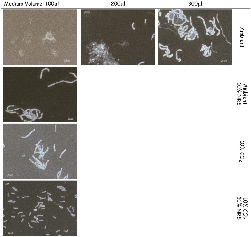
The basic medium used to test capsule and toxin induction was high glucose DMEM supplanted with glutamine, pyruvate and non-essential amino acids, hence sDMEM. We first tested the effect of growth media volume on capsule production in a 96-well tissue culture microplate format. This was done to allow higher-throughput testing of growth conditions and genetic manipulations. We inoculated B. anthracis Vollum spores (5x10⁵ CFU/ml) into 100μl, 200μl and 300μl sDMEM at 37°C in ambient atmosphere for 24h. No capsule production could be detected in 100μl sDMEM (Fig 1). However, increasing the culture volume resulted in partial (200μl) or full (300μl) capsule production (Fig 1). These findings could be possibly explained by higher CO2 concentrations reached in the well when bacteria are grown in larger volumes, due to a lower surface area to volume ratio, limiting gas exchange. This is coupled with a larger volume of multiplying and respiring bacteria, further increasing CO2 concentrations. This hypothesis is supported by the finding that growth in 10% CO2 atmosphere does induce capsule production in 100μl sDMEM (Fig 1). Having established non-capsule inducing growth conditions for our experiments, we next tested the effect of adding normal rabbit serum (NRS) to growth media. NRS was shown to induce capsule production, regardless of CO2 enrichment in the incubator’s atmosphere, with all the bacteria grown with NRS encapsulated in ambient or 10% CO2 atmosphere (Fig 1).
Fig 1. The effect of growth conditions on capsule induction by B. anthracis Vollum.
Spores were seeded into 100μl, 200μl or 300μl of sDMEM and incubated at 37°C in an ambient atmosphere (upper panel) for 24h. To examine the effect of serum or CO2 on capsule production, spores were seeded into 100μl sDMEM or sDMED-NRS (supplemented with 10% NRS). Samples were incubated at 37°C in under ambient or 10% CO2 atmosphere (see right hand side legend) for 24h. Capsule was imaged by India ink negative staining (capsule presence forms a typical bright outer layer).
The role of atxA on capsule production and toxin secretion under different growth conditions
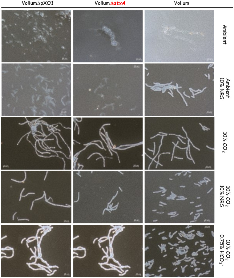
To determine the role of atxA on capsule and toxin production, we compared capsule production of the wild type Vollum strain with the ΔpXO1 or ΔatxA mutants (Table 1) following growth in sDMEM under the different growth conditions (NRS/CO2 presence). None of the strains produced capsule under ambient atmosphere in un-supplemented sDMEM (Fig 2). Addition of 10% NRS, under ambient atmosphere, induced capsule production by the wild type Vollum strain but not by the atxA null mutants (both ΔpXO1 or ΔatxA, Fig 2). Under 10% CO2, all three strains produced capsule. We therefore conclude that the response to NRS is atxA dependent, as lacking either the atxA gene or the entire pXO1 plasmid abrogates this response (Fig 2). These results also demonstrate the presence of another, atxA independent mechanism, responding to CO2. However, the effect of NRS in the presence of CO2 on capsule production under these conditions could not be resolved.
Fig 2. Capsule production of the Vollum wild type, ΔpXO1 or ΔatXA mutants under different growth conditions.
Spores of the wild type and different mutants (top panel) were seeded into 100μl of sDMEM. The same strains were grown in sDMEM supplemented with 10% NRS (second row). The same conditions or supplemented with 0.75% HCO3- were applied in the presence of 10% CO2 (lower three rows) for 24h. Capsule was imaged by India ink negative staining (capsule presence forms a typical bright outer layer).
As atxA, the major virulence regulator, regulates toxin expression (LF, EF and PA), we tested their secretion into growth medium after growth, both for the Vollum and the VollumΔatxA mutant (the ΔpXO1 mutant lacks the entire set of toxin genes and is therefore irrelevant for such a test). Toxins secretion was determined by ELISA for the most abundant component, PA. For the VollumΔatxA mutant, PA was undetectable, both under 10% CO2, with 10% NRS and in the presence of both. These results confirm the role of atxA in toxin expression regulation (Table 2). For the wild type Vollum, no toxins were induced in sDMEM alone coupled with under ambient atmosphere. The addition of 10% NRS alone induced high PA expression. sDMEM without NRS under 10% CO2 again did not induce PA expression (Table 2). No PA secretion could be detected in the atxA null mutant under any of the tested growth conditions. Our finding that 10% CO2 atmosphere in itself does not activate atxA-dependent toxin expression is surprising, since it was repeatedly demonstrated such activation is achievable by HCO3- addition. We therefore tested both capsule production and toxin expression in sDMEM supplemented with 0.75% HCO3- under 10% CO2 atmosphere. These atmospheric conditions were chosen since HCO3- in aqueous media is in equilibrium with CO2 and H2O, dramatically increasing the levels of the soluble CO2 in the medium even under ambient atmosphere, and we sought to expose our experimental controls to conditions as similar as possible.
Table 2. Protective antigen secretion (μg/ml) of Vollum or VollumΔatxA under different growth conditions.
| VollumΔatxA | Vollum | Atmosphere | Supplements | |
|---|---|---|---|---|
| PA μg/ml | - | - | Ambient | - |
| - | +++ | Ambient | 10% NRS | |
| - | - | 10% CO2 | - | |
| - | +++ | 10% CO2 | 10% NRS | |
| - | +++ | 10% CO2 | 0.75% HCO3- |
- <0.1, + 0.1–0.5, ++ 0.5–1.5, +++ 1.5–10
According to our previous findings, capsule production is CO2 dependent. Therefore, we found that the above tested strains were encapsulated under the conditions applied (Fig 2). PA secretion was tested only for Vollum and VollumΔatxA. Supplementing the sDMEM with 0.75% HCO3- induced PA secretion by the Vollum strain but not by the VollumΔatxA mutant. This indicates that HCO3- induces toxin secretion in an atxA dependent manner (Table 2). These findings were validated by using a previously reported VollumΔpXO2 chimera in which we substituted the genomic bclA gene (which is an exosporium glycoprotein non-essential for spore formation or virulence) with a CAP operon altered to be regulated by the PAprom (Table 1, S1 Appendix).
The effect of short incubations on capsule and toxins production in response the different growth conditions
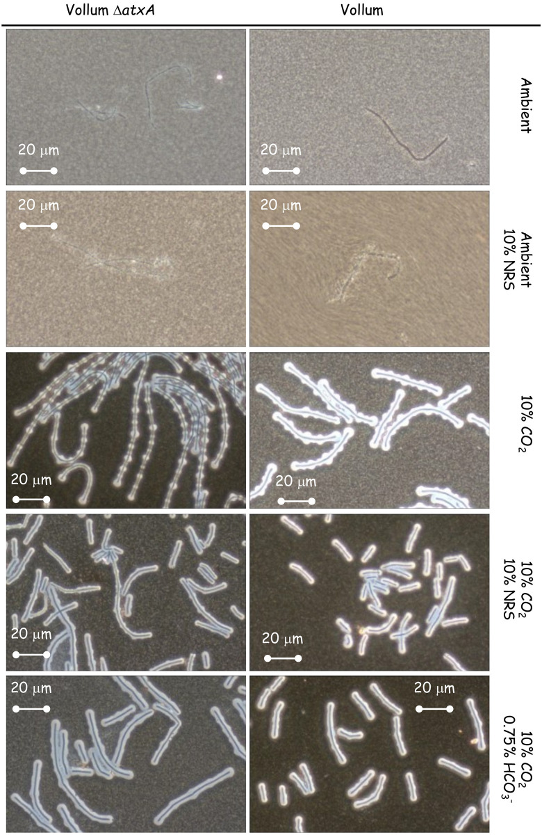
Aerobic growth affects different parameters of the liquid medium such as pH and O2/CO2 concentrations, especially when the bacteria reach high concentration (CFU/ml). As was shown previously [26], capsule production and toxin secretion can be detected as early as 2-5h of growth in sDMEM under 10% CO2 atmosphere. To minimize changes in media conditions resulting from bacterial growth, we examined capsule production and PA secretion after 5h growth in different growth conditions. Vollum growth under ambient atmosphere did not result in any capsule accumulation or toxin secretion following 24h incubation (Fig 2, Table 2). This was true also for a short (5h) incubation (Fig 3, Table 3). Supplementing the media with 10% NRS induced capsule production and PA secretion following 24h incubation under ambient atmosphere (Fig 2, Table 2). A shorter (5h) incubation induced measurable PA secretion (Table 3) but little or no capsule production (Fig 3). This PA secretion is AtxA dependent, as deletion of the atxA gene resulted in no PA accumulation following 5h (Table 3) or 24h incubation (Table 2). Incubating the bacteria in 10% CO2 atmosphere for 5h resulted in capsule production (Fig 3), similarly to that observed following a 24h incubation (Figs 1 and 2). This capsule accumulation was AtxA independent and was not significantly affected by the addition of 10% NRS or HCO3- (Fig 3). PA secretion was not induced by 10% CO2 in itself, but required addition of NRS or HCO3- (Table 3), however unlike the 24h incubation (Table 2), the amount of PA secreted in response to HCO3- was significantly less (~10%) than of that induced by NRS (Table 3). Under all conditions the PA secretion was AtxA dependent.
Fig 3. Effect of deleting atxA on capsule production after a short (5h) incubation under different growth conditions.
Spores of the wild type and ΔatxA mutant (top panel) were seeded into 100μl of sDMEM as is or supplemented with 10% NRS and incubated at 37°C in an ambient or 10% CO2 atmosphere (as indicated on the right) for 24h. Capsule was imaged by India ink negative staining (capsule presence forms a typical bright outer layer).
Table 3. PA secretion (μg/ml) by Vollum and VollumΔatxA following 5h growth in different conditions.
| VollumΔatxA | Vollum | Atmosphere | Supplements | |
|---|---|---|---|---|
| PA μg/ml | - | - | Ambient | - |
| - | +++ | Ambient | 10% NRS | |
| - | - | 10% CO2 | - | |
| / | +++ | 10% CO2 | 10% NRS | |
| / | + | 10% CO2 | 0.75% HCO3- |
- <0.1, + 0.1–0.5, ++ 0.5–1.5, +++ 1.5–10
Role of acpA and acpB in response to different growth conditions
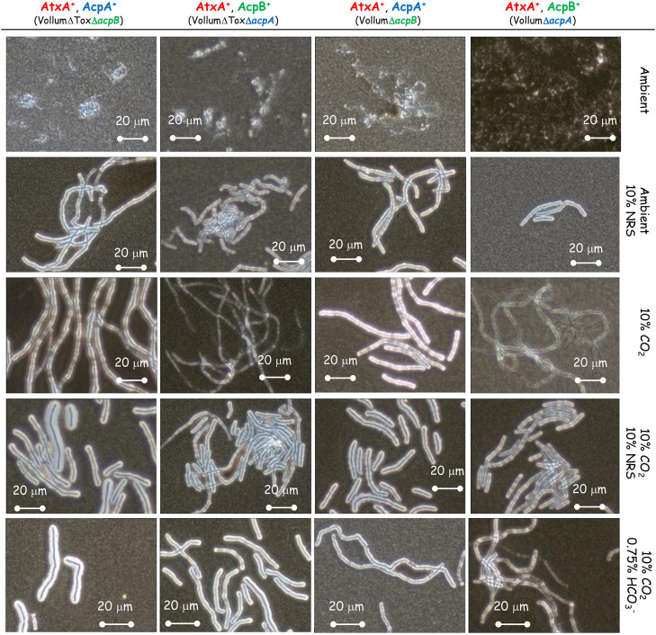
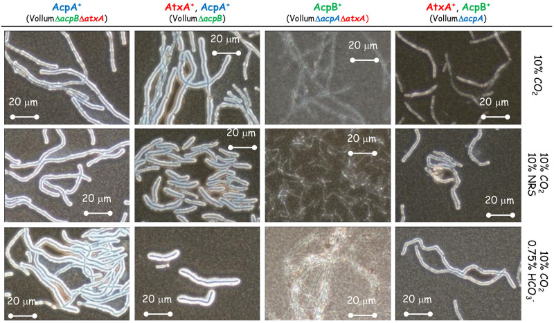
Toxin production is induced in an atxA dependent manner in response to HCO3- or NRS, while capsule production is also induced by a CO2 enriched (10%) atmosphere. Capsule production is known to be regulated by two regulatory proteins, AcpA and AcpB. We therefore tested the effect of deleting each of these genes on capsule production in response to different growth conditions. The complete coding region of acpA or acpB was deleted independently in the background of wild type Vollum or the toxin deficient mutant—VollumΔTox (VollumΔpagΔcyaΔlef Table 1). As was previously shown for the wild type Vollum strain (Fig 2), none of these mutants produced capsule following growth in sDMEM under ambient atmosphere (Fig 4). The presence of either acpA or acpB is sufficient for capsule production in sDMEM supplemented with 10% NRS, regardless to the presence or absence of 10% CO2 atmosphere (Fig 4). However, in the absence of NRS, only AcpA expressing mutants (lacking acpB) produce significant capsule when grown in 10% CO2 atmosphere. Adding 0.75% HCO3- to sDMEM induced capsule production in the presence of either acpA or acpB. Mutants lacking acpA (expressing only acpB), did not produce significant capsule in 10% CO2 atmosphere. To examine the role of atxA in these processes, we deleted the atxA gene in the background of our VollumΔToxΔacpA or ΔacpB mutants. Deleting atxA in the VollumΔToxΔacpA, expressing acpB, abolished capsule production under all tested conditions (Fig 5). However, deleting atxA in the VollumΔToxΔacpB, expressing acpA, did not affect capsule accumulation, compared to the AtxA expressing mutant. This finding indicates that acpA operates in an AtxA independent manner (Fig 5).
Fig 4. The effect of absence of acpA or acpB on capsule production in response to 10% NRS under ambient or 10% CO2 atmosphere.
Spores of the ΔacpA or acpB mutants (top panel) were seeded into 100μl of sDMEM as is or supplemented with0.75% HCO3- or 10% NRS and incubated at 37°C in an ambient or 10% CO2 atmosphere (as indicated on the right) for 24h. Capsule was imaged by India ink negative staining (capsule presence forms a typical bright outer layer).
Fig 5. Effect of AtxA on capsule production in the presence of either AcpA or AcpB.
Spores of the different mutants (top panel) were seeded into 100μl of sDMEM as is or supplemented with 0.75% HCO3- or 10% NRS and incubated at 37°C in 10% CO2 atmosphere (as indicated on the right) for 24h. Capsule was imaged by India ink negative staining (capsule presence forms a typical bright outer layer).
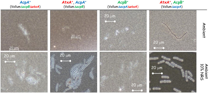
As we demonstrated (Fig 2), capsule production could be induced in ambient atmosphere by adding 10% NRS to sDMEM. This induction is AtxA dependent, since no capsule production was detected in the VollumΔatxA mutant under these conditions (Fig 2). Since AcpA dependent capsule production in 10% CO2 atmosphere was AtxA independent, we tested the role of AtxA on AcpA dependent capsule production in response to 10% NRS in ambient atmosphere. As 10% NRS induced capsule production of VollumΔacpB under ambient atmosphere (Figs 4 and 6) we tested the effect of atxA deletion on capsule production under these conditions. Unlike the CO2 induction, under ambient atmosphere, AcpA dependent capsule production in response to NRS is AtxA dependent (Fig 6).
Fig 6. Effect of AtxA on capsule production in response to 10% NRS in ambient atmosphere.
Spores of the different mutants (top panel) were seeded into 100μl of sDMEM as is or supplemented with 10% NRS and incubated at 37°C in ambient atmosphere (as indicated on the right) for 24h. Capsule was imaged by India ink negative staining (capsule presence forms a typical bright outer layer).
Gas content of the supplemented and basic DMEM media under the different growth conditions
Gas content was determined by Abbott iSTAT blood gas analyzer. The different media were incubated at 37°C in ambient or 10% CO2 atmosphere for 4 h and analyzed using the EC8+ cartridge. The results shown in Table 4 indicate that the main parameter that is affected by the 10% CO2 is the soluble CO2 (PCO2) rather than HCO3-. Unlike the HCO3- levels that are higher under all growth conditions from those considered normal for human blood, the PCO2 levels in the presence of 10%CO2 are well within or very close to normal values.
Table 4. Gas analysis of the sDMEM media under the different growth conditions.
| 10% CO2 | - | + | + | - | + | Normal arterial | Normal venous |
|---|---|---|---|---|---|---|---|
| Supplement | - | - | 10% FCS | 10% FCS | 0.75% HCO3 1:1* | ||
| pH | 8.0 | 7.691 | 7.447 | 8.149 | 7.972 | 7.35–7.45 | 7.31–7.41 |
| PCO2 (mmHg) | 18.4 | 34.9 | 58.1 | 14.2 | 34.3 | 35–45 | 41–51 |
| PO2 (mmHg) | 230 | 148 | 126 | 173 | 183 | 80–100 | 35–40 |
| HCO3 (mmol/L) | 48.7 | 42.2 | 40.1 | 49.5 | 79.2 | 22–26 | 22–26 |
| sO2 (%) | 100 | 100 | 99 | 100 | 100 | 95–100 | / |
*at the end of the 4h incubation the media was diluted 1:1 in sDMEM prior to the analysis since the undiluted media was off scale.
Discussion
Successful invasion requires the pathogen regulate its virulence factors in a way that will maximize their effect on host defense mechanisms. The trigger for such activation is usually host derived. This can be biological (such as proteins) of physical (pH or temperature for example). B. anthracis naturally infects humans following spore inhalation, contact with broken skin or ingestion of undercook contaminated meat. These routes present different environmental conditions [2]. It was previously demonstrated that toxin secretion and capsule production could be induced by growing the bacteria in culture media supplemented with HCO3- or serum (10–50%) in a CO2 enriched (5–15%) atmosphere [5, 16, 18, 19, 27–29]. HCO3- /CO2 condition were commonly used to study atxA, acpA and acpB regulation and their effect on toxin and capsule biosynthetic genes [16, 18, 30]. Since these conditions always included these two components, it was concluded that atxA was induced in response to CO2, regulating the induction of acpA and acpB. Although in some reports, capsule production was shown to be AtxA dependent [16, 31], the fact that ΔpXO1 variants are encapsulated contradicts this finding, alluding to additional, AtxA independent regulation of the process [19, 27]. The use of sDMEM as growth media enabled the examination of the effect of CO2, HCO3- and serum on these processes.
The parameter of soluble CO2 is influenced by multiple parameters, such as surface area to volume ratio and aerobic bacterial growth. Therefore, normal growth conditions were set to 100μl media/well (96 well tissue culture plate) for 24h at 37°C under ambient atmosphere (Fig 1). This baseline enabled testing the effect of different supplements and/or growth conditions on capsule (Fig 1) or toxin (Fig 2, Table 2) induction. Capsule production is induced by the addition of 10% NRS or growth under a 10% CO2 atmosphere (Fig 1). The serum capsule induction (under ambient atmosphere) is atxA dependent (Fig 2), since there was no significant capsule production in mutants that do not express AtxA (VollumΔatxA and VollumΔpXO1). Alternatively, capsule production in response to CO2 enriched atmosphere is AtxA independent, as there is no significant difference in capsule production, under these conditions, between AtxA expressing and atxA null mutants (Fig 2). Toxin secretion, as determined by measuring PA media concentrations, is serum dependent (Table 2), as PA could be detected only in NRS supplemented sDMEM regardless of CO2 enrichment. Adding HCO3- induced toxin secretion in an atxA dependent manner, similarly to serum (Fig 2, Table 2), with PA expressed by the wild type Vollum and not by the atxA null mutant.
We also found differences in the speed B. anthracis reacts to the different stimuli. This was done by examining toxin secretion and capsule production after a short incubation (5h). HCO3- was not as robust as NRS in inducing toxin secretion. Examining PA concentrations after 5h incubation in sDMEM supplemented with 0.75% HCO3- revealed about 1/10 of the concentration compared to that measured after 24h incubation (Fig 5, Table 4). However, NRS induction yielded similar concentrations at these two timepoints. Testing the effect of NRS on capsule production, reveals that 5h incubation under ambient atmosphere, inducing significant PA secretion, does not result in significant capsule production. Growth under a 10% CO2 atmosphere induced capsule production at 5h even in the absence of supplemented NRS or HCO3- (Table 4). Hence, Serum seemed more effective then HCO3- in inducing toxin secretion, with two process being AtxA dependent. The AtxA independent induction of capsule production by 10% CO2 appeared more effective then AtxA dependent serum induction (Fig 3).
Two major regulators; AcpA and AcpB control capsule biosynthesis by promoting transcription of acpB,C,A,D,E operon. acpA was shown to be regulated by AtxA (activated by NRS), while also depicting an additional atxA independent activation capacity by CO2 and HCO3-). This independent activation could be direct or mediated by an as yet unknown, additional promotor, in turn responding to a CO2 ingredient. We found that deleting acpA causes the bacteria to produce significantly less capsule in response to CO2, while maintaining its ability to respond to NRS or HCO3- (Fig 4). Deletion of acpB did not have any effect on capsule production under all tested conditions (Fig 4), supporting our previous in vivo data, which showed no effect on virulence [17]. A double deletion of atxA and acpA or acpB revealed that AcpB activity is strictly AtxA dependent under all the conditions tested (Fig 5). AcpA activity is not affected by the absence of atxA in the presence of CO2 (Fig 5) but is nulled in response to NRS under ambient atmosphere (Fig 6).
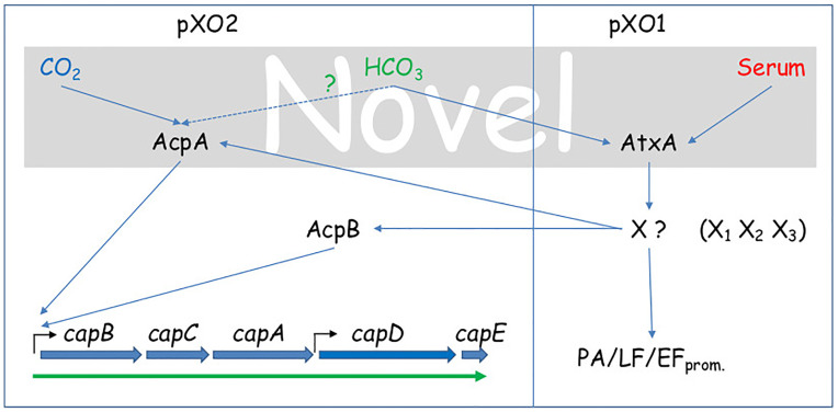
Our findings delineate the following regulation cascade; CO2 induces capsule production by activating of acpA in an AtxA independent manner. Serum activates the AtxA dependent cascade, inducing toxin secretion and eventually capsule production, by activating acpA and acpB (Fig 7). The order of the processes can be deduced from the lack of capsule production following 5h growth in NRS supplemented sDMEM under ambient atmosphere (Fig 3). HCO3- induces toxin secretion through the AtxA cascade, but in a less efficient manner (compared to NRS, Table 3). Direct activation of capsule production by HCO3- in an AtxA independent manner could not be eliminated, since even under ambient atmosphere, it modifies the levels of soluble CO2 (PCO2) and possibly induces capsule production via AcpA (Table 4). In terms of the initial infection stages of pathology, this differential regulation of toxins and capsule has a logical role. Inhalational and cutaneous infections involve spore phagocytosis by local innate immune cells and their migration to a draining lymph node. While in route (and within the phagocytic cell), the spore needs to germinate and produce the protective capsule. During this stage, toxin production could prove counterproductive, as it disrupts normal cellular function, possibly arresting the cell and preventing it from reaching the lymph node. Once there, toxin production is desirable, possibly enhancing bacterial release from the cells in the lymph node, to the blood stream, promoting pathogenesis. These results are supported by the transcription analysis of B. anthracis Sterne strain (pXO1+ pXO2-) in RAW 264.7 cells [32]. This analysis indicated that significant transcription induction of toxin genes was observed only in the late stage of infection in parallel to bacterial growth. The pathway sensing serum and CO2 remains to be elucidated and requires more research. Such a pathway may prove common to other pathogens as well as possibly providing additional therapeutic targets for intervention.
Fig 7. Proposed regulatory scheme for CO2, Serum and HCO3- regulation of capsule production and toxin secretion.
The three environmental signals that were studied are color coded; CO2 in blue, HCO3- in green and serum in red. The gray background indicates the novel part of the regulation pathway proposed based on this work.
Supporting information
(DOCX)
Data Availability
All relevant data are within the paper and its Supporting information files.
Funding Statement
The author(s) received no specific funding for this work.
References
- 1.Dixon TC, Meselson M, Guillemin J, Hanna PC. Anthrax. New England Journal of Medicine. 1999;341(11):815–26. doi: 10.1056/NEJM199909093411107 . [DOI] [PubMed] [Google Scholar]
- 2.Hanna P. Anthrax pathogenesis and host response. Curr Top Microbiol Immunol. 1998;225:13–35. Epub 1997/12/05. doi: 10.1007/978-3-642-80451-9_2 . [DOI] [PubMed] [Google Scholar]
- 3.Swartz MN. Recognition and management of anthrax—an update. N Engl J Med. 2001;345(22):1621–6. Epub 2001/11/13. doi: 10.1056/NEJMra012892 . [DOI] [PubMed] [Google Scholar]
- 4.Candela T, Fouet A. Bacillus anthracis CapD, belonging to the gamma-glutamyltranspeptidase family, is required for the covalent anchoring of capsule to peptidoglycan. Mol Microbiol. 2005;57(3):717–26. Epub 2005/07/28. doi: 10.1111/j.1365-2958.2005.04718.x . [DOI] [PubMed] [Google Scholar]
- 5.Candela T, Mock M, Fouet A. CapE, a 47-amino-acid peptide, is necessary for Bacillus anthracis polyglutamate capsule synthesis. J Bacteriol. 2005;187(22):7765–72. Epub 2005/11/04. doi: 10.1128/JB.187.22.7765-7772.2005 . [DOI] [PMC free article] [PubMed] [Google Scholar]
- 6.Heine HS, Shadomy SV, Boyer AE, Chuvala L, Riggins R, Kesterson A, et al. Evaluation of Combination Drug Therapy for Treatment of Antibiotic-Resistant Inhalation Anthrax in a Murine Model. Antimicrob Agents Chemother. 2017;61(9). Epub 2017/07/12. doi: 10.1128/AAC.00788-17 . [DOI] [PMC free article] [PubMed] [Google Scholar]
- 7.Candela T, Fouet A. Poly-gamma-glutamate in bacteria. Molecular Microbiology. 2006;60(5):1091–8. doi: 10.1111/j.1365-2958.2006.05179.x [DOI] [PubMed] [Google Scholar]
- 8.Wu R, Richter S, Zhang RG, Anderson VJ, Missiakas D, Joachimiak A. Crystal structure of Bacillus anthracis transpeptidase enzyme CapD. J Biol Chem. 2009;284(36):24406–14. Epub 2009/06/19. doi: 10.1074/jbc.M109.019034 . [DOI] [PMC free article] [PubMed] [Google Scholar]
- 9.Richter S, Anderson VJ, Garufi G, Lu L, Budzik JM, Joachimiak A, et al. Capsule anchoring in Bacillus anthracis occurs by a transpeptidation reaction that is inhibited by capsidin. Mol Microbiol. 2009;71(2):404–20. Epub 2008/11/20. doi: 10.1111/j.1365-2958.2008.06533.x . [DOI] [PMC free article] [PubMed] [Google Scholar]
- 10.Klimpel KR, Arora N, Leppla SH. Anthrax toxin lethal factor contains a zinc metalloprotease consensus sequence which is required for lethal toxin activity. Mol Microbiol. 1994;13(6):1093–100. Epub 1994/09/01. doi: 10.1111/j.1365-2958.1994.tb00500.x . [DOI] [PubMed] [Google Scholar]
- 11.Tang W-J, Guo Q. The adenylyl cyclase activity of anthrax edema factor. Molecular Aspects of Medicine. 2009;30(6):423–30. doi: 10.1016/j.mam.2009.06.001 [DOI] [PMC free article] [PubMed] [Google Scholar]
- 12.Liu S, Moayeri M, Leppla SH. Anthrax lethal and edema toxins in anthrax pathogenesis. Trends Microbiol. 2014;22(6):317–25. Epub 2014/04/02. doi: 10.1016/j.tim.2014.02.012 . [DOI] [PMC free article] [PubMed] [Google Scholar]
- 13.Ebrahimi CM, Kern JW, Sheen TR, Ebrahimi-Fardooee MA, van Sorge NM, Schneewind O, et al. Penetration of the blood-brain barrier by Bacillus anthracis requires the pXO1-encoded BslA protein. J Bacteriol. 2009;191(23):7165–73. Epub 2009/10/13. doi: 10.1128/JB.00903-09 . [DOI] [PMC free article] [PubMed] [Google Scholar]
- 14.Uchida I, Hornung JM, Thorne CB, Klimpel KR, Leppla SH. Cloning and characterization of a gene whose product is a trans-activator of anthrax toxin synthesis. J Bacteriol. 1993;175(17):5329–38. Epub 1993/09/01. doi: 10.1128/jb.175.17.5329-5338.1993 [DOI] [PMC free article] [PubMed] [Google Scholar]
- 15.Dai Z, Sirard JC, Mock M, Koehler TM. The atxA gene product activates transcription of the anthrax toxin genes and is essential for virulence. Mol Microbiol. 1995;16(6):1171–81. Epub 1995/06/01. doi: 10.1111/j.1365-2958.1995.tb02340.x [DOI] [PubMed] [Google Scholar]
- 16.Drysdale M, Bourgogne A, Hilsenbeck SG, Koehler TM. atxA controls Bacillus anthracis capsule synthesis via acpA and a newly discovered regulator, acpB. J Bacteriol. 2004;186(2):307–15. Epub 2004/01/02. doi: 10.1128/JB.186.2.307-315.2004 . [DOI] [PMC free article] [PubMed] [Google Scholar]
- 17.Sittner A, Bar-David E, Glinert I, Ben-Shmuel A, Schlomovitz J, Levy H, et al. Role of acpA and acpB in Bacillus anthracis capsule accumulation and toxin independent pathogenicity in rabbits. Microb Pathog. 2021;155:104904. Epub 2021/05/01. doi: 10.1016/j.micpath.2021.104904 . [DOI] [PubMed] [Google Scholar]
- 18.Fouet A. AtxA, a Bacillus anthracis global virulence regulator. Res Microbiol. 2010;161(9):735–42. Epub 2010/09/25. doi: 10.1016/j.resmic.2010.09.006 . [DOI] [PubMed] [Google Scholar]
- 19.Levy H, Glinert I, Weiss S, Sittner A, Schlomovitz J, Altboum Z, et al. Toxin-independent virulence of Bacillus anthracis in rabbits. PLoS One. 2014;9(1):e84947. Epub 2014/01/15. doi: 10.1371/journal.pone.0084947 . [DOI] [PMC free article] [PubMed] [Google Scholar]
- 20.Fouet A, Mock M. Differential influence of the two Bacillus anthracis plasmids on regulation of virulence gene expression. Infect Immun. 1996;64(12):4928–32. Epub 1996/12/01. doi: 10.1128/iai.64.12.4928-4932.1996 . [DOI] [PMC free article] [PubMed] [Google Scholar]
- 21.Bertin M, Chateau A, Fouet A. Full expression of Bacillus anthracis toxin gene in the presence of bicarbonate requires a 2.7-kb-long atxA mRNA that contains a terminator structure. Res Microbiol. 2010;161(4):249–59. Epub 2010/04/03. doi: 10.1016/j.resmic.2010.03.003 . [DOI] [PubMed] [Google Scholar]
- 22.Vietri NJ, Marrero R, Hoover TA, Welkos SL. Identification and characterization of a trans-activator involved in the regulation of encapsulation by Bacillus anthracis. Gene. 1995;152(1):1–9. Epub 1995/01/11. doi: 10.1016/0378-1119(94)00662-c . [DOI] [PubMed] [Google Scholar]
- 23.Levy H, Weiss S, Altboum Z, Schlomovitz J, Glinert I, Sittner A, et al. Differential Contribution of Bacillus anthracis Toxins to Pathogenicity in Two Animal Models. Infect Immun. 2012;80(8):2623–31. Epub 2012/05/16. doi: 10.1128/IAI.00244-12 . [DOI] [PMC free article] [PubMed] [Google Scholar]
- 24.Levy H, Fisher M, Ariel N, Altboum Z, Kobiler D. Identification of strain specific markers in Bacillus anthracis by random amplification of polymorphic DNA. FEMS Microbiol Lett. 2005;244(1):199–205. doi: 10.1016/j.femsle.2005.01.039 . [DOI] [PubMed] [Google Scholar]
- 25.Levy H, Weiss S, Altboum Z, Schlomovitz J, Rothschild N, Glinert I, et al. The effect of deletion of the edema factor on Bacillus anthracis pathogenicity in guinea pigs and rabbits. Microb Pathog. 2012;52(1):55–60. Epub 2011/10/25. doi: 10.1016/j.micpath.2011.10.002 . [DOI] [PubMed] [Google Scholar]
- 26.Makdasi E, Laskar O, Glinert I, Alcalay R, Mechaly A, Levy H. Rapid and Sensitive Multiplex Assay for the Detection of B. anthracis Spores from Environmental Samples. Pathogens. 2020;9(3). Epub 2020/03/04. doi: 10.3390/pathogens9030164 . [DOI] [PMC free article] [PubMed] [Google Scholar]
- 27.Welkos SL. Plasmid-associated virulence factors of non-toxigenic (pX01-) Bacillus anthracis. Microb Pathog. 1991;10(3):183–98. Epub 1991/03/01. doi: 10.1016/0882-4010(91)90053-d . [DOI] [PubMed] [Google Scholar]
- 28.Chiang C, Bongiorni C, Perego M. Glucose-dependent activation of Bacillus anthracis toxin gene expression and virulence requires the carbon catabolite protein CcpA. J Bacteriol. 2011;193(1):52–62. Epub 2010/10/26. doi: 10.1128/JB.01656-09 . [DOI] [PMC free article] [PubMed] [Google Scholar]
- 29.McCall RM, Sievers ME, Fattah R, Ghirlando R, Pomerantsev AP, Leppla SH. Bacillus anthracis Virulence Regulator AtxA Binds Specifically to the pagA Promoter Region. J Bacteriol. 2019;201(23). Epub 2019/10/02. doi: 10.1128/JB.00569-19 . [DOI] [PMC free article] [PubMed] [Google Scholar]
- 30.Guignot J, Mock M, Fouet A. AtxA activates the transcription of genes harbored by both Bacillus anthracis virulence plasmids. FEMS Microbiol Lett. 1997;147(2):203–7. Epub 1997/02/15. doi: 10.1111/j.1574-6968.1997.tb10242.x [DOI] [PubMed] [Google Scholar]
- 31.Drysdale M, Bourgogne A, Koehler TM. Transcriptional analysis of the Bacillus anthracis capsule regulators. J Bacteriol. 2005;187(15):5108–14. Epub 2005/07/21. doi: 10.1128/JB.187.15.5108-5114.2005 . [DOI] [PMC free article] [PubMed] [Google Scholar]
- 32.Bergman NH, Anderson EC, Swenson EE, Janes BK, Fisher N, Niemeyer MM, et al. Transcriptional profiling of Bacillus anthracis during infection of host macrophages. Infect Immun. 2007;75(7):3434–44. Epub 2007/05/02. doi: 10.1128/IAI.01345-06 . [DOI] [PMC free article] [PubMed] [Google Scholar]
Associated Data
This section collects any data citations, data availability statements, or supplementary materials included in this article.
Supplementary Materials
(DOCX)
Data Availability Statement
All relevant data are within the paper and its Supporting information files.


