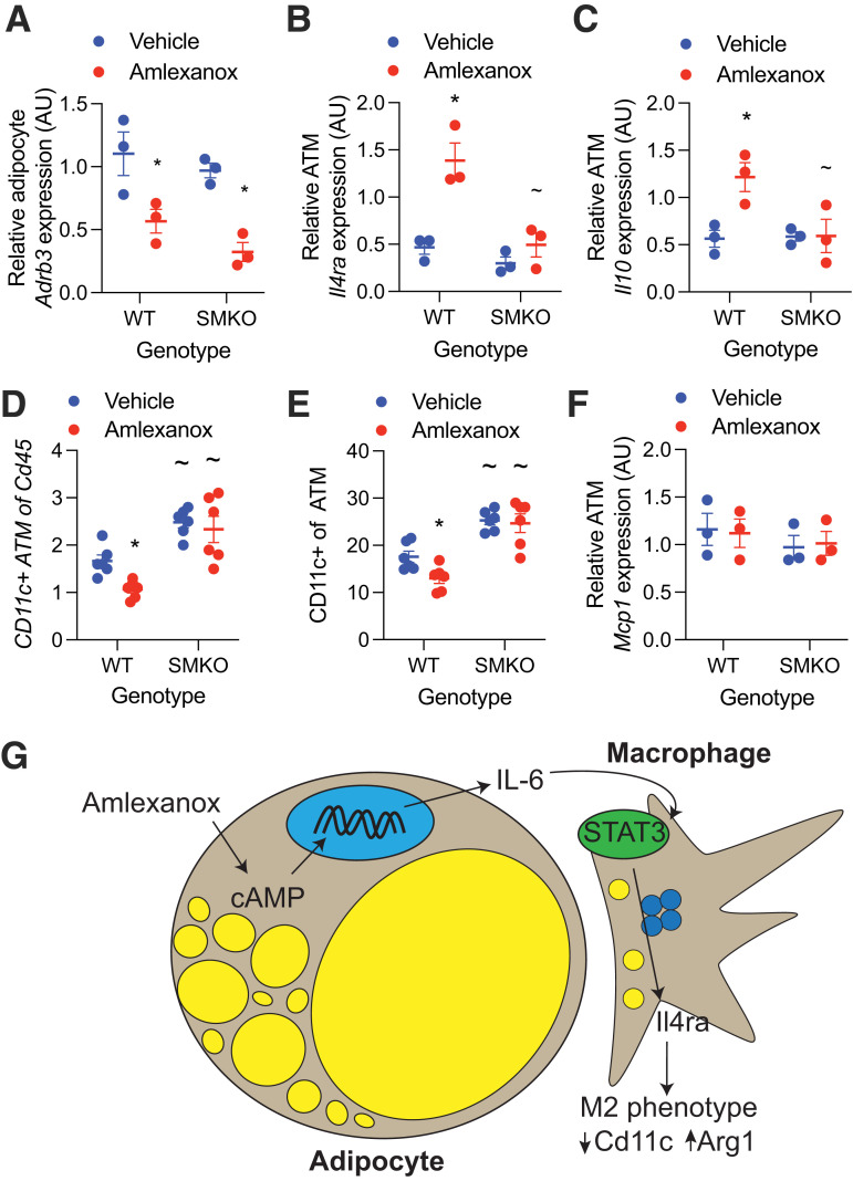
Figure 4.
In vivo sensitization of macrophages to IL-4 by amlexanox requires STAT3. A–C: Obese male mice aged 20–24 weeks were treated with 25 mg/kg amlexanox or vehicle control. After 4 h, the eWAT was collected and digested with collagenase (n = 3 per group). A: Quantitative PCR analysis of Adrb3 expression in mature adipocytes from epididymal fat. B and C: Quantitative PCR analysis of gene expression in epididymal ATMs (Cd45+, F4/80+, Cd11b+, and Cd3−) isolated by FACS from SVCs. D and E: FACS analysis of SVCs isolated from the epididymal fat of obese male mice aged 20–24 weeks treated with 25 mg/kg amlexanox or vehicle control for 52 h (n = 6 per group). Macrophages defined as CD45+, F4/80+, CD11b+, and Cd3− cells. D: CD11c+ macrophages as a percentage of CD45+ cells. E: CD11c+ macrophages as a percentage of total macrophages. F: Quantitative PCR analysis of Mcp1 expression in mature adipocytes from epididymal fat (n = 3 per group). G: Schematic of oral amlexanox treatment and activation of this adipocyte-to-macrophage communication axis. Statistical significance determined by post hoc analysis after significant two-way ANOVA. *P < 0.05 vehicle vs. amlexanox within genotype; ∼P < 0.05 WT vs. KO within treatment group. AU, arbitrary unit.

