



Abstract
Background
Despite an increasing number of patients with congenital heart disease (CHD) reaching reproductive age, the fertility of these patients remains undescribed. Therefore, the aim of the study was to evaluate the fertility in men and women with CHD by estimating the risk of infertility and comparing the birth rates, proportions of individuals becoming parents or remaining childless, and the number of children per parent with unaffected individuals.
Methods and Results
The study population consisted of individuals born between 1977 and 2000. Information on CHD, infertility, and live born children were obtained from the Danish health registries. Hazard ratios for infertility were analyzed using a Cox regression model. Differences of proportions and birth rates were calculated and compared between groups. Among 1 385 895 individuals, a total of 8679 (0.6%) were diagnosed with CHD. Men and women with simple or moderate CHD had no increased risk of infertility when compared with the reference population. Estimates for complex CHD groups were too imprecise for evaluation. Individuals with CHD were more often childless with consequently lower birth rates compared with unaffected individuals. However, those becoming parents had the same number of children as the reference population.
Conclusions
Men and women with simple or moderate CHD had the same risk of infertility as the reference population. Despite patients with CHD more often being childless, those becoming parents had the same number of children as parents without CHD. The current findings increase the knowledge regarding fertility in the CHD population.
Keywords: congenital heart disease, fertility, infertility, live births, reproductive health
Subject Categories: Epidemiology
Nonstandard Abbreviations and Acronyms
- AMH
anti‐Müllerian hormone
- CRS
Civil Registration System
- DMBR
Danish Medical Birth Registry
- DNPR
Danish National Patient Register
Clinical Perspective.
What Is New?
With this nationwide cohort study, we are the first to evaluate the risk of being diagnosed with infertility in men and women with congenital heart disease compared with unaffected men and women.
In both sexes, we found no increased risk of infertility when comparing individuals with simple or moderate heart defects with unaffected individuals; complex heart defect groups diagnosed with infertility were too small for firm conclusions.
Regardless of defect severity, men and women with congenital heart disease were more often childless, but those who became parents had the same number of children as unaffected individuals.
What Are the Clinical Implications?
Our study contributes with reassuring information to patients with congenital heart disease of simple or moderate complexity who may have concerns about their fertility.
Today, most patients with congenital heart disease (CHD) reach childbearing age, and birth rates are consequently increasing in this patient group. 1 , 2 , 3 , 4 Despite infertility being recognized as a global public health issue by the World Health Organization, 5 , 6 it remains unknown whether infertility is more common in patients with CHD compared with unaffected individuals. Fertility is mentioned in some, but not all, international guidelines 7 , 8 , 9 that describe impaired fertility in women with Fontan circulation. In women with other cardiac defects, it is stated that their fertility is most likely comparable with the female background population; however, these claims are unreferenced. 7 Although several case studies report higher rates of menstrual abnormalities 10 , 11 , 12 , 13 , 14 , 15 and spontaneous abortions 16 , 17 , 18 , 19 , 20 , 21 , 22 , 23 , 24 , 25 , 26 in women with severe heart defects, less is known about women with more simple lesions. More than one‐third of 505 Dutch and Belgian women with a septal defect were found to be diagnosed with a menstrual cycle disorder. 11 Furthermore, we previously published a cohort study with the unexpected findings that women with atrial septal defects had a higher risk of receiving fertility treatment compared with a matched reference cohort, 26 suggesting that fertility may also be impaired in women with less severe heart defects.
Knowledge of fertility in men with CHD is nearly nonexistent. Only erectile dysfunction has been mentioned as an impaired outcome related to their reproductive health. 27 , 28 , 29 A compromised blood flow to the male genitalia, as well as hypoxemia, may potentially also affect the ongoing production of semen and, therefore, the ability to reproduce. Although factors other than fertility influence family size today, lower birth rates in men and women with CHD, in comparison with reference populations, may also point toward impaired fertility. 2 , 30 , 31 , 32 , 33 , 34
Despite indications of fertility issues and reports of fertility concerns among young adults, 27 , 35 , 36 , 37 our knowledge is limited. With this nationwide cohort study, we therefore aimed to evaluate the fertility in both men and women with CHD compared with the background population by first evaluating the risk of being diagnosed with infertility and, second, comparing the rates of live born children, the proportions of individuals becoming parents or remaining childless, and the average number of children per parent.
METHODS
Data Availability Statement
The data set underlying this article is not publicly available because of national data security legislation on sensitive personal data. L.F.U. had full access to the data set and takes responsibility for its integrity and the data analysis. Researchers may apply for access to data from Statistics Denmark. Further information is available at https://www.dst.dk/en/TilSalg/Forskningsservice.
Study Population
We identified all individuals born alive in Denmark between January 1, 1977, and December 31, 2000. Since 1968, all residents in Denmark have been given a personal identification number from the Danish Civil Registration System (CRS), enabling linkage between all national registries. 38 The identification number includes information on date of birth and sex. The CRS also contains information on vital status, date of death, emigration, and disappearance as well as personal identification numbers for spouse(s) and children.
A flowchart of the establishment of the study population is shown in the Figure. For the final population, we excluded individuals diagnosed with syndromes related to both CHD and potentially impaired reproductive health. We further excluded individuals living in Greenland (a part of the Kingdom of Denmark), as Greenlandic women are not registered in the Danish In Vitro Fertilisation (IVF) register used to obtain information on infertility. To ensure that the study population consisted of individuals at risk of infertility, we started follow‐up at the age of 18 years and excluded those with infertility, competing risks (procedure codes for sterilization, hysterectomy, or bilateral oophorectomy), emigration (living outside Denmark for >6 months), death, or disappearance before this age. A diagnosis of infertility before age 18 is considered a result from surgical procedures or cancer treatment rather than unsuccessful pregnancy attempts. The study population was then followed until first registration with infertility, sterilization (and hysterectomy and bilateral oophorectomy in women), emigration, death, disappearance, or censoring at the end of follow‐up, whichever event came first.
Figure 1. Flowchart of the establishment of the study population of individuals born between January 1, 1977, and December 31, 2000.
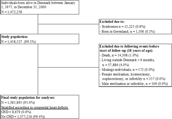
CHD indicates congenital heart disease.
Use of data from Statistics Denmark was approved by the Danish Data Protection Agency (approval No. P‐2020‐208). Ethical approval was waived because of the nature of the study with anonymized and untraceable data.
Assessment of Congenital Heart Defects
Identification of individuals with a congenital heart defect was possible using the DNPR (Danish National Patient Register), which contains information on all inpatient and outpatient contacts at all Danish hospitals. 39 From 1977 until the end of 1993, the International Classification of Diseases, Eighth Revision (ICD‐8) codes for CHD were used (746–747, except 746.7, 747.5–747.9 [not specific for CHD]), and thereafter the updated International Classification of Diseases, Tenth Revision (ICD‐10) was available (Q20–Q26, except for Q26.5 and Q26.6 [not specific to CHD]). Individuals with patent ductus arteriosus (ICD‐8 code 74709, ICD‐10 code DQ25.0) were included if born after gestational week 37. To increase the validity of the registered heart defect diagnosis, only main diagnoses given at 1 of the Danish university hospitals were accepted. If >1 heart defect was registered, the most severe defect was chosen. We studied CHD overall and subdivided it into simple, moderate, and complex CHD inspired by the guidelines from the European Society of Cardiology 8 (Table S1). Individuals who underwent correction of their heart defect were identified in the DNPR using ICD‐8 30 , 31 , 32 , 33 and ICD‐10 procedure codes related to surgical and catheter‐based repair.
Assessment of Infertility and Live Births
If pregnancy is not achieved after 1 year of unprotected, active sexual activity, the couple is per definition infertile 40 and can be referred for tax‐financed assessment and medically assisted reproduction treatment at a public fertility clinic in Denmark. 41 , 42 , 43 The only requirements are age <41 years in women and childlessness. In private fertility clinics, self‐paid treatment is offered for all women up to age 46 years regardless of parity. Today, around 50% of all treatments are performed at public clinics. In Denmark, lesbian and single women have free access to medically assisted reproduction treatment under the same requirements as heterosexual couples.
The DNPR and IVF register hold information on all individuals who are infertile and seek help in the Danish health care system. 39 , 44 Since 1994, registration of all fertility treatments (both public and private) in the IVF register is mandatory by law. The IVF register contains detailed information on each treated woman, including indication for treatment (male or female indication), type of treatment, pregnancy outcomes, and information on her partner, if any. Before 1994, information on individuals who were infertile was only registered in the DNPR. The DNPR contains information on both diagnosis codes for infertility and procedure codes for fertility treatment. In the present study, we classified men and women as infertile using 2 different definitions of infertility. In model 1, we ignored the indication for infertility diagnosis or treatment (male or female factor) as no causes are identified in 10% to 20% of couples who are infertile and 25% to 40% of cases are attributed to combined factors. 45 In model 2, we restricted the definition of infertility and considered only women as infertile if a female factor was registered as the indication and likewise with a male factor in men. Accordingly, women were classified as infertile in model 1 if they were registered in the IVF register or registered in the DNPR with 1 of the ICD‐10 diagnosis codes (N97 [female infertility], N97.0 [female infertility associated with anovulation], N97.1 [female infertility of tubal origin], N97.2 [female infertility of uterine origin], N97.3 [female infertility of cervical origin], N97.4 [female infertility associated with a male factor], N97.8 [female infertility of other origin], N97.8B [female infertility of ovarian origin], or N97.9 [female infertility of unspecified origin]) or registered with ICD‐10 procedure codes (BJFL0 [fertility treatment], BJFL00 [IVF treatment], BJFL01‐01B [micro‐insemination], BJFL04 [treatment with cryopreserved embryos], BJFL05 [treatment with donor egg], BJFL09 [fertility treatment, unspecified], or BJFL3 [insemination]). In model 2, women were classified as infertile if the following female factor‐specific codes were registered: N97.0, N97.1, N97.2, N97.3, N97.8B, or BJFL05. Single women (considered when ICD‐10 code Z60.2 “living alone” was registered without an infertility diagnosis) and women with a female partner were not classified as infertile in the present study.
Likewise, men were classified as infertile in model 1 if they were registered in the IVF register or registered in the DNPR with ICD‐10 diagnosis code N46 (male factor infertility) or if their wife was registered with the aforementioned DNPR diagnoses or procedures presented for model 1 in women. As women are the ones being treated and therefore more likely to be registered with infertility (regardless of indication), this increased the chance of identifying infertile couples erroneously not registered with male factor in the registries. Using information on marital status from the CRS register on all men in the study population, married couples were identified, allowing for identification of the female partner in the DNPR. In model 2, men were classified as infertile if they were registered with N46 (male factor infertility) or their wife was registered with N97.4 (female infertility associated with male factor) or if codes for treatment with donor semen were used (BJFL00A, BJFL03, BJFL31).
Using the DMBR (Danish Medical Birth Registry), we further identified all live born children born to men and women in the study population and obtained birth dates for each child.
Covariates
The year of birth of the study population was retrieved from the birthdate available from the CRS and categorized into 3 birth periods covering 1977 to 1984, 1985 to 1992, and 1993 to 2000. The 3 categories correspond to the age groups 18 to 25 years, 26 to 33 years, and 34 to 41 years. From the DMBR, information on the study participants' gestational age, birth weight, and their mother's age at birth were available. If gestational age was <22 weeks or >45 weeks, it was encoded as missing. Preterm birth was defined as birth before gestational week 37. Birth weight was converted into Z scores using reference material by Maršál et al 46 and Olsen et al. 47 This was done to account for differences in gestational ages between groups. Z scores express the number of SDs the birth weights deviate from the expected birth weight based on sex and gestational age. Information on socioeconomic status was obtained from Statistics Denmark. Based on level of income and employment, Danish individuals are annually categorized into a main source of income ranging from “owner of business” and “chiefs executive or employee with high income” to “employee with low income” and “student.” We retrieved this information for both the study participant and for the mother of the study participant and categorized them into high, middle, low, or other socioeconomic status based on the highest category observed during follow‐up. Lastly, information on congenital genital abnormalities was retrieved from the DNPR using the ICD‐8 codes 752.82 and 752.83 and ICD‐10 codes DQ50‐55. Covariates were categorized or kept continuous as shown in Table 1. Potential confounding factors were identified using directed acyclic graphs based on reviews of the current literature. 48 We adjusted for maternal socioeconomic status and maternal age at birth as well as the time period the study participant was born to account for improved diagnostics over time.
Table 1.
Background Characteristics Among Men and Women and Their Mothers According to CHD
| Men | Women | |||
|---|---|---|---|---|
| n=711 847 (51.4%) | n=674 048 (48.6%) | |||
| CHD | No CHD | CHD | No CHD | |
| n=4342 (0.6%) | n=707 505 (99.4%) | n=4337 (0.6%) | n=669 711 (99.4%) | |
| Study participant's characteristics | ||||
| Surgical or catheter‐based repair, n (%) | 1522 (35.1) | 1652 (34.0) | ||
| Period of birth, n (%) | ||||
| 1977–1984 (n=424 445) | 1035 (23.8) | 216 987 (30.7) | 1097 (25.3) | 205 326 (30.7) |
| 1985–1992 (n=454 788) | 1403 (32.3) | 232 243 (32.8) | 1286 (29.7) | 219 856 (32.8) |
| 1993–2000 (n=506 662) | 1904 (43.9) | 258 275 (36.5) | 1954 (45.0) | 244 529 (36.5) |
| Preterm birth, n (%) | ||||
| Yes | 426 (9.8) | 35 980 (5.1) | 406 (9.4) | 29 665 (4.4) |
| No | 3574 (82.3) | 607 862 (85.9) | 3583 (82.6) | 579 749 (86.6) |
| Missing | 342 (7.9) | 63 663 (9.0) | 348 (8.0) | 60 297 (9.0) |
| Mean birth weight Z score (SD) | −0.1 (1.3) | 0.0 (1.1) | −0.5 (1.3) | −0.3 (1.1) |
| Missing | 371 (8.5) | 66 228 (9.4) | 374 (8.6) | 62 504 (9.3) |
| Congenital genital abnormalities, n (%) | 236 (5.4) | 17 795 (2.5) | 23 (0.5) | 2058 (0.3) |
| Socioeconomic status* | ||||
| High | 547 (12.6) | 114 933 (16.2) | 446 (10.3) | 99 620 (14.9) |
| Middle | 1939 (44.7) | 344 538 (48.7) | 1714 (39.5) | 291 767 (43.6) |
| Low | 1576 (36.3) | 205 231 (29.0) | 1934 (44.6) | 243 991 (36.4) |
| Other | >275 (>6.3)† | 41 715 (5.9) | >238 (>5.5)† | 33 282 (5.0) |
| Missing | <5 (<0.1)† | 1088 (0.2) | <5 (<0.1)† | 1051 (0.2) |
| Maternal characteristics | ||||
| Maternal socioeconomic status* | ||||
| High | 1513 (34.8) | 259 672 (36.7) | 1521 (35.1) | 244 343 (36.5) |
| Middle | 2543 (58.6) | 413 563 (58.5) | >2518 (>58.1)† | 392 986 (58.7) |
| Low | 270 (6.2) | 31 206 (4.4) | 282 (6.5) | 29 426 (4.4) |
| Other | 7 (0.2) | 1036 (0.1) | <5 (<0.1)† | 905 (0.1) |
| Missing | 9 (0.2) | 2028 (0.3) | 11 (0.3) | 2051 (0.3) |
| Maternal age at birth, y | ||||
| <20 | 138 (3.2) | 22 788 (3.2) | 155 (3.6) | 21 375 (3.2) |
| 20–24 | 940 (21.6) | 162 435 (23.0) | 976 (22.5) | 154 125 (23.0) |
| 25–29 | 1706 (39.3) | >276 080 (>39.0)† | 1651 (38.1) | 261 569 (39.1) |
| 30–34 | 1154 (26.6) | 180 265 (25.5) | 1123 (25.9) | 170 479 (25.5) |
| ≥35 | 404 (9.3) | 65 932 (9.3) | 432 (10.0) | 62 163 (9.3) |
| Missing | 0 | <5 (0.0)† | 0 | 0 |
CHD indicates congenital heart disease.
Based on the highest value of socioeconomic status available during the follow‐up period.
According to the Danish Health Data Authority, numbers <5, including missing data, were masked to ensure unidentifiable data.
Statistical Analysis
Using Cox regression analyses, we estimated hazard ratios (HRs) with 95% CIs for being infertile in men and women with CHD compared with unaffected men and women. Age was used as the underlying time scale, which ensured comparison between groups with the exact same ages. Analyses were performed for each sex separately and fitted with robust standard errors to account for clustering of siblings. As clarified in the Methods section (“Assessment of Infertility and Live Births”), we performed 2 separate models based on different definitions of infertility. In addition, 2 subanalyses were performed. First, we repeated the analyses with CHD divided into simple, moderate, and complex disease. As the study population consisted of individuals born between 1977 and 2000 and followed until 2018, ages ranged from 18 to 41 years. Therefore, second, we repeated all analyses restricted to men and women aged 34 to 41 years at the end of the study to evaluate individuals at the end of their reproductive age period. The proportional hazards assumption was evaluated by means of log minus log survival plots comparing estimated survivors' curves for different categories of covariates included in the models.
Among individuals aged 34 to 41 years, we further calculated male and female birth rates for both CHD and non‐CHD groups by dividing the total numbers of live born children per 1000 individuals divided by the 41‐year period of observation. This was calculated only for the oldest age group because this group was closest to having completed their reproductive age. For both patients with CHD and unaffected individuals, we calculated the proportions of individuals either becoming parents or remaining childless during follow‐up. Differences between proportions were tested by means of Z tests. We further calculated the average number of children per parent in CHD and non‐CHD groups, and age at first birth was retrieved from subtracting the birth dates for the child from the child's parent.
All statistics were performed by using Stata 16.1 MP software (StataCorp, College Station, TX).
RESULTS
From January 1, 1977, to December 31, 2000, 1 472 258 individuals were born alive in Denmark. After exclusion of individuals with syndromes (n=12 225) or living in Greenland (n=1506) and individuals with death, emigration, disappearance, competing risks or infertility before the age of 18 years (n=72 632), the final study population consisted of 1 385 895 individuals (95.0%). Of these, 8679 (0.6%) were diagnosed with a congenital heart defect (4342 men and 4337 women). When divided into subgroups, 50.9% had a simple defect, 34.1% a moderate defect, 7.0% a complex defect, and 8.0% had unspecified defects (Table S1).
Table 1 shows characteristics of the study participants and their mothers according to CHD for each sex separately. Heart defect repair (catheter‐based or open‐heart surgery) was performed in 35.1% of the men and 34.0% of women. Compared with the reference cohorts, men and women with CHD were more often born preterm, and they were of lower socioeconomic status. Congenital genital malformations were more frequently diagnosed in men with CHD compared with unaffected men, with hypospadias as the most frequent malformation. In terms of maternal characteristics, mothers of men and women with CHD were more often of lower socioeconomic status. Birth weight Z scores and maternal age at birth did not differ between groups.
As expected, the total numbers of men and women who were infertile identified during follow‐up differed according to the definition of infertility used and across birth periods, with 10.1% diagnosed with infertility using the restrictive definition (model 2) among study participants aged 34 to 41 years. Data are available in Table S2.
Infertility
Men and women with CHD were followed for 9.4 (SD 6.5) years on average in comparison with 10.6 (SD 6.6) years in unaffected individuals. Crude and adjusted HRs with 95% CIs for infertility in men and women according to congenital heart defects are presented in Table 2. In both models, men and women with CHD had the same risk of infertility compared with the reference group of unaffected individuals. The results persisted when restricting to the individuals aged 34 to 41 years, thus with the longest follow‐up time (Table S3). When dividing CHD into subgroups, no increased risk of infertility was observed for men and women with simple or moderate defects (Table S4). In individuals with complex defects, there was a tendency of a higher risk of infertility, although the numbers were considerably small and compatible with no association.
Table 2.
Crude and Adjusted HRs for Infertility Among Men and Women Born 1977 to 2000 According to CHD
| Men | Women | |||
|---|---|---|---|---|
| CHD | No CHD | CHD | No CHD | |
| n=4342 | n=707 505 | n=4337 | n=669 711 | |
| Model 1* | ||||
| Number of individuals who were infertile (n=81 344) | 194 | 36 696 | 236 | 44 218 |
| Crude HR (95% CI) | 1.04 (0.90–1.20) | 1.00 (Reference) | 0.99 (0.87–1.13) | 1.00 (Reference) |
| Adjusted HR (95% CI)† | 1.04 (0.90–1.19) | 1.00 (Reference) | 1.00 (0.88–1.14) | 1.00 (Reference) |
| Model 2‡ | ||||
| Number of individuals who were infertile (n=63 061) | 141 | 25 381 | 206 | 37 333 |
| Crude HR (95% CI) | 1.09 (0.92–1.28) | 1.00 (Reference) | 1.02 (0.89–1.18) | 1.00 (Reference) |
| Adjusted HR (95% CI)† | 1.09 (0.92–1.29) | 1.00 (Reference) | 1.03 (0.90–1.19) | 1.00 (Reference) |
CHD indicates congenital heart disease; and HR, hazard ratio.
Model 1: men and women were classified as infertile when diagnosed with infertility regardless of indication.
Adjustment for birth period, maternal socioeconomic status, and maternal age at birth.
Model 2: women were classified as infertile when a female factor was the indication for fertility treatment, and men were classified as infertile if a male factor was registered.
Live Births
Assessing women aged 34 to 41 years, a total number of 1679 live born children were born to women with CHD (n=1097) compared with 341 590 children to unaffected women (n=205 326), yielding a lower birth rate for women with CHD (37.3 versus 40.6 children per 1000 women; birth rate ratio, 0.92 [95% CI, 0.86–0.99]). In the female group with CHD, 73.6% were mothers compared with 80.4% in the female reference population (Table 3), with the fewest mothers among women with complex CHD (66.7%). Thus, women with CHD were more often childless compared with unaffected women (6.7% points difference [95% CI, 4.2%–9.5%]). However, those becoming mothers had the same number of children (2.1 children, SD 0.8), and this was irrespective of the CHD severity. Overall, the median age at first child was the same for all groups, but when assessing the CHD severity, women with complex CHD were younger at first child compared with unaffected women.
Table 3.
Number of Men and Women With and Without Children, Mean Number of Children Per Parent, and Age at First Child According to CHD Overall and Subgroups
| Individuals born 1977 to 1984 (N=425 445) | Childless, n (%) | Parent, n (%) | Mean number of children per parent (SD) | Median age at first child (IQI), y |
|---|---|---|---|---|
| Women with no CHD (n=205 326) | 40 210 (19.6) | 165 116 (80.4) | 2.1 (0.8) | 28.0 (25.0–30.0) |
| Women with CHD (n=1097) | 290 (26.4) | 807 (73.6) | 2.1 (0.8) | 27.4 (25.0–30.0) |
| Simple (n=542) | 141 (26.0) | 401 (74.0) | 2.1 (0.8) | 27.0 (25.0–30.0) |
| Moderate (n=452) | 122 (27.0) | 330 (73.0) | 2.0 (0.8) | 28.0 (25.0–30.0) |
| Complex (n=45) | 15 (33.3) | 30 (66.7) | 2.2 (0.8) | 26.2 (23.1–28.9) |
| Unspecific (n=58) | 12 (20.7) | 46 (79.3) | 2.1 (0.8) | 27.8 (24.8–30.6) |
| Men with no CHD (n=216 987) | 69 702 (32.1) | 147 285 (67.9) | 2.0 (0.8) | 29.0 (26.0–32.0) |
| Men with CHD (n=1035) | 382 (36.9) | 653 (63.1) | 2.0 (0.8) | 29.0 (26.0–32.0) |
| Simple (n=412) | 141 (34.2) | 271 (65.8) | 1.9 (0.8) | 29.0 (26.0–32.0) |
| Moderate (n=499) | 185 (37.1) | 314 (62.9) | 1.9 (0.8) | 29.0 (26.0–32.0) |
| Complex (n=51) | 24 (47.1) | 27 (52.9) | 2.0 (0.8) | 28.4 (24.7–30.9) |
| Unspecific (n=73) | 32 (43.8) | 41 (56.2) | 2.0 (0.8) | 27.8 (25.5–31.4) |
CHD indicates congenital heart disease, and IQI, interquartile interval.
Among men aged 34 to 41 years, those diagnosed with CHD had a lower birth rate when compared with unaffected men (birth rate ratio, 0.92 [95% CI, 0.85–1.00]). A total number of 1277 live born children were identified in male patients (n=1035), corresponding to a birth rate of 30.1 children per 1000 men. The birth rate for unaffected men was 32.6. Men with CHD were more often childless (4.8% points difference [95% CI, 1.9%–7.7%]), and among men with complex heart defects, only approximately half of them (52.9%) had children (Table 3). The mean number of children and age at first child did not differ between groups.
DISCUSSION
In this cohort study, we investigated the risk of infertility in men and women with CHD in comparison with men and women without CHD. We found no increased risk of being diagnosed with infertility in either men or women registered with CHD of simple or moderate complexity. We were unable to come to a conclusion on individuals with complex defects because of the low number of cases. Results persisted when restricting to the oldest individuals aged 34 to 41 years with the longest follow‐up time. Although birth rates were lower for men and women with CHD, those becoming parents during follow‐up had the same number of children when compared with unaffected men and women.
To our knowledge, we are the first to estimate the risk of infertility in both men and women with CHD compared with men and women without CHD. Our results provide encouraging information to patients with simple or moderate lesions and contribute to our scarce knowledge on fertility in this population. In 2019, we evaluated women born with atrial septal defects and surprisingly found that they had a higher risk of receiving fertility treatment compared with a matched reference cohort. 26 One central point is that the DNPR was the only registry used in the previous study to identify women who were infertile, and in this registry, only procedure codes for fertility treatment were included. Consequently, cases were undoubtedly missed. Patients with CHD are probably more likely to seek fertility treatment at public hospitals with CHD expertise (and therefore registered in the DNPR) in contrast to heart‐healthy women, who are perhaps more likely to choose private fertility clinics. As these private treatments are only registered in the IVF registry, the prevalence of receiving fertility treatment in the reference cohort was likely more underestimated than that of women with atrial septal defects, and this might explain our findings of an increased risk of receiving fertility treatment in women with atrial septal defects. Furthermore, only women who already had given birth were included in the previous study. This may induce selection bias as both having a heart defect and being infertile may affect the probability of having given birth.
Other studies consist of several case series evaluating pregnancy and birth outcomes in women with various CHD diagnoses. 16 , 17 , 18 , 19 , 20 , 21 , 22 , 23 , 24 , 25 , 49 Based on replies from questionnaires and high rates of miscarriage, impaired fertility is consistently reported in women with Fontan circulation. 17 , 22 , 23 , 24 , 49 This is further supported by the well‐described risk of menstrual abnormalities in women with complex or cyanotic heart disease, 10 , 11 , 12 , 13 , 14 , 15 which is associated with irregular ovulation or complete anovulation. In most studies, hypoxemia appears to be a central risk factor. 10 , 12 , 16 , 19 , 21 , 22 , 24 Recently, Matsushita et al published a novel study assessing the levels of anti‐Müllerian hormone (AMH) in 43 women with complex heart defects. 15 The authors found significantly lower AMH levels when compared with age‐matched healthy controls. As AMH is a biomarker reflecting the ovarian reserve, low AMH levels in these patients may indicate an impaired ovarian function. Interestingly, Matsushita et al found no association between AMH levels and saturation levels. As proposed, this may point toward reduced cardiac output being a central risk factor of impaired fertility, at least in women. In addition, it remains unknown whether damage to the gonads occurs already in utero in some cases. In the present study, we unfortunately had too few registered with infertility among individuals with complex CHD to firmly evaluate their estimates. Of the women with complex CHD, 1 of 3 had no children, whereas nearly half of the men were childless. Some may have actively chosen a life without children, whereas some women with high‐risk conditions (modified World Health Organization classification IV) may have been advised against pregnancy. 7 We cannot reject a higher risk of infertility among individuals not embarking on pregnancy. However, among men and women becoming parents, we observed no difference in the number of children born, irrespective of the CHD severity. This may indicate that men and women with CHD who desire to have children have no impaired fertility or at least have no difficulties leading to a diagnosis of infertility. Although an overall lower birth rate was expected in men and women with complex defects, individuals with more simple lesions were also more often childless compared with individuals without CHD. This truly emphasizes the importance of patient education in early adolescence on topics such as relationships, sex, fertility, and pregnancy to reduce potential anxiety and concerns in young adults. This might prevent some of them from (unnecessarily) avoiding relationships and future children. Pointing in the opposite direction, however, Lammers et al found German women with CHD (n=25 585) to have a slightly higher number of pregnancies on average than women in the background population. 20 Nevertheless, transition programs for adolescents with CHD into adulthood have an important task to increase the empowerment of young adults, and for those with no lifelong follow‐up required, a general education of family doctors and nurses may help the patients toward living a life like everyone else as much as possible.
Our study has several strengths but also some limitations. Using the Danish registries, we had access to large, individual‐level data and were able to include >8600 individuals diagnosed with CHD. However, we had no information on clinical parameters, such as saturation levels, hemodynamics, and drug therapy. Several of these aspects would have been interesting to examine regarding their impact on fertility.
The DNPR was used to identify individuals with CHD. Because the registry's purpose is primarily administrative, the validity of diagnoses is essential when performing epidemiological studies. A previous validation study found a positive predictive value of 90% if only including diagnoses given at 1 of the Danish university hospitals with specialized cardiac centers. 50 We applied this approach to the present study knowing it may have been at the expense of having some milder cases not included in the CHD group. The prevalence of CHD was 0.6%, which is lower than previously reported in Danish populations (0.7% and 1.2%). 51 , 52 This is likely explained by the aforementioned approach in addition to the exclusion of individuals with certain syndromes as well as death before the age of 18 years. The DNPR also contains information on infertility diagnoses, which we combined with the IVF registry. Among men and women aged 34 to 41 years, the prevalence of infertility ranged between 10.1% and 12.6% depending on the definition of infertility used. This is lower than the 14% to 16% reported in a large, UK population‐based study with primary care data and a European population survey. 53 , 54 The follow‐up of individuals, in particular men, not beyond their reproductive age period may account for some of the missing data. In addition, we unfortunately lacked information on unmarried couples erroneously not registered together in the IVF registry. This may have resulted in some missed cases among men. Lastly, we cannot exclude that some of the childless men and women had difficulties conceiving but never sought help. However, our clinical impression is that pregnancy is a heartfelt wish for many patients.
With access to data on mothers to our study participants, we were able to adjust for maternal socioeconomic status and maternal age at birth, which we considered to be important confounding factors in the association between CHD and infertility. Adjustment for parity was omitted to avoid collider stratification bias, which is at risk when conditioning on a common effect of the exposure and outcome. A life with CHD may influence one's parity, and, naturally, parity is determined by one's ability to become pregnant and hold a pregnancy to term. Instead, we evaluated parity by comparing birth rates and proportions of individuals either becoming a parent or remaining childless in the study period. Important to notice is the fact that men and women with CHD, predominantly those with severe complexity, had a higher mortality rate than the reference populations and were therefore followed on average 1 year less. We accounted for this disparity between groups by including death as a competing risk in the Cox model.
In Denmark, the free and equal health care system ensures early diagnosis and intervention in children presenting with CHD and therefore improves their long‐term outcomes. Results from the present study may therefore only be generalized to populations with similar health care systems.
This is the first nationwide cohort study to evaluate the risk of infertility in both men and women diagnosed with CHD. In both sexes, we found no evidence for an increased risk of infertility when comparing individuals with simple and moderate CHD with individuals without CHD. For men and women with complex defects, we could neither reject nor confirm an association because of imprecise estimates. Regardless of defect severity, birth rates were lower than rates from the background population, but patients with CHD who had children reached the same number of children compared with unaffected parents. Naturally, we cannot reject a higher risk of infertility among individuals not engaging in relationships or couples not embarking on pregnancy or seeking fertility assistance. Still, our findings contribute with accurate data to CHD specialists providing prepregnancy counseling and may therefore reduce some of the potential concerns of living with CHD.
Sources of Funding
This work was supported by Novo Nordisk Foundation (NNF17SA0030576). The funders had no role in study design, analysis, decision to publish, or preparation of the manuscript.
Disclosures
None.
Supporting information
Tables S1–S4
Supplemental Material is available at https://www.ahajournals.org/doi/suppl/10.1161/JAHA.122.027409
For Sources of Funding and Disclosures, see page 9.
References
- 1. Marelli AJ, Mackie AS, Ionescu‐Ittu R, Rahme E, Pilote L. Congenital heart disease in the general population: changing prevalence and age distribution. Circulation. 2007;115:163–172. doi: 10.1161/CIRCULATIONAHA.106.627224 [DOI] [PubMed] [Google Scholar]
- 2. Bottega N, Malhamé I, Guo L, Ionescu‐Ittu R, Therrien J, Marelli A. Secular trends in pregnancy rates, delivery outcomes, and related health care utilization among women with congenital heart disease. Congenit Heart Dis. 2019;14:735–744. doi: 10.1111/chd.12811 [DOI] [PubMed] [Google Scholar]
- 3. Hayward RM, Foster E, Tseng ZH. Maternal and fetal outcomes of admission for delivery in women with congenital heart disease. JAMA Cardiol. 2017;2:664–671. doi: 10.1001/jamacardio.2017.0283 [DOI] [PMC free article] [PubMed] [Google Scholar]
- 4. Thompson JL, Kuklina EV, Bateman BT, Callaghan WM, James AH, Grotegut CA. Medical and obstetric outcomes among pregnant women with congenital heart disease. Obstet Gynecol. 2015;126:346–354. doi: 10.1097/AOG.0000000000000973 [DOI] [PMC free article] [PubMed] [Google Scholar]
- 5. Boivin J, Bunting L, Collins JA, Nygren KG. International estimates of infertility prevalence and treatment‐seeking: potential need and demand for infertility medical care. Hum Reprod. 2007;22:1506–1512. doi: 10.1093/humrep/dem046 [DOI] [PubMed] [Google Scholar]
- 6. https://www.who.int/health‐topics/infertility#tab=tab_1 (Accessed May 11th 2022).
- 7. Regitz‐Zagrosek V, Roos‐Hesselink JW, Bauersachs J, Blomström‐Lundqvist C, Cífková R, De Bonis M, Lung B, Johnson M, Kintscher U, Kranke P, et al. 2018 ESC guidelines for the management of cardiovascular diseases during pregnancy. Eur Heart J. 2018;39:3165–3241. doi: 10.1093/eurheartj/ehy340 [DOI] [PubMed] [Google Scholar]
- 8. Baumgartner H, De Backer J, Babu‐Narayan SV, Budts W, Chessa M, Diller GP, Lung B, Kluin J, Lang IM, Meijboom F, et al. 2020 ESC guidelines for the management of adult congenital heart disease. Eur Heart J. 2021;42:563–645. doi: 10.1093/eurheartj/ehaa554 [DOI] [PubMed] [Google Scholar]
- 9. Stout KK, Daniels CJ, Aboulhosn JA, Bozkurt B, Broberg CS, Colman JM, Crumb SR, Dearani JA, Fuller S, Gurvitz M, et al. 2018 AHA/ACC guideline for the management of adults with congenital heart disease: a report of the American College of Cardiology/American Heart Association task force on clinical practice guidelines. Circulation. 2019;139:e698–e800. [DOI] [PubMed] [Google Scholar]
- 10. Canobbio MM, Rapkin AJ, Perioff JK, Lin A, Child JS. Menstrual patterns in women with congenital heart disease. Pediatr Cardiol. 1995;16:12–15. doi: 10.1007/BF02310327 [DOI] [PubMed] [Google Scholar]
- 11. Drenthen W, Hoendermis ES, Moons P, Heida KY, Roos‐Hesselink JW, Mulder BJM, Van Dijk APJ, Vliegen HW, Sollie KM, Berger RMF, et al. Menstrual cycle and its disorders in women with congenital heart disease. Congenit Heart Dis. 2008;3:277–283. doi: 10.1111/j.1747-0803.2008.00202.x [DOI] [PubMed] [Google Scholar]
- 12. Vigl M, Kaemmerer M, Niggemeyer E, Nagdyman N, Seifert‐Klauss V, Trigas V, Bauer U, Schneider KTM, Berger F, Hess J, et al. Sexuality and reproductive health in women with congenital heart disease. Am J Cardiol. 2010;105:538–541. doi: 10.1016/j.amjcard.2009.10.025 [DOI] [PubMed] [Google Scholar]
- 13. Drenthen W, Pieper PG, Van Der Tuuk K, Roos‐Hesselink JW, Hoendermis ES, Voors AA, Mulder BJM, Van Dijk APJ, Yap SC, Vliegen HW, et al. Fertility, pregnancy and delivery in women after biventricular repair for double outlet right ventricle. Cardiology. 2008;109:105–109. doi: 10.1159/000105550 [DOI] [PubMed] [Google Scholar]
- 14. Canobbio MM, Mair DD, Rapkin AJ, Perloff JK, George BL. Menstrual patterns in females after the Fontan repair. Am J Cardiol. 1990;66:238–240. doi: 10.1016/0002-9149(90)90600-6 [DOI] [PubMed] [Google Scholar]
- 15. Matsushita K, Miyazaki A, Miyake M, Izumi C, Matsutani H, Shimada M, Fujiwara K, Hiraku D. Reduced ovarian function in women with complex congenital heart disease. Int J Cardiol. 2022;7:100317. doi: 10.1016/j.ijcchd.2021.100317 [DOI] [PMC free article] [PubMed] [Google Scholar]
- 16. Siu SC, Sermer M, Colman JM, Alvarez AN, Mercier LA, Morton BC, Kells CM, Bergin L, Kiess MC, Marcotte F, et al. Prospective multicenter study of pregnancy outcomes in women with heart disease. Circulation. 2001;104:515–521. doi: 10.1161/hc3001.093437 [DOI] [PubMed] [Google Scholar]
- 17. Cauldwell M, Steer PJ, Bonner S, Asghar O, Swan L, Hodson K, Head CEG, Jakes AD, Walker N, Simpson M, et al. Retrospective UK multicentre study of the pregnancy outcomes of women with a Fontan repair. Heart. 2018;104:401–406. doi: 10.1136/heartjnl-2017-311763 [DOI] [PubMed] [Google Scholar]
- 18. Roos‐Hesselink JW, Ruys TPE, Stein JI, Thilén U, Webb GD, Niwa K, Kaemmerer H, Budts W, Maggioni AP, et al. Outcome of pregnancy in patients with structural or ischaemic heart disease: results of a registry of the European Society of Cardiology. Eur Heart J. 2013;34:657–665. doi: 10.1093/eurheartj/ehs270 [DOI] [PubMed] [Google Scholar]
- 19. Therrien J, Barnes I, Somerville J. Outcome of pregnancy in patients with congenitally corrected transposition of the great arteries. Am J Cardiol. 1999;84:820–824. doi: 10.1016/S0002-9149(99)00444-0 [DOI] [PubMed] [Google Scholar]
- 20. Lammers AE, Diller GP, Lober R, Möllers M, Schmidt R, Radke RM, De‐Torres‐Alba F, Kaleschke G, Marschall U, Bauer UM, et al. Maternal and neonatal complications in women with congenital heart disease: a nationwide analysis. Eur Heart J. 2021;42:4252–4260. doi: 10.1093/eurheartj/ehab571 [DOI] [PubMed] [Google Scholar]
- 21. Khairy P, Ouyang DW, Fernandes SM, Lee‐Parritz A, Economy KE, Landzberg MJ. Pregnancy outcomes in women with congenital heart disease. Circulation. 2006;113:517–524. doi: 10.1161/CIRCULATIONAHA.105.589655 [DOI] [PubMed] [Google Scholar]
- 22. Cauldwell M, Von Klemperer K, Uebing A, Swan L, Steer PJ, Babu‐Narayan SV, Gatzoulis MA, Johnson MR, et al. A cohort study of women with a Fontan circulation undergoing preconception counselling. Heart. 2016;102:534–540. doi: 10.1136/heartjnl-2015-308788 [DOI] [PubMed] [Google Scholar]
- 23. Zentner D, Kotevski A, King I, Grigg L, D'Udekem Y. Fertility and pregnancy in the Fontan population. Int J Cardiol. 2016;208:97–101. doi: 10.1016/j.ijcard.2016.01.180 [DOI] [PubMed] [Google Scholar]
- 24. Presbitero P, Somerville J, Stone S, Aruta E, Spiegelhalter D, Rabajoli F. Pregnancy in cyanotic congenital heart disease. Outcome of mother and fetus. Circulation. 1994;89:2673–2676. doi: 10.1161/01.CIR.89.6.2673 [DOI] [PubMed] [Google Scholar]
- 25. Hardee I, Wright L, McCracken C, Lawson E, Oster ME. Maternal and neonatal outcomes of pregnancies in women with congenital heart disease: a meta‐analysis. J Am Heart Assoc. 2021;10:e017834. doi: 10.1161/JAHA.120.017834 [DOI] [PMC free article] [PubMed] [Google Scholar]
- 26. Udholm S, Udholm L, Nyboe C, Kesmodel US, Hjortdal VE. Pregnancy outcome in women with atrial septal defect: associated with in vitro fertilisation and pre‐eclampsia. Open Heart. 2019;6:e001148. doi: 10.1136/openhrt-2019-001148 [DOI] [PMC free article] [PubMed] [Google Scholar]
- 27. Opić P, Roos‐Hesselink JW, Cuypers JAAC, Witsenburg M, Van Den Bosch A, Van Domburg RT, Bogers AJJC, Utens EMWJ. Sexual functioning is impaired in adults with congenital heart disease. Int J Cardiol. 2013;168:3872–3877. doi: 10.1016/j.ijcard.2013.06.029 [DOI] [PubMed] [Google Scholar]
- 28. Cook SC, Arnott LM, Nicholson LM, Cook LR, Sparks EA, Daniels CJ. Erectile dysfunction in men with congenital heart disease. Am J Cardiol. 2008;102:1728–1730. doi: 10.1016/j.amjcard.2008.08.017 [DOI] [PubMed] [Google Scholar]
- 29. Vigl M, Hager A, Bauer U, Niggemeyer E, Wittstock B, Köhn FM, Hess J, Kaemmerer H. Sexuality and subjective wellbeing in male patients with congenital heart disease. Heart. 2009;95:1179–1183. doi: 10.1136/hrt.2008.156695 [DOI] [PubMed] [Google Scholar]
- 30. Chien SJ, Lin YJ, Lo MH, Huang CF, Yang YH. Congenital heart diseases impair female fertility. Front Pediatr. 2021;9:687276. doi: 10.3389/fped.2021.687276 [DOI] [PMC free article] [PubMed] [Google Scholar]
- 31. Kernell K, Sydsjö G, Bladh M, Nielsen NE, Josefsson A. Congenital heart disease in men ‐ birth characteristics and reproduction: a national cohort study. BMC Pregnancy Childbirth. 2014;14:187. doi: 10.1186/1471-2393-14-187 [DOI] [PMC free article] [PubMed] [Google Scholar]
- 32. Josefsson A, Kernell K, Nielsen NE, Bladh M, Sydsjo G. Reproductive patterns and pregnancy outcomes in women with congenital heart disease – a Swedish population‐based study. Acta Obs Gynecol Scand. 2011;90:659–665. doi: 10.1111/j.1600-0412.2011.01100.x [DOI] [PubMed] [Google Scholar]
- 33. Bygstad E, Pedersen LCVM, Pedersen TAL, Hjortdal VE. Tetralogy of Fallot in men: quality of life, family, education, and employment. Cardiol Young. 2012;22:417–423. doi: 10.1017/S1047951111001934 [DOI] [PubMed] [Google Scholar]
- 34. Pedersen LM, Pedersen TAL, Ravn HB, Hjortdal VE. Outcomes of pregnancy in women with tetralogy of Fallot. Cardiol Young. 2008;18:423–429. doi: 10.1017/S1047951108002345 [DOI] [PubMed] [Google Scholar]
- 35. Horner T, Liberthson R, Jellinek MS. Psychosocial profile of adults with complex congenital heart disease. Mayo Clin Proc. 2000;75:31–36. doi: 10.4065/75.1.31 [DOI] [PubMed] [Google Scholar]
- 36. Reid GJ, Siu SC, McCrindle BW, Irvine MJ, Webb GD. Sexual behavior and reproductive concerns among adolescents and young adults with congenital heart disease. Int J Cardiol. 2008;125:332–338. doi: 10.1016/j.ijcard.2007.02.040 [DOI] [PubMed] [Google Scholar]
- 37. Nakamura M, Kita S, Kikuchi R, Hirata Y, Shindo T, Shimizu N, Inuzuka R, Oka A, Kamibeppu K. A qualitative assessment of adolescent girls' perception of living with congenital heart disease: focusing on future pregnancies and childbirth. J Pediatr Nurs. 2018;38:e12–e18. doi: 10.1016/j.pedn.2017.11.003 [DOI] [PubMed] [Google Scholar]
- 38. Pedersen CB. The Danish civil registration system. Scand J Public Health. 2011;3:22–25. doi: 10.1177/1403494810387965 [DOI] [PubMed] [Google Scholar]
- 39. Lynge E, Sandegaard JL, Rebolj M. The Danish National Patient Register. Scand J Public Health. 2011;39:30–33. [DOI] [PubMed] [Google Scholar]
- 40. https://www.cdc.gov/reproductivehealth/infertility/index.htm (Accessed May 25th 2022).
- 41. Riis Jølving L, Erb K, Mertz Nørgård B, Fedder J, Due LM. The Danish National Register of assisted reproductive technology: content and research potentials. Eur J Epidemiol. 2021;36:445–452. doi: 10.1007/s10654-021-00742-8 [DOI] [PubMed] [Google Scholar]
- 42. Mohr S, Koch L. Transforming social contracts: the social and cultural history of IVF in Denmark. Reprod Biomed Soc Online. 2016;2:88–96. doi: 10.1016/j.rbms.2016.09.001 [DOI] [PMC free article] [PubMed] [Google Scholar]
- 43. https://ugeskriftet.dk/files/scientific_article_files/2021‐11/v04210371_web.pdf (Accessed May 27th 2022).
- 44. Andersen AN, Westergaard HB, Olsen J. The Danish in vitro fertilisation (IVF) register. Dan Med Bull. 1999;46:357–360. [PubMed] [Google Scholar]
- 45. https://www.eshre.eu/Europe/Factsheets‐and‐infographics (factsheet on infertility, August 2021, European Society of Human Reproduction and Embryology) (Accessed May 27th 2022).
- 46. Maršál K, Persson PH, Larsen T, Lilja H, Selbing A, Sultan B. Intrauterine growth curves based on ultrasonically estimated foetal weights. Acta Paediatr Int J Paediatr. 1996;85:843–848. doi: 10.1111/j.1651-2227.1996.tb14164.x [DOI] [PubMed] [Google Scholar]
- 47. Olsen IE, Groveman SA, Lawson ML, Clark RH, Zemel BS. New intrauterine growth curves based on United States data. Pediatrics. 2010;125:e214–e224. doi: 10.1542/peds.2009-0913 [DOI] [PubMed] [Google Scholar]
- 48. Williams TC, Bach CC, Matthiesen NB, Henriksen TB, Gagliardi L. Directed acyclic graphs: a tool for causal studies in paediatrics. Pediatr Res. 2018;84:487–493. doi: 10.1038/s41390-018-0071-3 [DOI] [PMC free article] [PubMed] [Google Scholar]
- 49. Drenthen W, Pieper PG, Roos‐Hesselink JW, Van Lottum WA, Voors AA, Mulder BJMM, van Dijk APJ, Vliegen HW, Sollie KM, Moons P, et al. Pregnancy and delivery in women after Fontan palliation. Heart. 2006;92:1290–1294. doi: 10.1136/hrt.2005.085407 [DOI] [PMC free article] [PubMed] [Google Scholar]
- 50. Agergaard P, Hebert A, Bjerre J, Sørensen KM, Olesen C, Østergaard JR. Children diagnosed with congenital cardiac malformations at the national university departments of pediatric cardiology: positive predictive values of data in the Danish National Patient Registry. Clin Epidemiol. 2011;3:61–66. doi: 10.2147/CLEP.S15627 [DOI] [PMC free article] [PubMed] [Google Scholar]
- 51. Garne E. Congenital heart defects—Occurrence, surgery and prognosis in a Danish county. Scand Cardiovasc J. 2004;38:357–362. doi: 10.1080/14017430410024379 [DOI] [PubMed] [Google Scholar]
- 52. Øyen N, Poulsen G, Boyd HA, Wohlfahrt J, Jensen PKA, Melbye M. National time trends in congenital heart defects, Denmark, 1977‐2005. Am Heart J. 2009;157:467–473. doi: 10.1016/j.ahj.2008.10.017 [DOI] [PubMed] [Google Scholar]
- 53. Dhalwani NN, Fiaschi L, West J, Tata LJ. Occurrence of fertility problems presenting to primary care: population‐level estimates of clinical burden and socioeconomic inequalities across the UK. Hum Reprod. 2013;28:960–968. doi: 10.1093/humrep/des451 [DOI] [PubMed] [Google Scholar]
- 54. Juul S, Karmaus W, Olsen J. Regional differences in waiting time to pregnancy: pregnancy‐based surveys from Denmark, France, Germany, Italy and Sweden. The European Infertility and Subfertility Study Group. Hum Reprod. 1999;14:1250–1254. [DOI] [PubMed] [Google Scholar]
Associated Data
This section collects any data citations, data availability statements, or supplementary materials included in this article.
Supplementary Materials
Tables S1–S4
Data Availability Statement
The data set underlying this article is not publicly available because of national data security legislation on sensitive personal data. L.F.U. had full access to the data set and takes responsibility for its integrity and the data analysis. Researchers may apply for access to data from Statistics Denmark. Further information is available at https://www.dst.dk/en/TilSalg/Forskningsservice.