

ABSTRACT
The current case report showcases an atypical symptomatic post-traumatic Axillary Web Syndrome in a 63-year-old Caucasian male patient with complete resolution of symptoms with no intervention. Axillary web syndrome is a condition where the skin area under the axilla becomes taut and on palpation there is a cord-like feeling similar to a guitar string, usually bound together as spider web appearance. The case report highlights the importance of appropriate physical examination and also the need for Community Diagnostic Centres and Point of Care Ultrasound services to help provide patients with timely diagnosis, reduce patient anxiety, and enhance patient experience and outcomes. The current case study is specifically useful for healthcare professionals working in primary care, especially in the National Health Service, where resources are already stretched to avoid unnecessary referrals, interventions and investigations. The case report is atypical since axillary web syndrome is typically and largely seen in cancer patients, specifically post breast cancer surgery in females and very rarely seen as a post traumatic presentation in acute setting.
Keywords: Axillary Web Syndrome, first contact practitioner, physiotherapy, breast cancer
INTRODUCTION
Axillary web syndrome (AWS) is a condition where the skin area under the axilla becomes taut and on palpation there is a cord-like feeling similar to a guitar string, usually bound together as spider web appearance [1]. Although AWS are primarily found in the axillary area it can extend down to the medial upper arm, antecubital fossa, forearm, wrist and up to the base of the thumb, and sometimes to the breast area, lateral chest wall, and the abdominal wall [2-4]. Although the exact pathophysiology behind AWS is not clear, the condition is commonly associated with sentinel lymph node biopsy (SLNB) or axillary lymph node dissection (ALND) in up to 85% of breast cancer patients [2].
Diagnosis of AWS is mostly a combination of subjective assessment, feeling of tight band-like structure or pulling sensation under the axilla especially when trying to reach end range abduction or flexion affecting active range of motion. On physical examination, one might find visible cord like structure at the axilla when the shoulder is in full abduction and on palpation of the area [2,5]. AWS can be asymptomatic or can cause pain, pulling sensation in the axilla, reduced function, reduced range of movement and neurological symptoms [4]. Few studies have reported favorable outcomes from various physiotherapy interventions which includes exercises, soft tissue mobilization, manual lymph drainage, and compression bandaging [6].
The current case report showcases an atypical symptomatic post-traumatic AWS in a male with complete resolution of symptoms with no intervention. To our knowledge there are only a handful of such cases reported in the English literature and hence can be useful for healthcare professionals working in primary care, especially in the National Health Service (NHS) where resources are already stretched to avoid unnecessary referrals, interventions and investigations.
CASE REPORT
This 63-year-old Caucasian male patient, was referred to the First Contact Practitioner (FCP) clinic in the primary care setting with the complaint of pain under the left axilla and reduced shoulder range of motion. The patient reported having a fall after tripping on a pavement and landing on the left knee and left elbow in January 2022. Two weeks later he noticed tenderness, swelling and ‘long hard mass’ under the left axilla on palpation. The patient visited Urgent Treatment Centre (UTC) in early February 2022 concerned about the hard mass appearance and although he was reassured there was nothing sinister to worry about, he was not given any specific diagnosis and was advised to see the GP if symptoms did not resolve in few weeks. The patient did not have any diagnostic tests at the UTC.
The patient’s continued to have aching in the axilla and reduced end range shoulder movement due to ‘hard mass’ under the axilla and hence made an appointment with his General Physician (GP) and subsequently was referred to the FCP clinic for diagnosis and action plan. The patient was seen as a face-to-face appointment in the FCP clinic and on assessment there was no visual swelling or skin color changes, however, cording was visible but very subtle (Figure 1).
Fig. 1.
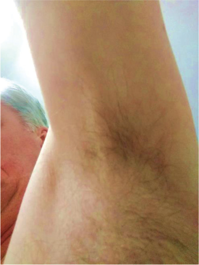
Cording at axilla.
The patient had pain free full range of motion in his neck, shoulders, and elbows except tightness at the right axilla area at the end range shoulder flexion with elbow in flexion. No neurological symptoms, radiating pain or loss of power in the upper limb was noted. On palpation cording under the right axilla was found with mild pain reported on palpation. The cording at this stage did not have any impact on patients’ activities of daily life or his quality of life. The patient was educated about the condition and a wait-and-see approach was taken. The patient was reviewed in 2 weeks and then again in 5 weeks, where the symptoms had completely resolved without any intervention.
DISCUSSION
The current case report aims to increase awareness of post-traumatic AWS, a rare presentation of AWS, otherwise commonly seen as a complication due to surgical intervention or post cancer treatment patients. Only a very few case reports have been published on non-cancer related AWS and even fewer on post-traumatic AWS in English language [7-9] Despite decades of research, the exact pathogenesis behind AWS is not fully understood, and there is no gold standard treatment protocol for this condition [6,10], however, this phenomenon has been attributed by some to possible lymphatic vessel thrombosis [11], fibrosis of the dilated lymphatics [12] and, fibroblastic proliferation around a subcutaneous lymphatic vessel [13]. High resolution Ultrasound Imaging (US) and Magnetic Resonance Imaging (MRI) suggests cord as trilaminar, presenting as fibrosis or a vessel and in some cases endoluminal thrombus, non-fatty lymphocele [14-17]. AWS has been hypothesized as disruption of intercosto-brachial nerves however earlier studies have refuted this and no direct correlation to the nerve was found [1,3]. However, AWS in non-cancer / non-surgical patients is poorly understood and there seems to be a void in the literature and the exact pathophysiology behind this phenomenon is unknown.
Regardless of the pathogenesis, physiotherapy, rehabilitation exercises, patient education and, manual therapy has been suggested by few as treatment options [18], but more high-quality studies are needed to either support or refute the role of physiotherapy / rehabilitation in AWS [18,19].
One of the major limitations of this case report is lack of imaging which could have confirmed the AWS and perhaps given an insight into the pathogenesis as well. This highlights the need for the Community Diagnostic Centres (CDC) in primary care, a part of the NHS long term plan [20] and Point of Care Ultrasound (POCUS) service as outlined in the Chartered Society of Physiotherapy guidance [21] as access to US scan in primary care setting could have helped in gaining instant imaging as extended examination, thereby reducing the need for unnecessary referral to UTC and re referral to GP’s thus reducing duplication of work, reducing resource waste, providing optimum care and reducing patient’s anxiety.
CONCLUSION
The current case report highlights the possibility of post-traumatic AWS presentation in outpatient clinics and that simple visual examination combined with palpation of the area can help identify AWS. It is important that the clinicians take time to assess the patient by exposing the area and looking for cording along the axilla. Without the visual feedback or palpation of the area the patients can be misdiagnosed as having shoulder pathology. The current case report also suggests a ‘wait and see’ approach to post-traumatic AWS alongside education as this can not only empower the patient but also help reduce fear, avoidance, beliefs and doctor shopping. Finally, the current case study also acknowledges the need for CDC and POCUS type services to help provide patients timely diagnosis, reduce patient anxiety, and enhance patient experience and outcomes.
Funding
Nothing to declare.
Statement of Conflict of Interest
None of the authors declares any relevant conflicts of interest.
Consent
Patient consent was obtained to use the pictures and for using the clinical details for publishing the case report. No patient identifiable information has been included in the case report.
REFERENCES
- 1.Moskovitz AH, Anderson BO, Yeung RS, et al. Axillary web syndrome after axillary dissection. Am J Surg. 2001;181(5):434–439. doi: 10.1016/S0002-9610(01)00602-X. [DOI] [PubMed] [Google Scholar]
- 2.Severeid K, Simpson J, Templeton B, et al. Lymphatic cording among patients with breast cancer or melanoma referred to physical therapy. Rehab Oncol. 2007;25(4):8–13. doi: 10.1097/01893697-200725040-00002. [DOI] [Google Scholar]
- 3.Bergmann A, Mendes VV, de Almeida Dias R, et al. Incidence and risk factors for axillary web syndrome after breast cancer surgery. Breast Cancer Res Treat. 2012;131(3):987–992. doi: 10.1007/s10549-011-1805-7. [DOI] [PubMed] [Google Scholar]
- 4.Yeung WM, McPhail SM, Kuys SS. A systematic review of axillary web syndrome (AWS) J Cancer Surviv. 2015;9(4):576–598. doi: 10.1007/s11764-015-0435-1. [DOI] [PubMed] [Google Scholar]
- 5.Figueira PVG, Haddad CAS, de Almeida Rizzi SKL, et al. Diagnosis of Axillary Web Syndrome in Patients After Breast Cancer Surgery: Epidemiology, Risk Factors, and Clinical Aspects: A Prospective Study. Am J Clin Oncol. 2018;41(10):992–996. doi: 10.1097/COC.0000000000000411. [DOI] [PubMed] [Google Scholar]
- 6.Dinas K, Kalder M, Zepiridis L, et al. Axillary web syndrome: Incidence, pathogenesis, and management. Curr Probl Cancer. 2019;43(6):100470. doi: 10.1016/j.currproblcancer.2019.02.002. [DOI] [PubMed] [Google Scholar]
- 7.Welsh P, Gryfe D. Atypical presentation of axillary web syndrome (AWS) in a male squash player: a case report. J Can Chiropr Assoc. 2016;60(4):294–298. [PMC free article] [PubMed] [Google Scholar]
- 8.Puentes Gutiérrez AB, García Bascones M, Puentes Gutiérrez R, et al. Síndrome axillary web idiopático [Idiopathic axillary web syndrome] Rehabilitacion (Madr) 2020;54(1):68–72. doi: 10.1016/j.rh.2019.10.002. [DOI] [PubMed] [Google Scholar]
- 9.Dündar Ahi E, Ozen S, Saraçgil Coşar SN. Idiopathic Axillary Syndrome: A Case Report on a Rare Entity. J Phys Med Rehab Sc. 2022;25(1) doi: 10.31609/jpmrs.2021-82996. [DOI] [Google Scholar]
- 10.Fukushima KF, Carmo LA, Borinelli AC, et al. Frequency and associated factors of axillary web syndrome in women who had undergone breast cancer surgery: a transversal and retrospective study. Springerplus. 2015;4:112. doi: 10.1186/s40064-015-0889-7. [DOI] [PMC free article] [PubMed] [Google Scholar]
- 11.Johansson K, Chong H, Ciornei CD, et al. Axillary Web Syndrome: Evidence for Lymphatic Origin with Thrombosis. Lymphat Res Biol. 2020;18(4):329–332. doi: 10.1089/lrb.2019.0074. [DOI] [PubMed] [Google Scholar]
- 12.Reedijk M, Boerner S, Ghazarian D, et al. A case of axillary web syndrome with subcutaneous nodules following axillary surgery. Breast. 2006;15(3):411–413. doi: 10.1016/j.breast.2005.09.005. [DOI] [PubMed] [Google Scholar]
- 13.Rashtak S, Gamble GL, Gibson LE, et al. From furuncle to axillary web syndrome: shedding light on histopathology and pathogenesis. Dermatology. 2012;224(2):110–114. doi: 10.1159/000337210. [DOI] [PubMed] [Google Scholar]
- 14.Leduc O, Fumière E, Banse S, et al. Identification and description of the axillary web syndrome (AWS) by clinical signs, MRI and US imaging. Lymphology. 2014;47(4):164–176. [PubMed] [Google Scholar]
- 15.Koehler LA, Hunter DW, Haddad TC, et al. Characterizing axillary web syndrome: ultrasonographic efficacy. Lymphology. 2014;47(4):156–163. [PMC free article] [PubMed] [Google Scholar]
- 16.Mullen LA, Harvey SC. Review of axillary web syndrome: What the radiologist should know. Eur J Radiol. 2019;113:66–73. doi: 10.1016/j.ejrad.2019.02.001. [DOI] [PubMed] [Google Scholar]
- 17.Koehler LA, Haddad TC, Hunter DW, et al. Axillary web syndrome following breast cancer surgery: symptoms, complications, and management strategies. Breast Cancer (Dove Med Press) 2018;11:13–19. doi: 10.2147/BCTT.S146635. [DOI] [PMC free article] [PubMed] [Google Scholar]
- 18.Lippi L, de Sire A, Losco L, et al. Axillary Web Syndrome in Breast Cancer Women: What Is the Optimal Rehabilitation Strategy after Surgery? A Systematic Review. J Clin Med. 2022;11(13):3839. doi: 10.3390/jcm11133839. [DOI] [PMC free article] [PubMed] [Google Scholar]
- 19.da Luz CM, Deitos J, Siqueira TC, et al. Management of Axillary Web Syndrome after Breast Cancer: Evidence-Based Practice. Rev Bras Ginecol Obstet. 2017;39(11):632–639. doi: 10.1055/s-0037-1604181. [DOI] [PMC free article] [PubMed] [Google Scholar]
- 20.Health Education England. NHS; 2022. Community Diagnostic Centres (CDCs) and their role, NHS choices. Available at: https://www.hee.nhs.uk/our-work/cancer-diagnostics/community-diagnostic-centres-cdc (Accessed: January 21, 2023) [Google Scholar]
- 21.CSP. Practice guidance for physiotherapists using point of care ultrasound (Pocus) in physiotherapy practice, The Chartered Society of Physiotherapy. 2022. Available at: https://www.csp.org.uk/publications/practice-guidance-physiotherapists-using-point-care-ultrasound-pocus-physiotherapy (Accessed: January 21, 2023)