Abstract
Background
Chronic obstructive pulmonary disease (COPD) is commonly diagnosed when the airflow limitation is well established and symptomatic. We aimed to identify individuals at risk of developing COPD according to the concept of pre-COPD and compare their clinical characteristics with 1) those who have developed the disease at a young age, and 2) the overall population with and without COPD.
Methods
The EPISCAN II study is a cross-sectional, population-based study that aims to investigate the prevalence of COPD in Spain in subjects ≥40 years of age. Pre-COPD was defined as the presence of emphysema >5% and/or bronchial thickening by computed chromatography (CT) scan and/or diffusing capacity of the lung for carbon monoxide (DLCO) <80% of predicted in subjects with respiratory symptoms and post-bronchodilator forced expiratory volume in 1 s/forced vital capacity (FEV1/FVC) >0.70. Young COPD was defined as FEV1/FVC <0.70 in a subject ≤50 years of age. Demographic and clinical characteristics were compared among pre-COPD, young COPD and the overall population with and without COPD.
Results
Among the 1077 individuals with FEV1/FVC <0.70, 65 (6.0%) were ≤50 years of age. Among the 8015 individuals with FEV1/FVC >0.70, 350 underwent both DLCO testing and chest CT scanning. Of those, 78 (22.3%) subjects fulfilled the definition of pre-COPD. Subjects with pre-COPD were older, predominantly women, less frequently active or ex-smokers, with less frequent previous diagnosis of asthma but with higher symptomatic burden than those with young COPD.
Conclusions
22.3% of the studied population was at risk of developing COPD, with similar symptomatic and structural changes to those with well-established disease without airflow obstruction. This COPD at-risk population is different from those that develop COPD at a young age.
Short abstract
Subjects fulfilling the definition of pre-COPD show similar symptomatic and structural changes to those with well-stablished disease. The fixed ratio of FEV1/FVC definition for COPD is missing an important group of patients that have significant disease. https://bit.ly/3B3FUO3
Introduction
Chronic obstructive lung disease (COPD) is a prevalent lung condition traditionally associated with cigarette smoking that usually remains underdiagnosed or is diagnosed in advanced stages of the disease process [1, 2]. Noteworthily, most patients are diagnosed in the sixth or seventh decade of life when symptoms are bothersome or exacerbations appear. We have recently described the prevalence of COPD in Spain, where the disease affects 11.8% of adults 40 years and older randomly selected from the general population [3]. 78% of COPD had not been diagnosed before [3]. The mean age of the COPD population was 65 years, an age when structural and functional changes in the lungs and other organs affected by the presence of COPD are mostly irreversible.
For this reason, it has been claimed that we should look at COPD “upstream in the river” [4] and a number of definitions of early COPD have been proposed [5], aiming to, on the one hand, raise attention to the early origins of the disease and, on the other hand, point out that we are arriving late to initiate a disease-modifying therapy for COPD [6] or a preventive measure such as smoking cessation.
However, the search for early identification of those patients at risk of developing COPD remains controversial. Attempts to define a Global Initiative for Chronic Obstructive Lung Disease (GOLD) “stage 0” based on the symptomatic and healthcare burden of smokers with normal spirometry failed to demonstrate to be an effective strategy [7]. Nevertheless, a number of cohort studies have found associations between respiratory symptoms [8] or low diffusing capacity of the lung for carbon monoxide (DLCO) [9] and the development of COPD. More recently, the concept of pre-COPD, that includes not only symptoms but also structural or functional abnormalities compatible with those found in COPD, has been proposed [10] as a risk marker of developing the disease.
A better knowledge of the natural history of the disease should clarify whether the development of COPD is a continuum that starts at young age in patients with symptoms and no airflow limitation, some of whom of them will progress to parenchymal abnormalities and airflow obstruction.
We aimed to identify patients at risk of developing COPD according to the concept of pre-COPD in a large cohort of well characterised patients taken from the general population, and compare their clinical characteristics with those who have developed the disease at a young age and with the overall population with or without COPD.
Methods
Population
The EPISCAN II study is a national, multicentre, cross-sectional, population-based epidemiological study aiming to investigate the prevalence and determinants of COPD in Spain. The protocol, fieldwork and methods have been described elsewhere [11]. Briefly, the fieldwork was conducted from April 2017 to February 2019 in 20 teaching hospitals throughout Spain. Subjects from the general population who were resident in the postal code areas nearest the participating hospitals were selected. The inclusion criteria were as follows: men or women aged 40 years or older with no physical or cognitive difficulties that would prevent them from completing spirometry or any of the study procedures. A randomised sample of 400 COPD and 400 non-COPD participants in the short visit from 12 preselected sites were invited to complete further testing in a long visit, according to quotas of age (10-year strata) and sex. Tests included single-breath DLCO and computed chromatography (CT) scan of the thorax. The study was approved by the ethics committees of each of the participating centres and all participants provided informed consent. The EPISCAN II protocol is registered at https://clinicaltrials.gov (NCT03028207) and at www.gsk-clinicalstudyregister.com/study/205932.
For the purposes of this pre-specified secondary objective of EPISCAN II, we defined early COPD as young COPD, meaning a post-bronchodilator forced expiratory volume in 1 s/forced vital capacity (FEV1/FVC) <0.70 in a subject younger than 50 years. Pre-COPD was defined according to the definition of Han et al. [10] as the presence of emphysema >5% and/or presence of bronchial thickening in the CT scan and/or DLCO <80% of predicted in subjects with respiratory symptoms and a post-bronchodilator FEV1/FVC >0.70. Bronchial thickening was considered when the measurement of airway thickness at the bronchiole level was greater than or equal to the highest quartile of the sample. Respiratory symptoms were considered as the presence of cough and/or phlegm production and/or dyspnoea defined as modified Medical Research Council (mMRC) score >0 for <80 years of age and >1 for those ≥80 years old) or a COPD Assessment Test (CAT) score >10.
Variables and procedures
Demographic information on sex, age, level of education, comorbidities, weight, height and smoking were collected. Forced spirometry was performed pre- and post-bronchodilation using a pneumotachograph (Vyntus Spiro, Carefusion, Germany), according to standardised procedures as previously described [5, 9]. Single-breath DLCO (MasterScreen Diffusion, Carefusion, Germany) was measured according to the American Thoracic Society (ATS)/European Respiratory Society recommendations [12], and adjusted by haemoglobin levels and atmospheric pressure. Global Lung Function Initiative equations were used as reference values [13]. The 6-min walk test was performed following ATS recommendations [14] and the BODE (body mass index, airflow obstruction, dyspnoea and exercise) index [15] was calculated accordingly. CT images were acquired during maximal inspiration, without contrast and with low-dose radiation, and 120 kV peak as the acquisition voltage. The images obtained underwent semi-automatic post-processing for determination of the percentage of emphysema, areas of extension, airway thickness, other measurements, and lung parenchyma attenuation and airway wall thickness, as previously described.
For the diagnosis of respiratory symptoms, the answers to the European Coal and Steel Community questionnaire were used [16]. The diagnosis of cough was considered when the participant answered yes to any of the cough-related questions of the questionnaire. Specific questions for dyspnoea and chronic bronchitis were included. The degree of dyspnoea was evaluated by the mMRC dyspnoea scale [17]. Health status was assessed by the CAT questionnaire [18]. The comorbidities were quantified by the Charlson and the COPD-specific comorbidity test (COTE) indices [19, 20]. Exacerbations in the previous year requiring the use of antibiotics and/or corticosteroids and the need for emergency visits or hospital admissions were recorded.
Statistical analysis
Categorical variables were presented as n (%), and continuous variables as mean±sd or median (interquartile range (IQR)), according to their distribution. The characteristics of the subgroups defined (pre-COPD and young COPD) have been compared using Student's t-test, the Mann–Whitney U-test or the χ2 test. In the case of the comparison between COPD, non-COPD and the subgroups defined, ANOVA and χ2 were used. Data were analysed with the Statistical Analysis System Enterprise Guide 7.15, considering a statistical significance (p-value) of 0.05 for all comparisons.
Results
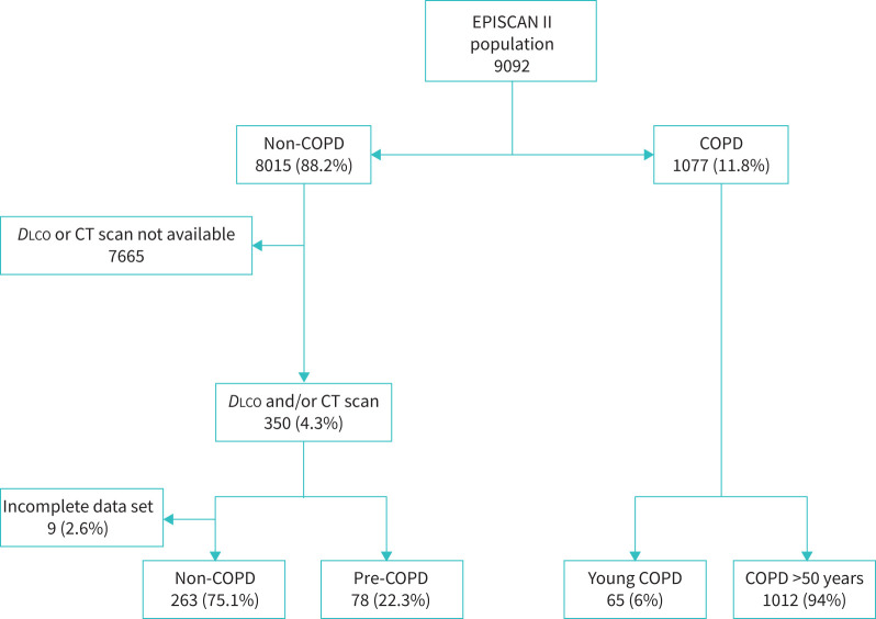
The EPISCAN II population included 9092 subjects who were able to perform a valid spirometry. Of those, 728 (8.0%) subjects underwent DLCO measurement and 668 (7.3%) chest CT scanning. As previously shown, 11.8% of the EPISCAN II population fulfilled criteria for COPD (figure 1).
FIGURE 1.
Flowchart of participants included in the analysis. DLCO: diffusing capacity of the lung for carbon monoxide; CT: computed tomography.
Prevalence of young COPD
Among the 1077 individuals with a post-bronchodilator FEV1/FVC <0.70, 65 were ≤50 years of age (6% of the COPD population). Individuals had a mean±sd age of 45.8±2.6 years and 65% of them were symptomatic, as previously defined by the presence of cough with phlegm production or dyspnoea or CAT ≥10.
Prevalence of pre-COPD
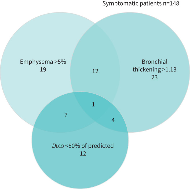
Among the 8015 individuals with a post-bronchodilator FEV1/FVC >0.70 in a valid spirometry, 350 (4.4%) underwent both DLCO testing and chest CT scanning. Of those, 148 (42.3%) were symptomatic, 51 (14.6%) had a DLCO <80% of predicted, 101 (28.9%) had >5% emphysema and 40 (11.4%) had bronchial diameter >1.13 mm (value of the highest quartile) on chest CT scan. 78 (22.3%) subjects fulfilled the pre-specified definition of pre-COPD (figure 2).
FIGURE 2.

Proportional Venn diagram of COPD trait sub-populations. DLCO: diffusing capacity of the lung for carbon monoxide.
Characteristics of pre-COPD versus young COPD
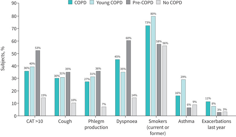
When comparing individuals with pre-COPD with those with young COPD, there were distinctions between these two (table 1): pre-COPD individuals were older, with median (IQR) age of 65 (54–72) versus 46 (43–48) years (p<0.0001) respectively, and less frequently active or ex-smokers (57.6% versus 80%, p=0.0002), but with higher symptomatic burden as per mMRC dyspnoea scale ≥1 (61.5% versus 35.4%, p=0.01) (table 1 and figure 3) or CAT (11.3 versus 9.1, p=0.03).
TABLE 1.
Differential demographic and clinical characteristics of young COPD (YC) and pre-COPD (PC) subjects compared to COPD (C) and non-COPD (NC) populations
| C | YC | PC | NC | p-value | |||||
| C versus YC | NC versus YC | C versus PC | NC versus PC | PC versus YC | |||||
| Subjects | 1012 | 65 | 78 | 263 | |||||
| General characteristics | |||||||||
| Age, years, mean±sd, median (interquartile range) | 67.8±9.9, 68.0 (59.0–75.0) | 45.8±2.6, 46.0 (43.0–48.0) | 63.5±11.6, 65.0 (54.0–72.0) | 59.3±10.1, 59.0 (51.0–67.0) | <0.0001 | <0.0001 | 0.003 | 0.0002 | <0.0001 |
| Females | 418 (41.3%) | 30 (46.2%) | 44 (56.4%) | 163 (62.0%) | 0.44 | 0.02 | 0.009 | 0.37 | 0.22 |
| BMI, kg·m−2 | 27.4±4.6 | 27.4±7.0 | 28.0±4.8 | 26.7±4.5 | 0.88 | 0.49 | 0.31 | 0.06 | 0.52 |
| Smoking status | 0.004 | <0.0001 | 0.009 | 0.59 | 0.0002 | ||||
| Active smokers | 301 (29.7%) | 32 (49.2%) | 14 (17.9%) | 58 (22.1%) | |||||
| Former smokers | 433 (42.8%) | 20 (30.8%) | 31 (39.7%) | 90 (34.2%) | |||||
| Never-smokers | 278 (27.5%) | 13 (20%) | 33 (42.3%) | 115 (43.7%) | |||||
| Smoking exposure, pack-years | 29.2±30.1 | 22.2±21.35 | 18.5±25.6 | 13.1±17.9 | 0.06 | 0.0005 | 0.002 | 0.04 | 0.35 |
| Education level | 0.15 | 0.03 | 0.23 | 0.27 | 0.56 | ||||
| No studies | 41 (4.1%) | 0 | 1 (1.3%) | 2 (0.8%) | |||||
| Primary education | 270 (26.7%) | 14 (21.5%) | 17 (21.8%) | 52 (19.8%) | |||||
| Secondary education | 243 (24.0%) | 19 (29.3%) | 17 (21.8%) | 45 (17.1%) | |||||
| University or vocational training | 454 (44.9%) | 31 (47.7%) | 42 (53.8%) | 164 (62.4%) | |||||
| Clinical characteristics | |||||||||
| mMRC dyspnoea scale | 0.14 | 0.0009 | 0.32 | <0.0001 | 0.01 | ||||
| Grade 0 | 499 (49.2%) | 42 (64.6%) | 30 (38.5%) | 223 (84.8%) | |||||
| Grade 1 | 355 (35.1%) | 18 (27.7%) | 36 (46.2%) | 36 (13.7%) | |||||
| Grade 2 | 113 (11.2%) | 3 (4.6%) | 9 (11.5%) | 3 (1.1%) | |||||
| Grade 3 | 39 (3.9%) | 2 (3.1%) | 3 (3.8%) | 1 (0.4%) | |||||
| Grade 4 | 6 (0.6%) | 0 | 0 | 0 | |||||
| CAT | 9.1±6.8 | 9.1±6.4 | 11.3±6.1 | 5.7±5.1 | 0.98 | <0.0001 | 0.004 | <0.0001 | 0.03 |
| Cough and phlegm# | 621 (66.3%) | 37 (58.7%) | 78 (100.0%) | 70 (28.0%) | 0.21 | <0.0001 | <0.0001 | <0.0001 | <0.0001 |
| Asthma | 163 (16.1%) | 19 (29.2%) | 5 (6.4%) | 24 (9.1%) | 0.006 | <0.0001 | 0.02 | 0.45 | 0.0003 |
| Charlson Comorbidity Index | 0.7±1.1 | 0.6±1.7 | 0.5±0.9 | 0.3±0.8 | 0.71 | 0.01 | 0.22 | 0.01 | 0.63 |
| COTE index | 1.2±2.4 | 1.4±2.8 | 1.7±2.8 | 0.9±2.1 | 0.44 | 0.12 | 0.10 | 0.01 | 0.62 |
| Exacerbations last year | 114 (11.3%) | 5 (7.7%) | 2 (2.6%) | 9 (3.4%) | 0.37 | 0.12 | 0.01 | 0.70 | 0.15 |
| Treatments | |||||||||
| Any respiratory treatment | 641 (63.3%) | 28 (43.1%) | 39 (50.0%) | 86 (32.7%) | 0.001 | 0.11 | 0.01 | 0.005 | 0.40 |
| Treatment with short-acting β-agonist | 251 (24.8%) | 17 (26.2%) | 4 (5.1%) | 8 (3.0%) | 0.80 | <0.0001 | <0.0001 | 0.37 | 0.0004 |
| Treatment with anticholinergics | 159 (15.7%) | 4 (6.2%) | 2 (2.6%) | 5 (1.9%) | 0.03 | 0.06 | 0.001 | 0.71 | 0.28 |
| Treatment with inhaled corticosteroids | 189 (18.7%) | 10 (15.4%) | 4 (5.1%) | 9 (3.4%) | 0.50 | 0.0002 | 0.002 | 0.48 | 0.03 |
Data are presented as mean±sd or n (%), unless otherwise stated. BMI: body mass index; mMRC: modified Medical Research Council; CAT: COPD Assessment Test; COTE: COPD-specific comorbidity test. #: European Coal and Steel Community questionnaire.
FIGURE 3.
Differential clinical characteristics among subjects classified as young COPD (n=65) or pre-COPD (n=78) compared to COPD (n=1012) and non-COPD (n=263) in the general population. Data on clinical characteristics were collected in >95% of individuals per group. “Exacerbations last year” accounts for at least one exacerbation requiring antibiotics, oral steroids or an emergency visit in the previous 12 months. CAT: COPD Assessment Test.
A previous diagnosis of asthma was reported by the patient in 29.2% versus 6.4% and median blood eosinophil count was higher (233 versus 158 cells per μL) in the young COPD group compared to pre-COPD respectively (table 2). However, only 5.1% of pre-COPD patients were receiving treatment with short-acting β-agonist or inhaled corticosteroids, whereas 26.2% and 15.4% of young COPD were receiving them, respectively. No statistically significant differences were found in the history of exacerbations in the previous year, that tended to be higher in the young COPD group compared to pre-COPD group (7.7% versus 2.6%, p=0.15) (figure 3).
Characteristics of pre-COPD compared to the overall COPD and non-COPD population
When comparing the patients fulfilling the criteria of pre-COPD with the overall COPD population and the non-COPD (and non-pre-COPD) population, pre-COPD patients were more frequently female than the COPD population, with younger age and were less frequently smokers or ex-smokers, but had similar symptomatic burden measured by mMRC dyspnoea (table 1 and figure 3). Pre-COPD and COPD had similar burdens of comorbidities with higher Charlson and COTE indices than the control population. Also, both pre-COPD and COPD patients had impaired exercise capacity measured by the 6-min walk test, with similar emphysema and lower DLCO (table 2). Pre-COPD also showed a higher bronchiole thickness than the control group. However, the pre-COPD group was similar to the control population without COPD in spirometric parameters, blood eosinophil counts, and history of asthma or use of respiratory medication.
TABLE 2.
Differential functional, inflammatory and imaging characteristics of young COPD (YC) and pre-COPD (PC) subjects compared to COPD (C) and non-COPD (NC) populations
| C | YC | PC | NC | p-value | |||||
| C versus YC | NC versus YC | C versus PC | NC versus PC | PC versus YC | |||||
| Subjects | 1012 | 65 | 78 | 263 | |||||
| Lung function | |||||||||
| FVC, % of predicted | 99.3±18.5 | 99.8±14.8 | 101.2±16.5 | 104.9±13.3 | 0.83 | 0.02 | 0.39 | 0.11 | 0.60 |
| FEV1, % of predicted | 80.6±18.8 | 80.3±16.2 | 103.6±18.0 | 105.3±13.7 | 0.08 | <0.0001 | <0.0001 | 0.33 | <0.0001 |
| 6MWT distance, m | 477.1±108.1 | 525.5±121.0 | 467.3.0±114.8 | 527.2±87.8 | 0.09 | 0.94 | 0.47 | <0.0001 | 0.07 |
| DLCO, % of predicted | 88.6±23.1 | 108.9±19.5 | 90.9±19.4 | 101.5±17.4 | 0.001 | 0.11 | 0.40 | <0.0001 | 0.001 |
| BODE index# | 1.5±1.2 | 1.1±0.8 | 1.4±0.9 | 0.9±0.4 | 0.13 | 0.30 | 0.24 | <0.0001 | 0.22 |
| BODEx index¶ | 1.4±1.0 | 1.3±0.9 | 1.2±0.6 | 0.9±0.3 | 0.19 | <0.0001 | 0.01 | <0.0001 | 0.31 |
| Biomarkers | |||||||||
| Eosinophils per μL | 191±123, 175 (101–249) | 233±169, 182 (168–227) | 158±95, 140 (89–215) | 159±120, 128 (88–198) | 0.22 | 0.02 | 0.03 | 0.98 | 0.02 |
| CRP, mg·dL−1 | 1.9±4.0, 0.4 (0.1–2.0) | 2.1±4.1, 0.2 (0.1–1.0) | 1.6±2.8, 0.6 (0.1–1.5) | 1.3±2.7, 0.4 (0.1–1.1) | 0.93 | 0.33 | 0.46 | 0.43 | 0.60 |
| Fibrinogen, g·L−1 | 3.9±0.9, 3.7 (3.3–4.5) |
3.3±0.8, 3.0 (2.7–4.0) | 3.9±1.1, 3.9 (3.2–4.5) | 3.7±0.8, 3.6 (3.1–4.3) | 0.02 | 0.07 | 0.995 | 0.10 | 0.04 |
| Imaging | |||||||||
| Emphysema >5% | 160 (59.7%) | 3 (27.3%) | 39 (50.0%) | 55 (20.9%) | 0.03 | 0.61 | 0.12 | <0.0001 | 0.15 |
| Bronchiole thickness, mm | 1.1±0.1, 1.1 (1.0–1.1) | 1.1±0.1, 1.1 (1.1–1.2) | 1.1±0.2, 1.1 (1.0–1.2) | 1.0±0.2, 1.1 (1.0–1.1) | 0.67 | 0.21 | 0.03 | 0.0001 | 0.69 |
Data are presented as mean±sd or mean±sd, median (interquartile range), unless otherwise stated. FVC: forced vital capacity; FEV1: forced expiratory volume in 1 s; 6MWT: 6-min walk test; DLCO: diffusing capacity of the lung for carbon monoxide; BODE: body mass index, obstruction, dyspnoea, exercise; BODEx: simplified BODE; CRP: C-reactive protein; IQR: interquartile range. #: out of 10; ¶: out of 9.
Characteristics of young COPD compared to the overall COPD and non-COPD populations
Patients with COPD younger than 50 years, compared to a population without criteria for COPD nor pre-COPD, were more frequently males, more frequently active or former smokers, with more symptomatic burden measured by mMRC dyspnoea and CAT scores, had more comorbidities measured by means of the Charlson and COTE indices, and suffered more exacerbations (table 1 and figure 3). They more frequently reported a past medical history of asthma and showed higher blood eosinophil counts than controls.
In comparison with the overall COPD population, young COPD patients had similar airflow limitation, and similar symptomatic and exacerbation burden, despite having better exercise capacity and therefore lower BODE index scores. Young COPD patients were less frequently treated with anticholinergics than the overall COPD population.
Discussion
Three important messages should be taken from this research: first, we have shown, for the first time, that 22.3% of a subsample of the general population would qualify for the definition of pre-COPD, and this has important implications in terms of symptoms and health status in this untreated population. Second, we have also shown that 6% of the population that fulfils the criteria for a diagnosis of COPD are younger than 50 years, but this young COPD population has similar symptomatic, exacerbation and comorbidity burden than the overall older COPD population. Third, the pre-COPD population appears different from those that develop COPD at a young age.
Interpretation of novel findings
In this population-based study we have identified, within a randomised subgroup who underwent CT scanning and DLCO measurement, that 22.3% of this population have symptoms, reduced exercise capacity, emphysema and small airway thickness similar to the population with COPD despite not fulfilling spirometric criteria for COPD. This population is likely to be at high risk of developing COPD and its consequences but we are not detecting them in a timely manner, which reflects a limitation of spirometry as a screening tool. Interestingly enough, we have also shown that 6% of the population with COPD are younger than 50 years and they have a well-established disease with similar health-related outcomes to the older COPD population. Noteworthily, it is not possible to assume that the pre-COPD condition evolves to young COPD, since pre-COPD subjects were older, predominantly women with less smoking burden despite similar symptomatic and DLCO or structural impairment to the young COPD population.
Previous studies
Martinez et al. [21] proposed the name “early COPD” for those patients younger than 50 years with chronic airflow limitation and evidence of structural or progressive functional impairment such as visual emphysema, air trapping and bronchial thickening. Soriano et al. [5] included the concept of disease activity as part of the concept of early COPD in an attempt to include exacerbations as part of the patient at risk profile. However, as GOLD update of 2022 underlines, early means “near the beginning of a process” and because COPD may start early in life and takes a long time to be clinically manifested, determining if someone really suffers “early” COPD is challenging; therefore, it is more appropriate to label them as “young COPD” [1].
A recent prospective, multicentre, case–control study [22, 23, 24] aimed to describe the characteristics of young COPD (defined only by age <50 years) that included smokers of >10 pack-years with or without post-bronchodilator FEV1/FVC <0.70, found that these young COPD patients already had moderate airflow limitation and were often symptomatic, used healthcare resources frequently, had air trapping and reduced diffusing capacity, and had frequent evidence of emphysema by CT (61%). Notably, less than half of cases (46%) had been previously diagnosed with COPD. These observations were reproduced in the ECLIPSE and COPDGene cohorts [23]. Çolak et al. [25] also found that among individuals under 50 years of age with 10 pack-years or greater of tobacco consumption from the general population, 15% fulfilled the criteria of early COPD. Individuals with early COPD more often had chronic respiratory symptoms and severe lung function impairment, and an increased risk of acute respiratory hospitalisations and early death. Depending on amount of smoking exposure, 24% of young adults in the general population with early COPD develop clinical COPD 10 years later [26].
Previous attempts to identify individuals at risk of developing COPD have rendered different results. The COPDgene study found that 43% of smokers with normal FEV1/FVC ratio had airwall thickening or emphysema on CT and 23% had mMRC dyspnoea scores ≥2, and these patients had reduced exercise capacity and increased exacerbation-like events [27]. In addition, in SPIROMICS, nearly 50% of smokers with normal spirometry had similar symptomatic burden to those with COPD with mild or moderate airflow limitation [28]. A proportion of these symptomatic smokers also showed airway wall thickening on CT similar to our results.
The importance of symptoms without obstruction led to the concept of “GOLD 0” or “COPD at risk”, and many studies have shown that a proportion ranging from 11.6% to 20.5% of these individuals develop COPD during follow-up [29, 30]. This at-risk status of GOLD 0 was initially considered but later abandoned, because the proportion of individuals that progressed to COPD was considered low and not different from those who were not considered as GOLD 0 [10]. Noteworthily, we have been more precise than the proposal of pre-COPD in defining a symptom threshold using CAT and mMRC scores adjusted by age, since we have previously shown that this may imply an impact on mortality [17].
Other markers of risk for developing COPD have been previously explored, like physiological measurements or imaging. A reduced single-breath DLCO (<80% of predicted) in active smokers with normal spirometry followed over 45 months was found to be associated to a higher incidence of GOLD-defined COPD compared to those with normal DLCO (22% versus 3%, respectively) [9]. Our population with pre-COPD had reduced DLCO to a similar extent as the whole COPD population, which supports the hypothesis that these individuals are more prone to develop GOLD-defined COPD.
Imaging is another way to identify patients at risk for developing GOLD-defined COPD. In different trials using CT scan for screening of lung cancer, smokers with no evidence of airflow limitation at baseline who developed COPD during follow-up had more emphysema on CT [31, 32]. In addition, increased airway thickness measured by CT in those trials was significantly (and independently from the presence of emphysema) associated with incident COPD [33]. In keeping with these previous findings, our data support the importance of imaging in the new category of pre-COPD, showing a similar extent of emphysema and increased airway thickness to the COPD population. We used 5% quantified emphysema as a threshold to determine risk, as previously shown by Lynch et al. [34].
We have included in our analysis the population with COPD who were younger than 50 years, assuming that lung growth and development reach their peak at around 20–25 years of age in men but at 15 years in women and begin to decline later [35]. In population-based studies, these younger individuals with COPD have more frequently a previous diagnosis of asthma, as we have found in our population. A “diagnosis of asthma” (not necessarily the disease) is frequently associated with abnormal lung development [36] and the latter is now a well-recognised cause of COPD [37].
Clinical implications
To identify subjects at risk of developing COPD and those with already established disease at early stages who are at risk of progressing may have important clinical implications. Very few therapeutic trials have been conducted in symptomatic individuals without airflow limitation. A recently published perspective by experts in the field highlighted the need for randomised controlled trials focused on young COPD or pre-COPD patients to reduce disease progression, providing innovative approaches to identifying and engaging potential study subjects [6]. Moreover, it has been recently suggested that this group of patients with symptoms and emphysema should be considered as having COPD despite not having evidence of airflow obstruction [38, 39].
According to our results, different strategies should be implemented to identify this population at risk, since they show clear differences from other populations. Pre-COPD populations are highly symptomatic individuals, predominantly women with less smoking exposure, whereas young COPD are patients with well-established disease at younger age have higher smoking burden and more frequently diagnosis of asthma.
Strengths and limitations
There are several strengths to this research, including novelty, an unbiased population approach, and the use of low-dose CT scanning and DLCO to characterise COPD beyond spirometry. It should be mentioned that similarities and differences in emphysema, DLCO and bronchial thickness found in the pre-COPD group were attributes used to define it. Moreover, a number of limitations should be considered. The main limitation of this study is the lack of longitudinal follow-up that could confirm that those fulfilling the criteria for pre-COPD are really at higher risk of developing COPD. Nevertheless, the data shown here underlie the importance and impact of the disease in young subjects taken from the general population, and of those with respiratory symptoms and associated functional and/or structural abnormalities. Additionally, it is possible that the small sample size could limit the magnitude of the differences found in some subanalyses between those with pre-COPD and young COPD due to potential type 1 and type 2 errors. Furthermore, given the population approach, the representativity of hospital-based populations and the most severe COPD patients is limited. Finally, it is possible that we may have underestimated the amount of emphysema by using low-dose CT [40]. Nevertheless, we think that this does not have a major impact in our findings.
Conclusions
We found that 22.3% of the studied population is at risk of developing COPD, with similar symptomatic and structural changes to those with well-stablished disease without any evidence of airway obstruction. This at-risk population is different from those that develop COPD at young age. Different strategies to tackle with these two early faces of COPD should be considered. In addition, our findings reflect that fixed ratio of FEV1/FVC definition for COPD is missing an important group of patients that have significant disease.
Supplementary material
Please note: supplementary material is not edited by the Editorial Office, and is uploaded as it has been supplied by the author.
Supplementary material 00334-2022.SUPPLEMENT (294.4KB, pdf)
Acknowledgements
The collaboration of Monica Sarmiento and Neus Canal from IQVIA, and Carolina Peña and José Julio Jiménez from GSK is explicitly acknowledged. EPISCAN II Scientific Committee: Borja G. Cosío, Ciro Casanova, Inmaculada Alfageme, Pilar de Lucas, Julio Ancochea, Marc Miravitlles, Juan José Soler-Cataluña, Francisco García-Río, José Miguel Rodríguez González-Moro, Guadalupe Sánchez and Joan B. Soriano. A list of Principal Investigators, collaborators, and participating centres can be found in the Supplementary material.
Provenance: Submitted article, peer reviewed.
This study is registered at www.clinicaltrials.gov with identifier number NCT03028207. Information on the GSK data sharing commitments and requesting access to anonymised individual participant data and associated documents can be found at www.clinicalstudydatarequest.com.
Ethics approval and consent to participate: The study was approved by the ethics committees of each of the participating centres, and all participants provided informed consent.
Author contributions: Study concept and design: B.G. Cosío, C. Casanova, M. Miravitlles, J.J. Soler-Cataluña, J.B. Soriano, F. García-Río, P. de Lucas, I. Alfageme, J.M. Rodríguez González-Moro, G. Sánchez and J. Ancochea. Data acquisition: J.J. Soler-Cataluña, J. Ancochea and B.G. Cosío. Analysis and interpretation of the data: M. Miravitlles, J.J. Soler-Cataluña, J.B. Soriano and B.G. Cosío. Drafting of the manuscript: B.G. Cosío and C. Casanova. Critical revision and approval for submission: M. Miravitlles, J.J. Soler-Cataluña, J.B. Soriano, F. García-Río, P. de Lucas, I. Alfageme, C. Casanova, J.M. Rodríguez González-Moro, G. Sánchez, J. Ancochea and B.G. Cosío. All authors have read and approved the final manuscript.
Conflict of interest: B.G. Cosío has received speaker or consulting fees from AstraZeneca, Boehringer Ingelheim, Chiesi, GlaxoSmithKline, Menarini, Novartis, Sanofi and TEVA, and research grants from Menarini, AstraZeneca and Boehringer Ingelheim.
Conflict of interest: C. Casanova has received speaker or consulting fees from AstraZeneca, Bial, Boehringer Ingelheim, Chiesi, GlaxoSmithKline, Menarini and Novartis, and research grants from GlaxoSmithKline, Menarini and AstraZeneca.
Conflict of interest: J.J. Soler-Cataluña has received speaker fees from AstraZeneca, Bial, Boehringer Ingelheim, Chiesi, Esteve, Ferrer, GlaxoSmithKline, Menarini, Novartis and Teva, and consulting fees from AstraZeneca, Bial, Boehringer Ingelheim, GlaxoSmithKline, Ferrer and Novartis.
Conflict of interest: J.B. Soriano has nothing to disclose. F. García-Río has received speaker or consulting fees from AstraZeneca, Boehringer Ingelheim, Chiesi, GlaxoSmithKline, Menarini, Novartis, Pfizer and Rovi, and research grants from Chiesi, Esteve, Gebro Pharma, GlaxoSmithKline, Menarini and TEVA.
Conflict of interest: P. de Lucas has nothing to disclose.
Conflict of interest: I. Alfageme has nothing to disclose.
Conflict of interest: J.M. Rodríguez González-Moro has nothing to disclose.
Conflict of interest: G. Sánchez is a GSK employee within the Medical Department.
Conflict of interest: J. Ancochea has received speaker or consulting fees from Actelion, Air Liquide, Almirall, AstraZeneca, Boehringer Ingelheim, Carburos Médica, Chiesi, Faes Farma, Ferrer, GlaxoSmithKline, InterMune, Linde Healthcare, Menarini, MSD, Mundipharma, Novartis, Pfizer, Roche, Rovi, Sandoz, Takeda and Teva.
Conflict of interest: M. Miravitlles has received speaker or consulting fees from AstraZeneca, Bial, Boehringer Ingelheim, Chiesi, Cipla, CSL Behring, Laboratorios Esteve, Gebro Pharma, Kamada, GlaxoSmithKline, Grifols, Menarini, Mereo Biopharma, Novartis, pH Pharma, Palobiofarma SL, Rovi, TEVA, Spin Therapeutics, Verona Pharma and Zambon, and research grants from Grifols.
Support statement: The EPISCAN II study was sponsored by GlaxoSmithKline. Funding information for this article has been deposited with the Crossref Funder Registry.
References
- 1.Halpin DMG, Criner GJ, Papi A, et al. Global initiative for the diagnosis, management, and prevention of chronic obstructive lung disease. The 2020 GOLD Science committee report on COVID-19 and chronic obstructive pulmonary disease. Am J Respir Crit Care Med 2021; 203: 24–36. doi: 10.1164/RCCM.202009-3533SO [DOI] [PMC free article] [PubMed] [Google Scholar]
- 2.Miravitlles M, Calle M, Molina J, et al. Spanish COPD Guidelines (GesEPOC) 2021: Updated Pharmacological treatment of stable COPD. Arch Bronconeumol 2022; 58: 69–81. doi: 10.1016/J.ARBRES.2021.03.005 [DOI] [PubMed] [Google Scholar]
- 3.Soriano JB, Alfageme I, Miravitlles M, et al. Prevalence and determinants of COPD in Spain: EPISCAN II. Arch Bronconeumol (Engl Ed) 2021; 57: 61–69. doi: 10.1016/J.ARBRES.2020.07.024 [DOI] [PubMed] [Google Scholar]
- 4.Lange P, Celli B, Agustí A, et al. Lung-function trajectories leading to chronic obstructive pulmonary disease. N Engl J Med 2015; 373: 111–122. doi: 10.1056/nejmoa1411532 [DOI] [PubMed] [Google Scholar]
- 5.Soriano JB, Polverino F, Cosio BG. What is early COPD and why is it important? Eur Respir J 2018; 52: 1801448. doi: 10.1183/13993003.01448-2018 [DOI] [PubMed] [Google Scholar]
- 6.Martinez FJ, Agusti A, Celli BR, et al. Treatment trials in young patients with COPD and Pre-COPD patients: time to move forward. Am J Respir Crit Care Med 2021; 205: 275–287. doi: 10.1164/rccm.202107-1663SO [DOI] [PMC free article] [PubMed] [Google Scholar]
- 7.Pompe E, Strand M, van Rikxoort EM, et al. Five-year progression of emphysema and air trapping at CT in smokers with and those without chronic obstructive pulmonary disease: results from the COPDGene study. Radiology 2020; 295: 218–226. doi: 10.1148/radiol.2020191429 [DOI] [PMC free article] [PubMed] [Google Scholar]
- 8.Allinson JP, Hardy R, Donaldson GC, et al. The presence of chronic mucus hypersecretion across adult life in relation to chronic obstructive pulmonary disease development. Am J Respir Crit Care Med 2016; 193: 662–672. doi: 10.1164/RCCM.201511-2210OC [DOI] [PMC free article] [PubMed] [Google Scholar]
- 9.Harvey BG, Strulovici-Barel Y, Kaner RJ, et al. Progression to COPD in smokers with normal spirometry/low DLCO using different methods to determine normal levels. Eur Respir J 2016; 47: 1888–1889. doi: 10.1183/13993003.00435-2016 [DOI] [PubMed] [Google Scholar]
- 10.Han MLK, Agusti A, Celli BR, et al. From GOLD 0 to pre-COPD. Am J Respir Crit Care Med 2021; 203: 414–423. doi: 10.1164/rccm.202008-3328PP [DOI] [PMC free article] [PubMed] [Google Scholar]
- 11.Alfageme I, de Lucas P, Ancochea J, et al. 10 years after EPISCAN: a new study on the prevalence of copd in spain – a summary of the EPISCAN II protocol. Arch Bronconeumol (Engl Ed) 2019; 55: 38–47. doi: 10.1016/j.arbres.2018.05.011 [DOI] [PubMed] [Google Scholar]
- 12.MacIntyre N, Crapo RO, Viegi G, et al. Standardisation of the single-breath determination of carbon monoxide uptake in the lung. Eur Respir J 2005; 26: 720–735. doi: 10.1183/09031936.05.00034905 [DOI] [PubMed] [Google Scholar]
- 13.Stanojevic S, Graham BL, Cooper BG, et al. Official ERS technical standards: Global Lung Function Initiative reference values for the carbon monoxide transfer factor for Caucasians. Eur Respir J 2017; 50: 1700010. doi: 10.1183/13993003.00010-2017 [DOI] [PubMed] [Google Scholar]
- 14.Crapo RO, Casaburi R, Coates AL, et al. ATS statement: guidelines for the six-minute walk test. Am J Respir Crit Care Med 2002; 166: 111–117. doi: 10.1164/AJRCCM.166.1.AT1102 [DOI] [PubMed] [Google Scholar]
- 15.Celli BR, Cote CG, Marin JM, et al. The body-mass index, airflow obstruction, dyspnea, and exercise capacity index in chronic obstructive pulmonary disease. N Engl J Med 2004; 350: 1005–1012. doi: 10.1056/NEJMOA021322 [DOI] [PubMed] [Google Scholar]
- 16.Cotes JE, Chinn DJ. Questionnaire of the ECSC on respiratory symptoms. Eur Respir J 1989; 2: 1021–1022. [PubMed] [Google Scholar]
- 17.Casanova C, Marin JM, Martinez-Gonzalez C, et al. Differential effect of modified Medical Research Council Dyspnea, COPD Assessment Test, and Clinical COPD Questionnaire for symptoms evaluation within the new GOLD staging and mortality in COPD. Chest 2015; 148: 159–168. doi: 10.1378/chest.14-2449 [DOI] [PubMed] [Google Scholar]
- 18.De Torres JP, Marin JM, Martinez-Gonzalez C, et al. Clinical application of the COPD assessment test: longitudinal data from the COPD History Assessment in Spain (CHAIN) Cohort. Chest 2014; 146: 111–122. doi: 10.1378/chest.13-2246 [DOI] [PubMed] [Google Scholar]
- 19.Charlson M, Szatrowski TP, Peterson J, et al. Validation of a combined comorbidity index. J Clin Epidemiol 1994; 47: 1245–1251. doi: 10.1016/0895-4356(94)90129-5 [DOI] [PubMed] [Google Scholar]
- 20.De Torres JP, Casanova C, Marín JM, et al. Prognostic evaluation of COPD patients: GOLD 2011 versus BODE and the COPD comorbidity index COTE. Thorax 2014; 69: 799–804. doi: 10.1136/THORAXJNL-2014-205770 [DOI] [PubMed] [Google Scholar]
- 21.Martinez FJ, Han MK, Allinson JP, et al. At the root: defining and halting progression of early chronic obstructive pulmonary disease. Am J Respir Crit Care Med 2018; 197: 1540–1551. doi: 10.1164/rccm.201710-2028PP [DOI] [PMC free article] [PubMed] [Google Scholar]
- 22.Global Initiative for Chronic Obtructive Lung Disease. Global strategy for prevention, diagnosis and management of COPD: 2023 report. https://goldcopd.org/2023-gold-report-2/ Date last accessed 29 November 2022.
- 23.Cosío BG, Pascual-Guardia S, Borras-Santos A, et al. Phenotypic characterisation of early COPD: a prospective case–control study. ERJ Open Res 2020; 6: 00047–02020. doi: 10.1183/23120541.00047-2020 [DOI] [PMC free article] [PubMed] [Google Scholar]
- 24.Borràs-Santos A, Garcia-Aymerich J, Soler-Cataluña JJ, et al. Determinants of the appearance and progression of early-onset chronic obstructive pulmonary disease in young adults. A case-control study with follow-up. Arch Bronconeumol (Engl Ed) 2019; 55: 312–318. [DOI] [PubMed] [Google Scholar]
- 25.Çolak Y, Afzal S, Nordestgaard BG, et al. Prevalence, characteristics, and prognosis of early chronic obstructive pulmonary disease the Copenhagen general population study. Am J Respir Crit Care Med 2020; 201: 671–680. doi: 10.1164/rccm.201908-1644OC [DOI] [PMC free article] [PubMed] [Google Scholar]
- 26.Colak Y, Afzal S, Nordestgaard BG, et al. Importance of early COPD in young adults for development of clinical COPD findings from the Copenhagen general population study. Am J Respir Crit Care Med 2021; 203: 1245–1256. doi: 10.1164/rccm.202003-0532OC [DOI] [PMC free article] [PubMed] [Google Scholar]
- 27.Regan EA, Lynch DA, Curran-Everett D, et al. Clinical and radiologic disease in smokers with normal spirometry. JAMA Intern Med 2015; 175: 1539–1549. doi: 10.1001/jamainternmed.2015.2735 [DOI] [PMC free article] [PubMed] [Google Scholar]
- 28.Woodruff PG, Barr RG, Bleecker E, et al. Clinical significance of symptoms in smokers with preserved pulmonary function. N Engl J Med 2016; 374: 1811–1821. doi: 10.1056/NEJMOA1505971 [DOI] [PMC free article] [PubMed] [Google Scholar]
- 29.Vestbo J, Prescott E, Lange P, et al. Association of chronic mucus hypersecretion with FEV1 decline and chronic obstructive pulmonary disease morbidity. Copenhagen City Heart Study Group. Am J Respir Crit Care Med 1996; 153: 1530–1535. doi: 10.1164/AJRCCM.153.5.8630597 [DOI] [PubMed] [Google Scholar]
- 30.Vestbo J, Lange P. Can GOLD Stage 0 provide information of prognostic value in chronic obstructive pulmonary disease? Am J Respir Crit Care Med 2002; 166: 329–332. doi: 10.1164/RCCM.2112048 [DOI] [PubMed] [Google Scholar]
- 31.Mohamed Hoesein FAA, Van Rikxoort E, Van Ginneken B, et al. Computed tomography-quantified emphysema distribution is associated with lung function decline. Eur Respir J 2012; 40: 844–850. doi: 10.1183/09031936.00186311 [DOI] [PubMed] [Google Scholar]
- 32.McAllister DA, Ahmed FS, Austin JHM, et al. Emphysema predicts hospitalisation and incident airflow obstruction among older smokers: a prospective cohort study. PLoS One 2014; 9: e93221. doi: 10.1371/JOURNAL.PONE.0093221 [DOI] [PMC free article] [PubMed] [Google Scholar]
- 33.Mohamed Hoesein FAA, De Jong PA, Lammers JWJ, et al. Airway wall thickness associated with forced expiratory volume in 1 second decline and development of airflow limitation. Eur Respir J 2015; 45: 644–651. doi: 10.1183/09031936.00020714 [DOI] [PubMed] [Google Scholar]
- 34.Lynch DA, Austin JHM, Hogg JC, et al. CT-definable subtypes of chronic obstructive pulmonary disease: a statement of the Fleischner society. Radiology 2015; 277: 192–205. doi: 10.1148/RADIOL.2015141579 [DOI] [PMC free article] [PubMed] [Google Scholar]
- 35.Kohansal R, Martinez-Camblor P, Agustí A, et al. The natural history of chronic airflow obstruction revisited: an analysis of the Framingham offspring cohort. Am J Respir Crit Care Med 2009; 180: 3–10. doi: 10.1164/RCCM.200901-0047OC [DOI] [PubMed] [Google Scholar]
- 36.Agusti A, Faner R. Lung function trajectories in health and disease. Lancet Respir Med 2019; 7: 358–364. doi: 10.1016/S2213-2600(18)30529-0 [DOI] [PubMed] [Google Scholar]
- 37.Martinez FD. Early-life origins of chronic obstructive pulmonary Disease. N Engl J Med 2016; 375: 871–878. doi: 10.1056/NEJMRA1603287 [DOI] [PubMed] [Google Scholar]
- 38.Barnes PJ, Vestbo J, Calverley PM. The pressing need to redefine “COPD”. Chronic Obstr Pulm Dis 2019; 6: 380–383. doi: 10.15326/jcopdf.6.5.2019.0173 [DOI] [PMC free article] [PubMed] [Google Scholar]
- 39.Lowe KE, Regan EA, Anzueto A, et al. COPDGene® 2019: Redefining the diagnosis of chronic obstructive pulmonary disease. Chronic Obstr Pulm Dis 2019; 6: 384–399. doi: 10.15326/jcopdf.6.5.2019.0149 [DOI] [PMC free article] [PubMed] [Google Scholar]
- 40.Wisselink HJ, Pelgrim GJ, Rook M, et al. Ultra-low-dose CT combined with noise reduction techniques for quantification of emphysema in COPD patients: An intra-individual comparison study with standard-dose CT. Eur J Radiol 2021; 138: 109646. doi: 10.1016/j.ejrad.2021.109646 [DOI] [PubMed] [Google Scholar]
Associated Data
This section collects any data citations, data availability statements, or supplementary materials included in this article.
Supplementary Materials
Please note: supplementary material is not edited by the Editorial Office, and is uploaded as it has been supplied by the author.
Supplementary material 00334-2022.SUPPLEMENT (294.4KB, pdf)


