Abstract

Keywords: cardiovascular, inward rectifier, neural, potassium channels, renal
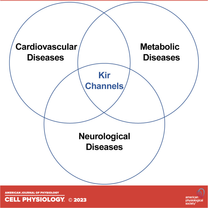
Inward rectifier potassium (Kir) currents were first recorded from frog skeletal muscle by Sir Bernard Katz in 1949 (1). Unlike the depolarization-activated, outwardly rectifying, delayed rectifier potassium (K+) currents of nerve cell membranes that were known at the time, Katz observed that these “anomalous” currents exhibited an unusual voltage dependency and preferentially carried K+ current inwardly. The field has experienced tremendous growth during the last 70 years, as we now know that those anomalous inward rectifier currents are carried by Kir channels, and that the Kir channel family is made up of 16 genes (termed KCNJx), and that the genes are expressed in a cell-type-specific manner in both excitable and nonexcitable cells. The encoded proteins play fundamentally important roles in diverse cellular, tissue, and organ processes, ranging from regulation of hormone secretion to maintaining the complex physiology of the heart, vascular system, nervous system, and kidneys. The discovery of disease-causing mutations and studies of transgenic rodent models has removed any doubt of their importance for human health. Some Kir channels are putative drug targets, and, with ongoing efforts to develop the pharmacology of the channel family (2–5), evaluating their therapeutic potential is becoming possible. To celebrate and summarize the current state of this exciting field, we have assembled leading experts in Kir channel biology for a Special Collection in the American Journal of Physiology - Cell Physiology (https://journals.physiology.org/topic/ajpcell-collections/inward-rectifying-k+channels).
In their review article, Drs. Conor McClenaghan and Colin Nichols (6) discuss the role of cardiovascular-specific, ATP-regulated Kir (KATP) channels in a newly described genetic disease called Cantu syndrome (CS). The KATP channels underlying CS are made up of the Kir6.1 (encoded by KCNJ8; Table 1) pore-forming subunits and regulatory sulfonylurea receptor 2B (SUR2B) subunits (encoded by ABCC9; Table 1) and are expressed primarily in arterial smooth muscle (ASM) cells. Gain-of-function (GOF) mutations in either subunit cause CS, which is characterized by diverse pathologies including excessive hair growth, facial dysmorphia, enlarged heart, reduced vascular resistance, and other disorders. New clinical evidence suggests that the treatment of patients with the nonspecific KATP channel inhibitor, glibenclamide, can reverse certain aspects of the disease. The authors discuss the challenges of this treatment strategy and critical need for developing inhibitory drugs that are specific for Kir6.1/SUR2B.
Table 1.
Human genes encoding inward rectifier K+ (Kir) channels and associated diseases
| Gene | Channel | Chrom. | Human diseases |
|---|---|---|---|
| KCNJ1 | Kir1.1 ROMK | Chr. 11 | Bartter’s syndrome |
| KCNJ2 | Kir2.1 | Chr. 17 | Andersen–Tawil syndrome; Short QT syndrome 3; Familial atrial fibrillation 9 |
| KCNJ3 | Kir3.1 GIRK1 | Chr. 2 | |
| KCNJ4 | Kir2.3 | Chr. 22 | |
| KCNJ5 | Kir3.4 GIRK4 | Chr. 11 | Long QT syndrome; Familial hyperaldosteronism type III |
| KCNJ6 | Kir3.2 GIRK2 | Chr. 21 | |
| KCNJ8 | Kir6.1 | Chr. 12 | Cantu syndrome |
| KCNJ9 | Kir3.3 GIRK3 | Chr. 1 | |
| KCNJ10 | Kir4.1 | Chr. 1 | EAST/SeSAME syndrome |
| KCNJ11 | Kir6.2 | Chr. 11 | Familial hyperinsulinemic hypoglycemia 2; Permanent neonatal diabetes mellitus 2; transient neonatal diabetes 3; maturity-onset diabetes of the young |
| KCNJ12 | Kir2.2 | Chr. 17 | |
| KCNJ13 | Kir7.1 | Chr. 2 | Snowflake vitreoretinal degeneration; Leber congenital amaurosis |
| KCNJ14 | Kir2.4 | Chr. 19 | |
| KCNJ15 | Kir4.2 | Chr. 21 | |
| KCNJ16 | Kir5.1 | Chr. 17 | Hypokalemic tubulopathy and deafness |
| KCNJ18 | Kir2.6 | Chr. 17 | Thyrotoxic periodic paralysis |
EAST/SeSAME (Epilepsy, Ataxia, Sensorineural deafness, and (a renal salt-wasting) Tubulopathy/Seizures, Sensorineural deafness, Ataxia, Mental disability, and Electrolyte imbalance).
Drs. Michael Davis, Kim, and Nichols (7) discuss the role of KATP channels in lymphatic vessel function and their untapped therapeutic potential for treating lymphedema. The authors critically evaluate evidence that lymphatic smooth muscle (LSM) cells primarily express Kir6.1/SUR2B KATP channels and discuss their role in regulating basal lymphatic vessel function and responses to vasodilatory agonists. More than half of patients with CS develop lymphedema, presumably due to overactive LSM KATP channels. In support of this idea, mice carrying a GOF mutation in KCNJ8 (Kir6.1) exhibit severe lymphatics dysfunction. Several transgenic mouse models carrying specific CS mutations have been generated and await investigation. The authors conclude with a careful review of evidence suggesting that lymphatic KATP channels might contribute to primary lymphedema caused by impaired lymphatic contraction, as well as secondary lymphedema in the setting of congestive heart failure, obesity, and metabolic syndrome.
Drs. William Coetzee and Show-Ling Shyng and colleagues (8) offer an authoritative review on the molecular mechanisms that maintain the proper level of cell surface KATP channels to support the normal physiology of insulin-secreting pancreatic beta cells, smooth muscle cells, and cardiomyocytes. In beta cells, KATP channels play well-established roles in regulating insulin secretion in response to the elevation of blood glucose following a meal. Channel cell surface expression is critically regulated by glucose abundance, as well as changes in intracellular energy homeostasis. Heritable GOF and loss-of-function (LOF) mutations in the genes that encode pancreatic Kir6.2 (KCNJ11; Table 1) and SUR1 (ABCC8; Table 1) KATP channels cause neonatal diabetes and congenital hyperinsulinism, respectively, and some of these mutations cause defects in channel trafficking. Vascular smooth muscle and cardiomyocyte Kir6.1/SUR2B KATP channels play essential roles in cardiovascular physiology and pathophysiology. Like that of Kir6.2/SUR1 channels of the pancreas, the cell surface expression of Kir6.1/SUR2B is dynamically regulated to meet the physiological demands of the tissues and organs these channels support. The authors describe in detail the molecular mechanisms that fine tune the membrane expression of KATP channels, including transcriptional regulation, protein folding, and assembly in the ER, anterograde protein trafficking, including a description of known membrane sorting and anchoring motifs found in KATP channels, clustering with other ion channels, endocytic recycling, and channel degradation pathways.
Drs. Wen-Hu Wang and Dao-Hong Lin (9) discuss how heteromeric Kir4.1/Kir5.1 channels regulate sodium (Na+) and potassium handling by the kidneys. Kir4.1/Kir5.1 comprises the major basolateral K+ conductance of most nephron segments and is essential for matching renal K+ and Na+ excretion to dietary intake of these ions. Kir5.1, which does not form functional channels on its own, is considered a regulatory subunit of Kir4.1, and confers heteromeric Kir4.1/Kir5.1 with unique single channel properties and regulation by intracellular pH, PIP2, and hepatocyte nuclear factor-1 transcription factor. Other Kir4.1/Kir5.1 regulatory mechanisms discussed include Src family kinases, caveolin-1, bradykinin type 2 receptor, angiotensin II type 2 receptor, and calcium-sensing receptor. A major focus of the review deals with the integrative physiology of Kir4.1/Kir5.1-dependent ion transport in the distal convoluted tubule and cortical collecting duct. They conclude with a discussion of how loss of function mutations in KCNJ10 or KCNJ16 cause renal electrolyte imbalances in humans; Table 1.
Drs. Alexander Staruschenko, Matthew Hodges, and Oleg Palygin (10) discuss the molecular expression patterns and functions of Kir5.1-containing channels in the nervous system and discuss a potential new role in epilepsy and other seizure disorders. The physiology of Kir5.1 in the nervous system is relatively poorly understood at present, but the identification of gene transcripts and proteins in various brain regions, the retina, and the inner ear hint at important functions in these tissues. Expression analysis of Kir5.1 in serotonergic brainstem neurons suggests not only that Kir5.1 may form heteromeric channels with Kir subunits other than Kir4.1, but also that these channels play important roles in respiratory CO2/H+ sensing in the brainstem. The latter idea has been confirmed in mice and rats by showing that deletion of Kir5.1 (KCNJ16) leads to augmented ventilatory responses to hypercapnia. KCNJ16-knockout rats exhibit stereotypical audiogenic seizures with cortical involvement in response to 10 kHz sound of 75 dB intensity. Repeated seizures are associated with high mortality, which is prevented by placing animals on a high-K+ diet. Diet supplement does not prevent seizure susceptibility, suggesting that knockout of KCNJ16 leads to altered neuronal excitability independently of plasma K+. Based on this and other work, the investigators propose that genetic screening for KCNJ16 mutations in patients with undiagnosed seizure disorders may lead to a better understanding and treatment of this patient population.
Dr. Kevin Wickman and colleagues (11) discuss the functions, signaling, regulation, pharmacology, and therapeutic potential of G protein-gated Kir channels, or GIRKs, expressed in the central nervous system (CNS). The GIRK channel subfamily comprises GIRK1 (Kir3.1), GIRK2 (Kir3.2), GIRK3 (Kir3.3), and GIRK4 (Kir3.4). The major GIRK subtype expressed in the brain appears to be heteromeric GIRK1/2, although GIRK3 and GIRK4 may also contribute to channel heterogeneity in some neurons. The authors discuss established and emerging molecular mechanisms of channel regulation by Gαβγ-proteins, PIP2, phosphorylation, regulators of G-protein signaling (RGS) proteins, intracellular Na+ ions, and cholesterol, and discuss evidence for the existence of GIRK channel “signalosomes.” They go into considerable detail discussing trafficking- and phosphorylation-dependent mechanisms underlying GIRK channel plasticity observed following animal exposure to drugs of abuse, psychostimulants, neuronal activity, aversive experiences, amyloid beta, N-methyl-d-aspartate (NMDA) receptor activation, with a special emphasis on GIRK3. The authors finish their article with an extensive review of GIRK channel pharmacology (activators and inhibitors) and challenges realizing the value of targeting GIRK channels in psychostimulant and alcohol abuse disorders and cognitive disorders such as Down syndrome and Alzheimer’s disease.
Drs. Katie Beverley and Bikash Pattnaik (12) provide a critical assessment of evidence supporting the molecular expression and putative functions of Kir channels in the eye. The best available evidence from analysis of Kir channel transcripts and proteins favors the notion that different cell types of the eye express a unique repertoire of Kir channel homomers and heteromers to support the complex integrative physiology of vision. Kir2.1 appears to be expressed in retinal bipolar cells and retinal pigmented epithelial (RPE) cells and contribute to retinal development and vascularization of the eye. Kir4.1 homomers and/or Kir4.1/Kir5.1 heteromers are expressed in retinal Muller glial cells where they help maintain K+ homeostasis required for proper vision, may be involved in the pathogenesis of glaucoma, and represent off targets of clinically used drugs associated with visual dysfunction. The best understood Kir channel involved in the visual system is Kir7.1, in part, because LOF mutations in KCNJ13; Table 1 cause Leber congenital amaurosis (LCA) and snowflake vitreoretinal degeneration (SVD), two rare forms of progressive blindness in humans. The loss of Kir7.1 function in RPE cells diminishes K+ recycling into the subretinal space and K+ buffering needed for optimal photoreceptor activity, leading to progressive photoreceptor degeneration, retinal detachment, and nystagmus. The authors conclude by highlighting the need to clarify which Kir channel subunits are expressed in various cell types, using a combination of subtype-specific pharmacology and “omics” approaches, and evaluate the therapeutic potential of Kir channels across development and in various disease states.
As part of this special collection, Beverley and Pattnaik (13) also present a detailed characterization of the Kir7.1 mutation, threonine 153 to isoleucine (T153I), which gives rise to LCA in humans. They found that this pore mutation has no effect on channel expression or localization to the plasma membrane, but rather prevents normal K+ conduction by distortion of the channel’s narrow inner pore.
Drs. Ciria Hernandez and Roger Cone and colleagues (14) discuss a new and exciting role of Kir7.1 in the central regulation of energy homeostasis. The authors begin with a detailed consideration of the Kir7.1 channel atomic structure and how known disease-causing mutations (e.g., LCA and SVD) and newly identified polymorphisms may impact channel structure and function. They go on to discuss their discovery that Kir7.1 plays a vital role in regulating hunger via the melanocortin type 4 receptor (MC4R) signaling pathway in the brain. MC4R is a member of the Gα-stimulatory family of G-protein coupled receptors that modulate feeding behavior and energy homeostasis in response to melanocortin agonists, such as α-melanocyte stimulating hormone (α-MSH) and agouti-related peptide (AgRP). The investigators found that α-MSH and AgRP induce membrane depolarization and hyperpolarization, respectively, through modulation of Kir7.1 activity. They conclude by raising the intriguing possibility that small-molecules targeting the MC4R-Kir7.1 coupling complex may offer novel therapeutic approaches for treating feeding and body weight disorders.
This collection on Kir channels started with eight authoritative reviews and one original paper covering the biology, physiology, and pharmacology of nine Kir channels. We hope that it will encourage additional submissions of either primary research articles or review articles. Notable contributions will be added to the collection.
GRANTS
This work was supported by Eunice Kennedy Shriver National Institute of Child Health and Human Development Grant R01HD099777 and National Institute of Diabetes and Digestive and Kidney Diseases Grant R01DK120821 (to J. S. Denton).
DISCLOSURES
Jerod Denton, PhD, and Eric Delpire, PhD, served as Guest Editors of the special collection in which this editorial appears but were not involved and did not have access to information regarding the peer-review process or final disposition of this editorial. An alternate editor oversaw the peer-review and decision-making process for this editorial.
AUTHOR CONTRIBUTIONS
J.S.D. and E.D. drafted manuscript; edited and revised manuscript; approved final version of manuscript.
ACKNOWLEDGMENTS
This editorial is part of the special collection “Inward Rectifying K+ Channels.” Dr. Jerod Denton and Dr. Eric Delpire served as Guest Editors of this collection.
REFERENCES
- 1. Katz B. Les constantes electriques de la membrane du muscle. Arch Sci Physiol 3: 285–300, 1949. [Google Scholar]
- 2. Denton JS, Pao AC, Maduke M. Novel diuretic targets. Am J Physiol Renal Physiol 305: F931–F942, 2013. doi: 10.1152/ajprenal.00230.2013. [DOI] [PMC free article] [PubMed] [Google Scholar]
- 3. Swale DR, Kharade SV, Denton JS. Cardiac and renal inward rectifier potassium channel pharmacology: emerging tools for integrative physiology and therapeutics. Curr Opin Pharmacol 15: 7–15, 2014. doi: 10.1016/j.coph.2013.11.002. [DOI] [PMC free article] [PubMed] [Google Scholar]
- 4. Kharade SV, Nichols C, Denton JS. The shifting landscape of KATP channelopathies and the need for 'sharper' therapeutics. Future Med Chem 8: 789–802, 2016. doi: 10.4155/fmc-2016-0005. [DOI] [PMC free article] [PubMed] [Google Scholar]
- 5. Weaver CD, Denton JS. Next-generation inward rectifier potassium channel modulators: discovery and molecular pharmacology. Am J Physiol Cell Physiol 320: C1125–C1140, 2021. doi: 10.1152/ajpcell.00548.2020. [DOI] [PMC free article] [PubMed] [Google Scholar]
- 6. McClenaghan C, Nichols CG. Kir6.1 and SUR2B in Cantú syndrome. Am J Physiol Cell Physiol 323: C920–C935, 2022. doi: 10.1152/ajpcell.00154.2022. [DOI] [PMC free article] [PubMed] [Google Scholar]
- 7. Davis MJ, Kim HJ, Nichols CG. KATP channels in lymphatic function. Am J Physiol Cell Physiol 323: C1018–C1035, 2022. doi: 10.1152/ajpcell.00137.2022. [DOI] [PMC free article] [PubMed] [Google Scholar]
- 8. Yang HQ, Echeverry FA, ElSheikh A, Gando I, Anez Arredondo S, Samper N, Cardozo T, Delmar M, Shyng SL, Coetzee WA. Subcellular trafficking and endocytic recycling of KATP channels. Am J Physiol Cell Physiol 322: C1230–C1247, 2022. doi: 10.1152/ajpcell.00099.2022. [DOI] [PMC free article] [PubMed] [Google Scholar]
- 9. Wang WH, Lin DH. Inwardly rectifying K+ channels 4.1 and 5.1 (Kir4.1/Kir5.1) in the renal distal nephron. Am J Physiol Cell Physiol 323: C277–C288, 2022. doi: 10.1152/ajpcell.00096.2022. [DOI] [PMC free article] [PubMed] [Google Scholar]
- 10. Staruschenko A, Hodges MR, Palygin O. Kir5.1 channels: potential role in epilepsy and seizure disorders. Am J Physiol Cell Physiol 323: C706–C717, 2022. doi: 10.1152/ajpcell.00235.2022. [DOI] [PMC free article] [PubMed] [Google Scholar]
- 11. Luo H, Marron Fernandez de Velasco E, Wickman K. Neuronal G protein-gated K+ channels. Am J Physiol Cell Physiol 323: C439–C460, 2022. doi: 10.1152/ajpcell.00102.2022. [DOI] [PMC free article] [PubMed] [Google Scholar]
- 12. Beverley KM, Pattnaik BR. Inward rectifier potassium (Kir) channels in the retina: living our vision. Am J Physiol Cell Physiol 323: C772–C782, 2022. doi: 10.1152/ajpcell.00112.2022. [DOI] [PMC free article] [PubMed] [Google Scholar]
- 13. Beverley KM, Shahi PK, Kabra M, Zhao Q, Heyrman J, Steffen J, Pattnaik BR. Kir7.1 disease mutant T153I within the inner pore affects K+ conduction. Am J Physiol Cell Physiol 323: C56–C68, 2022. doi: 10.1152/ajpcell.00093.2022. [DOI] [PMC free article] [PubMed] [Google Scholar]
- 14. Hernandez C, Gimenez L, Dahir N, Cone R. Kir7.1 channels: elucidating structure from function as a novel player in energy homeostasis control. Am J Physiol Cell Physiol, 2023. doi: 10.1152/ajpcell.00335.2022. [DOI] [PMC free article] [PubMed] [Google Scholar]


