Introduction
Trichophyton (T.) benhamiae is considered an emergent zoophilic dermatophyte, with more cases being reported from various countries around the world. We hereby present two cases of tinea barbae by T. benhamiae.
Case Presentation
Case 1
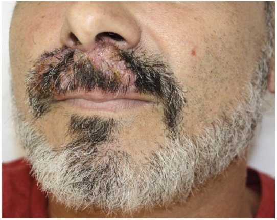
A 48-year-old man attended the emergency department with a 1-month history of facial lesions treated with ciclopirox and mupirocin ointment. He had a healthy pet dog. On examination, he had extensive impetiginized crusts all over the nasolabial triangle. Removal of the crusts revealed erythematous, vegetating plaques on the nasolabial folds (Figure 1).
Figure 1.

Case 1: clinical image of tinea barbae caused by Trichophyton benhamiae. Erythematous vegetating plaques on the nasolabial folds, devoid of hair in some areas.
Case 2
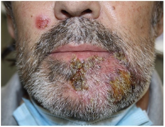
Another 50-year-old man, owner of a healthy dog, came to the outpatient clinic complaining of a two-week facial rash previously treated with topical clobetasol and gentamicin without improvement. On examination he had an erythematous plaque on his chin, with some pustules and erosions covered by serous-hematic crust, and a 2–3 cm nodule in the plaque’s border (Figure 2). Some of his closest family members were being treated for tinea corporis.
Figure 2.

Case 2: clinical picture of tinea barbae caused by Trichophyton benhamiae. Erythematous plaques in the chin and right cheek, with erosions and pustules, with a 2–3 cm nodule in the chin plaque border.
Scales were gathered for fungal culture. In both cases, T. benhamiae was identified by MALDI-TOF (matrix-assisted laser desorption/ionization time-of-flight) mass spectrometry analysis. Terbinafine 250 mg daily for three months completely cleared the lesions in both patients.
Conclusions
T. benhamiae, previously known as Arthroderma (A.) benhamiae, is nowadays a species on its own according to the latest dermatophyte taxonomy, based on the analysis of the internal transcribed spacer (ITS) ribosomal DNA region [1,2].
Every year, more cases of T. benhamiae are being reported worldwide particularly among children. This zoophilic dermatophyte is mainly transmitted by guinea pigs, and seldom by other infected animals like rabbits, cats, dogs and even a fox [3]. Our patients were both adults and only had contact with their pet dogs, which were apparently unaffected; however, we have no information about their veterinary evaluation. Retrospectively our patients couldn’t remember being near a guinea pig, which can be silent carriers of T. benhamiae [4]. We haven’t found studies about T. benhamiae colonization in dogs.
Clinically, it usually causes highly inflammatory tinea corporis and faciei which can be confused with impetigo, delaying a correct diagnosis [5]. There are scattered reports of kerion celsi and onychomycosis [3,5]. To the best of our knowledge, only one case of tinea barbae by T. benhamiae has been previously reported by Braun et al in 2013, a 24-year-old male in which the authors identified A. benhamiae by PCR in the patient and in his guinea pig [6].
Identification of T. benhamiae requires molecular methods due to its similarity to other fungal species in standard cultures. Yellow subtype of this fungus grows in colonies that may be diagnosed as Microsporum canis, and the unusual white subtype is usually identified as T. mentagrophytes. Polymerase chain reaction (PCR) of the ITS region and MALDI-TOF both allow for a correct diagnosis [7].
Treatment is akin to that of other dermatophyte infections. If the infection covers an extensive area or hair follicles are affected, oral treatment is preferred, terbinafine being the first choice [3,5].
Tinea barbae by T. benhamiae seems to be rare. Previous contact with animals, especially guinea pigs, and inflammatory lesions on physical examination should prompt the diagnosis of T. benhamiae infection. Molecular diagnostic methods like PCR and MALDI-TOF are necessary to ensure correct identification of this emergent dermatophyte.
Footnotes
Funding: None.
Competing Interests: None.
Authorship: All authors have contributed significantly to this publication.
References
- 1.Shiraki Y, Hiruma M, Matsuba Y, et al. A case of tinea corporis caused by Arthroderma benhamiae (teleomorph of Tinea mentagrophytes) in a pet shop employee. J Am Acad Dermatol. 2006;55(1):153–154. doi: 10.1016/j.jaad.2005.05.048. [DOI] [PubMed] [Google Scholar]
- 2.de Hoog GS, Dukik K, Monod M, et al. Toward a Novel Multilocus Phylogenetic Taxonomy for the Dermatophytes. Mycopathologia. 2017;182(1–2):5–31. doi: 10.1007/s11046-016-0073-9. [DOI] [PMC free article] [PubMed] [Google Scholar]
- 3.Tan J, Liu X, Gao Z, Yang H, Yang L, Wen H. A case of Tinea Faciei caused by Trichophyton benhamiae: First report in China. BMC Infect Dis. 2020;20(1):1–5. doi: 10.1186/s12879-020-4897-z. [DOI] [PMC free article] [PubMed] [Google Scholar]
- 4.Berlin M, Kupsch C, Ritter L, Stoelcker B, Heusinger A, Gräser Y. German-wide analysis of the prevalence and the propagation factors of the zoonotic dermatophyte trichophyton benhamiae. J Fungi. 2020;6(3):1–11. doi: 10.3390/jof6030161. [DOI] [PMC free article] [PubMed] [Google Scholar]
- 5.Nenoff P, Uhrlaß S, Krüger C, et al. T Trichophyton species of Arthroderma benhamiae - a new infectious agent in dermatology. J Dtsch Dermatol Ges. 2014;12(7):571–581. doi: 10.1111/ddg.12390. [DOI] [PubMed] [Google Scholar]
- 6.Braun SA, Jahn K, Westermann A, Bruch-Gerharz D, Reifenberger J. Tinea barbae profunda durch Arthroderma benhamiae. Der Hautarzt. 2013;64(10):720–722. doi: 10.1007/s00105-013-2646-6. [DOI] [PubMed] [Google Scholar]
- 7.Sabou M, Denis J, Boulanger N, et al. Molecular identification of Trichophyton benhamiae in Strasbourg, France: A 9-year retrospective study. Med Mycol. 2018;56(6):723–734. doi: 10.1093/mmy/myx100. [DOI] [PubMed] [Google Scholar]


