

Introduction
Colored sweat is a sporadic disorder, which can be due to apocrine, true eccrine and pseudo-ecrrine chromhidrosis [1]. Pseudo-chromhidrosis is characterized by excretion of normal colorless sweat, which becomes colored following contact with products of chromogenic microbial or extrinsic chemicals on the skin surface [2]. There are very few case reports of pseudo-chromhidrosis [1–4]. Hereby, we present three sporadic case of red, black and pink pseudo-chromhidrosis.
Case Presentation
Case 1. An apparently healthy 10-year-old female presented with reddish discoloration of both palms for last 7 days. Her brother was also suffering from the similar complaint. Clinical examination revealed palmar creases with reddish secretion (Figure 1). Skin biopsy was negative for lipofuscin granules around eccrine orifices which ruled out apocrine chromhidrosis. Clinical diagnosis of pseudo-chromhidrosis was made.
Figure 1.
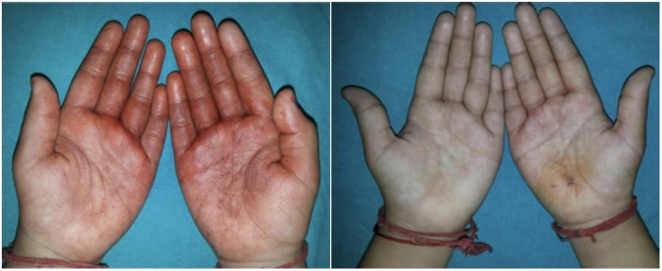
Pre-treatment and post-treatment photographs of patients of red pseudo-chromhidrosis.
Figure 2.

Pre-treatment and post-treatment photographs of patients of pink pseudo-chromhidrosis.
Case 2. A 7-year-old boy presented with progressive darkening of both the palms since last 5 days. Skin biopsy was performed which supported the diagnosis of pseudo-chromhidrosis.
Case 3. A 36-year-old female, known case of rheumatoid arthritis presented with 10 days history of pink discoloration of both palms which aggravated with exertion. She did not give consent for biopsy. Clinical diagnosis of pseudo-chromhidrosis was made.
In all the three cases, color fade on rubbing with absolute alcohol. All patients denied intake of food, vitamin supplements, use of cosmetics or dyes that could have caused such discoloration. There was no specific odor or bleeding from any site. Psychological assessment was normal and family history was insignificant for all patients. Routine biochemical tests, gram staining, fungal scrapping and staining revealed no abnormality. All patients were prescribed oral erythromycin and topical clindamycin which resulted in complete relief with no recurrence for all patients.
Conclusions
Chromhidrosis, ie colored sweat can be produced by eccrine and apocrine glands. Eccrine chromhidrosis due to intrinsic factors is known as true eccrine chromhidrosis and secondary to extrinsic factors is known as pseudo-chromhidrosis. In pseudo-chromhidrosis, sweat produced is colorless but it becomes colored because of chromogen, which include chromogenic bacteria, chemicals, paints, dyes and self-tanning products [2].
Apocrine chromhidrosis is due to presence of increased amount of lipofuscin granules in apocrine glands and their excretion in sweat produces colored sweat [5]. While the diagnosis of eccrine chromhidrosis depends on detailed patient history and exclusion of ingestion of pigments, diagnosis of pseudo-chromhidrosis is based on exclusion of chromhidrosis and successful treatment with antibiotics or antiseptic scrub in this case. Thus, it is very important to distinguish between apocrine or eccrine chromhidrosis and pseudo-chromhidrosis as there is difference in management with different types. Pseudo-chromhidrosis can be treated with topical or systemic antibiotics and cessation of offending agents.
Although, pseudo-chromhidrosis does not constitute a health issue, it may cause psychological stress and social embarrassment. Thus, dermatologist must be aware of the various colors of chromhidrosis in order to determine its actual cause as pseudo-chromhidrosis is easily treatable with antiseptic scrub, topical, systemic antibiotics [2]. On detail literature scan, red pseudo-chromhidrosis has never been reported on palms and pink pseudo-chromhidrois has been once been reported in the literature.
Footnotes
Funding: None.
Competing Interests: None.
Authorship: All authors have contributed significantly to this publication.
References
- 1.Nair PA, Kota RK, Surti NK, Diwan NG, Gandhi SS. Yellow pseudochromhidrosis in a young female. Indian Dermatol Online J. 2017;8(1):42–44. doi: 10.4103/2229-5178.198778. [DOI] [PMC free article] [PubMed] [Google Scholar]
- 2.Koley S, Mandal RK. Red and black pseudochromhidrosis. Indian J Dermatol. 2016;61(4):454–457. doi: 10.4103/0019-5154.185733. [DOI] [PMC free article] [PubMed] [Google Scholar]
- 3.LeFeber WP, Golitz LE. Green foot. Pediatr Dermatol. 1984;2(1):38–40. doi: 10.1111/j.1525-1470.1984.tb00439.x. [DOI] [PubMed] [Google Scholar]
- 4.Mapare A, Tapre V, Khandelwal A. Apocrine chromhidrosis over dorsum of foot. Journal of Dental and Medical Sciences. 2012;2(3):33–34. [Google Scholar]
- 5.Singal A, Thami GP. Red pseudochromhidrosis of the neck. Clin Exp Dermatol. 2004;29(5):548–549. doi: 10.1111/j.1365-2230.2004.01567.x. [DOI] [PubMed] [Google Scholar]