

Case Presentation
A 19-year-old female presented with cicatricial acneiform lesions bilaterally on cheeks and subcutaneous nodular lesions on the bilaterally proximal aspect of the upper and lower extremities (Figure 1, A–E). She was previously diagnosed with acne vulgaris and treated with topical 2% erythromycin and isotretinoin 0.05 % gel for one year. Dermoscopic examination of the facial lesions revealed multiple linear and branching vessels over translucent yellowish-orange globular structures on scar-like areas (Figure 1, A and B). An incisional biopsy was performed from the atrophic plaque on the right arm. The histologic specimen shown lobular panniculitis accompanied by core crumbs, focal fibrin thrombi, focal hyaline necrosis, and lipomembranous changes (Figure 1G). An additional punch biopsy from the facial lesion reported a granuloma formation, perivascular and perifollicular inflammation accompanied by calcification (Figure 1H). Laboratory tests revealed positivity for double stranded DNA and antinuclear antibodies (ANA) homogeneously positive with a titer of 1:320. Anti-SS-A, anti-SS-B and anti-CCP were negative. Serum complement C3 and C4 levels were within normal limits. Based on the clinical, histopathological and laboratory findings, a diagnosis of cutaneous lupus erythematosus was made. The patient was referred to the Rheumatology department and treated with a daily regimen of azathioprine 100 mg, hydroxychloroquine 400 mg, prednisolone 5 mg and indomethacin 25 mg. Topical tacrolimus 0.03% ointment and tretinoin 0.025 % cream were also administered for the treatment of the cutaneous lesions on her face.
Figure 1.
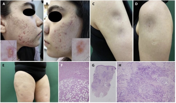
(A,B) Multiple bizarre cicatricial acneiform lesions on cheeks with multiple linear and branching vessels over translucent yellowish-orange globular structure on scar-like areas, dermoscopically. (C) Multiple subcutaneous atrophic nodular lesions on the right arm. (D) Subcutaneous atrophic nodular lesion on the left arm. (E) Multiple subcutaneous nodular lesions on the lower proximal extremity. (F) Septal thickening and lobular inflammation rich in histiocytes (H&E, ×200). (G,H) Periadnexial inflammation with focal granulomatous reaction (H&E, ×40, ×200).
Teaching Point
Lupus erythematosus is one of the major imitators in dermatology and may present with various cutaneous manifestations such as symmetrical confluent erythema and edema overlying the malar cheeks, erythema and edema of the hands, symmetric erythematous eruption of non-indurated macules and papules, scaly annular lesions or papulo-squamous plaques, exfoliative erythroderma, discoid scaly purplish macule or papules, hyperkeratotic/verrucous, bullous, urticarial or mucosal lesions [1]. To date, acneiform presentation and granulomatous formation as seen in our patient have been rarely reported both in cutaneous and systemic lupus erythematous [2–7] Hence, lupus erythematous should be definitely included in the differential diagnosis of refractory acne lesions, and in these cases dermoscopy should be considered mandatory for the diagnosis of difficult-to-treat acneiform eruptions.
Footnotes
Funding: None.
Competing Interests: None.
Authorship: All authors have contributed significantly to this publication.
References
- 1.Walling HW, Sontheimer RD. Cutaneous lupus erythematosus: issues in diagnosis and treatment. Am J Clin Dermatol. 2009;10(6):365–381. doi: 10.2165/11310780-000000000-00000. [DOI] [PubMed] [Google Scholar]
- 2.Sitohang IBS, Rheza AM, Sirait SP, Fitri EM, Suseno LS. Acne Vulgaris Mimicking Cutaneous Lupus Erythematosus in an Adolescent: Report of a Rare Case. Case Rep Dermatol. 2021;13(1):69–74. doi: 10.1159/000511530. [DOI] [PMC free article] [PubMed] [Google Scholar]
- 3.Mohanty B, Kumar B. Systemic lupus erythematosus camouflaging: As refractory acne in a young girl. J Family Med Prim Care. 2019;8(1):276–279. doi: 10.4103/jfmpc.jfmpc_376_18. [DOI] [PMC free article] [PubMed] [Google Scholar]
- 4.Deruelle-Khazaal R, Ségard M, Cottencin-Charrière AC, Carotte-Lefebvre I, Thomas P. Lésions acnéiformes révélatrices d’un lupus érythémateux chronique [Chronic lupus erythematosus presenting as acneiform lesions] Ann Dermatol Venereol. 2002;129(6–7):883–885. [PubMed] [Google Scholar]
- 5.Vieira ML, Marques ERMC, Leda YLA, Noriega LF, Bet DL, Pereira GAAM. Chronic cutaneous lupus erythematosus presenting as atypical acneiform and comedonal plaque: case report and literature review. Lupus. 2018;27(5):853–857. doi: 10.1177/0961203317726377. [DOI] [PubMed] [Google Scholar]
- 6.Farias DF, Gondim RM, Redighieri IP, Muller H, Petri V. Comedonic lupus: a rare presentation of discoid lupus erythematosus. An Bras Dermatol. 2011;86(4 Suppl 1):S89–S91. doi: 10.1590/s0365-05962011000700023. [DOI] [PubMed] [Google Scholar]
- 7.Henostroza-Inga K, Torres-Ibérico R, Atamari-Anahui N, Lipa-Chancolla R. Dermatitis granulomatosa neutrofílica en empalizada como presentación inicial de lupus eritematoso sistémico [Palisaded neutrophilic granulomatous dermatitis as the initial presentation of systemic lupus erythematosus] Bol Med Hosp Infant Mex. 2021;78(6):652–656. doi: 10.24875/BMHIM.21000035. [DOI] [PubMed] [Google Scholar]