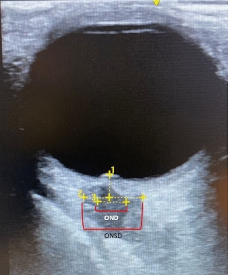
Figure 1. Right ocular ultrasonography in transverse plane.
Papilla protrusion is shown which is suggestive of papilledema indicated by #1. A widened optic nerve sheath diameter (ONSD) of 7.6 mm is indicated by #2 and #3 shows the optic nerve diameter (OND). Both findings are suggestive of elevated intracranial pressure. Not pictured were the results of the ultrasonography of the left eye but were comparable in terms of widened optic nerve sheath diameter and papilledema.

