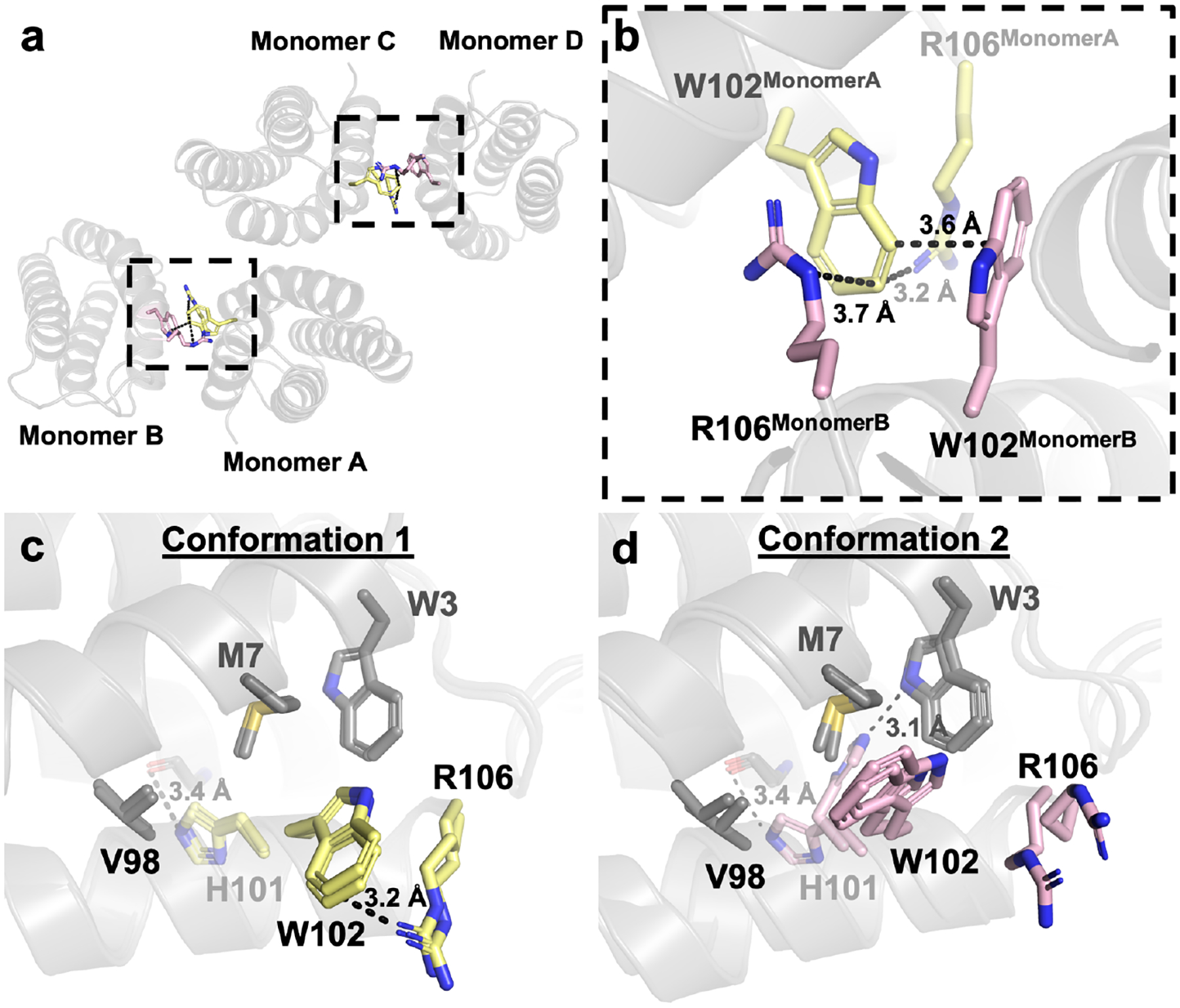
Figure 4.

(a) Crystallographic interface of ApoCyt. (b) Cation-p interactions between W102 and R106 that help stabilize the crystallographic interface. (c) Conformation 1 of the redesigned ApoCyt heme pocket. (d) Conformation 2 of the redesigned heme pocket. Residues that vary between the two conformations are highlighted in yellow and magenta, respectively.
