Background:
Three-dimensional imaging can be used to obtain objective assessments of facial morphology that is useful in a variety of clinical settings. The VECTRA H1 is unique in that it is relatively inexpensive, handheld, and does not require standardized environmental conditions for image capture. Although it provides accurate measurements when imaging relaxed facial expressions, the clinical evaluation of many disorders involves the assessment of facial morphology when performing facial movements. The aim of this study was to assess the accuracy and reliability of the VECTRA H1, specifically when imaging facial movement.
Methods:
The accuracy, intrarater, and interrater reliability of the VECTRA H1 were assessed when imaging four facial expressions: eyebrow lift, smile, snarl, and lip pucker. Fourteen healthy adult subjects had the distances between 13 fiducial facial landmarks measured at rest and the terminal point of each of the four movements by digital caliper and by the VECTRA H1. Intraclass correlation and Bland–Altman limits of agreement were used to determine agreement between measures. The agreement between measurements obtained by five different reviewers was evaluated by intraclass correlation to determine interrater reliability.
Results:
Median correlation between digital caliper and VECTRA H1 measurements ranged from 0.907 (snarl) to 0.921 (smile). Median correlation was very good for both intrarater (0.960–0.975) and interrater reliability (0.997–0.999). The mean absolute error between modalities, and both within and between raters was less than 2 mm for all movements tested.
Conclusion:
The VECTRA H1 met acceptable standards for the assessment of facial morphology when imaging facial movements.
Takeaways
Question: Does the VECTRA H1 3D imaging system provide accurate and reliable measurements of facial morphology when imaging facial movements or expressions?
Findings: The VECTRA H1 met acceptable standards for accuracy and reliability when imaging facial movements.
Meaning: The relatively low cost and portability of the VECTRA H1 3D imaging system make it a feasible option for integration into clinical practice to provide quantitative assessment of facial morphology.
INTRODUCTION
Three-dimensional (3D) imaging systems can be used to objectively assess facial morphology in a variety of research and clinical settings. Some examples include the evaluation of congenital craniofacial dysmorphisms1 and for the early screening of genetic anomalies,2 along with pretreatment planning and treatment monitoring in orthodontics3 and facial aesthetic surgery.4,5 Compared to two-dimensional (2D) imaging, 3D methods have been shown to be more accurate and reliable, especially when assessing complex facial movements such as smiling and oral synkinesis.6,7 For example, when images are taken face-on, 2D imaging is inherently prone to underestimating amplitude in the anteroposterior plane, which may be clinically relevant depending on the purpose of the assessment.6 With significant improvements in technology in recent years, 3D imaging devices have largely replaced 2D systems.8
Numerous 3D imaging systems are widely available; however, many are expensive, lack mobility, and require specialized environmental conditions such as a dedicated room or standardized lighting.9 These limitations make integration into clinical practice difficult and may prevent accurate imaging of patients who are hospitalized or immobile.10 The VECTRA H1 is a relatively affordable handheld device capable of capturing 3D facial images without specific environmental requirements. The system relies on the capture of three consecutive images that are then stitched together to generate a 3D model. This process could theoretically introduce image distortion and increased error in facial morphology measurements, especially when attempting to image facial movements or clinical populations.
Although previous studies have shown that the VECTRA H1 is accurate and reliable, the system has only been validated to image immobile human faces at rest.8,10,11 Furthermore, the manufacturer recommends that images are captured, whereas the patient maintains a relaxed facial expression with their gaze fixed straight ahead and mouth closed to ensure accuracy. This limits the utility of the VECTRA H1 in the clinical evaluation of conditions affecting facial function such as facial nerve palsy, as assessment typically relies on observation while the patient performs a variety of facial movements or expressions.12 Therefore, the purpose of this study was to assess the accuracy and reliability of the VECTRA H1, specifically when imaging facial movements.
METHODS
Population
Approval was obtained from the University of Alberta Ethics Board (Pro00092593) before data collection. Written informed consent was obtained from all subjects. A sample of 14 healthy adult subjects without facial nerve pathology (five women; nine men; 26 ± 7 years) were recruited. Subjects were excluded if they had previously been diagnosed with a condition affecting facial nerve integrity. Five novice medical reviewers were recruited to participate in the study as “raters” to evaluate the outcomes.
3D Imaging System
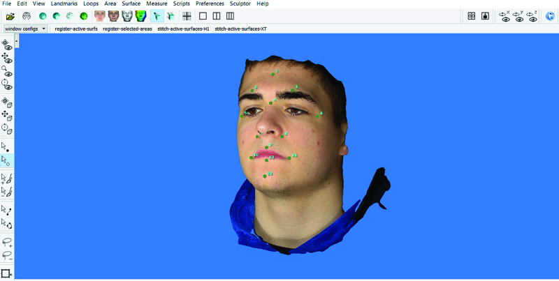
The VECTRA H1 has a built-in image “splitter” that accounts for two perspectives with each image captured creating a single stereophotograph. The VECTRA H1 also has a laser alignment system to assist with image capture precision. For each facial movement, a series of three stereophotographs were taken according to the manufacturer’s specifications at the terminal point of the facial movement being tested. The first image was obtained holding the camera approximately 30 cm below mid-face angled up at the face from a point 45 degrees to the subject’s right side with the alignment laser directed at the midpoint between the zygoma and cheilion (Fig. 1). (See Video [online], which displays the operator view of image acquisition demonstrating the three stereophotograph series and laser alignment functionality.) The second image was taken from directly in front of the subject with the alignment laser directed at the midpoint of the subject’s philtrum (Fig. 1). (See Video [online].) The third image was taken from approximately 30 cm below mid-face angled up at the subject’s face from a point 45 degrees to the subject’s left with the alignment laser directed at the midpoint between the zygoma and cheilion on the left side (Fig. 1). (See Video [online].) The time taken to capture an image is comparable to a standard handheld camera. (See Video [online].) VECTRA H1 software was then used to merge the three stereophotographs, generating the 3D image (VECTRA, Canfield Scientific Inc, Fairfield, N.J.) (Fig. 2).
Fig. 1.
Visual representation of how the lateral and frontal images are captured using the VECTRA H1. Images taken from the Canfield Imaging VECTRA H1 User Guide (http://canfieldupgrade.com/assets/media/VECTRA-H1-User-Guide.pdf). Used with permission from Canfield Scientific, Inc.
Fig. 2.
Operator view of the final three-dimensional image created after stereophotograph merging.
Video 1. displays the operator view of image acquisition demonstrating the three stereophotograph series and laser alignment functionality.) The second image was taken from directly in front of the subject with the alignment laser directed at the midpoint of the subject’s philtrum.
Data Collection
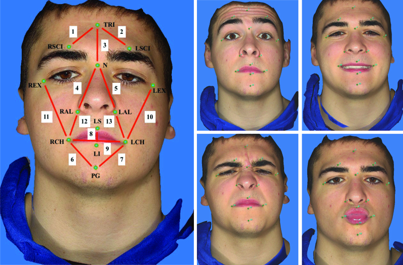
Each subject had 13 fiducial facial landmarks physically marked on their face with a small dot from a fine tip marker approximately 2 mm in diameter (Table 1; Fig. 3). Physical anthropometric measurements of 13 distances between the midpoint of the marked dots at these landmarks were taken at rest and at the end point of each facial movement using a 77-mm digital caliper (General Tools Ultra Tech) (Fig. 3). Participants were instructed to perform each facial movement to a comfortable degree with their gaze fixed straight ahead. Participants were given an opportunity to practice each facial movement before measurement and frequent breaks were provided to prevent fatigue and maintain consistency during caliper measurements. A single researcher conducted all direct facial measurements using a single digital caliper. Landmarks and linear distances were selected based on standard anthropometric measures described in the literature.13,14 The facial movements tested were rest, eyebrow lift, open mouth smile, snarl, and lip pucker. These movements were chosen because they are the primary movements assessed by the Sunnybrook Facial Grading System.15 Eye closure was excluded due to the difficulty in obtaining accurate caliper measurements in the ocular region. Following completion of the 13 anthropometric measurements, the subject was asked to hold the same facial movement, whereas three stereophotographs were obtained with the VECTRA H1 to generate a single 3D image of the facial movement. The three stereographs were then repeated to generate a second 3D image of each facial movement to be used in the intrarater reliability portion of the study. Using VECTRA H1 software, facial markers were applied to the 3D images directly over the facial landmarks previously marked on the subject’s face with fine tip marker. The VECTRA H1 software was then used to measure the distances between facial markers, the same distances measured using the digital caliper.
Table 1.
Facial Markers and Corresponding Coding
| Markers | Coding | Distances |
|---|---|---|
| Lower Trichion | TRI | 1. TRI-RSCI |
| Right eyebrow | RSCI | 2. TRI-LSCI |
| Left eyebrow | LSCI | 3. TRI-N |
| Nasion | N | 4. N-RAL |
| Right lateral canthus | REX | 5. N-LAL |
| Left lateral canthus | LEX | 6. RCH-PG |
| Right alae | RAL | 7. LCH-PG |
| Left alae | LAL | 8. LS-LI |
| Labiale superioris | LS | 9. RCH-LCH |
| Right cheilion | RCH | 10. LEX-LCH |
| Left cheilion | LCH | 11. REX-RCH |
| Labiale inferioris | LI | 12. RAL-RCH |
| Pogonion | PG | 13. LAL-LCH |
Fig. 3.
Facial landmarks and facial movements used for analysis. Facial markers and distances measured (A) and facial movements tested including eyebrow lift (B), smile (C), snarl (D) and lip pucker (E). Green circles refer to facial landmarks described in Table 1. Red lines and corresponding numbering refer to the 13 facial distances measured.
Agreement between digital caliper measurements and measurements obtained using VECTRA H1 software were compared to assess the accuracy of the VECTRA H1 3D imaging system. To assess intrarater reliability, each facial movement was captured by 3D imaging twice by the same researcher and then VECTRA H1 software facial distance measurements were compared between images. Interrater reliability was assessed between the novice medical reviewers who had no previous experience using the VECTRA H1 3D imaging system. The same 13 facial landmarks were placed on the subject’s face using a fine tip marker and the subject was asked to repeat the facial movement protocol. Each reviewer took three stereophotographs of the subject according to manufacturer specifications while the five facial movements were performed to generate one 3D image per facial movement for each rater. Using VECTRA H1 software, facial markers were applied to each 3D image directly over the facial landmarks that had been marked on the subject’s face with fine tip marker, and the same 13 distances between landmarks were measured. The agreement between distances measured by each reviewer was assessed to determine interrater reliability of the VECTRA H1.
Data Analysis
The averaged VECTRA H1 measurements from the two sets of 3D images were used when determining the agreement between VECTRA H1 and digital caliper measurements. To evaluate accuracy, the agreement between measurements obtained using the VECTRA H1 camera were compared to the measurements obtained using the direct caliper for each facial distance using intraclass correlation (ICC). The mean absolute error between modalities was also calculated by subtracting the mean measurement value obtained using the VECTRA H1 from the mean value obtained using the direct caliper for each facial distance. The technical error of measurement (TEM) was also calculated to facilitate more accurate comparison of the results with similar studies in the literature. It is calculated using the following formula: TEM = √ (ΣD2)/2N, where D is the difference between digital caliper and VECTRA H1 measurements and N represents the number of individuals measured.11 Finally, Bland–Altman limits of agreement were used to determine the limits of agreement of the different measurement methods. Limits of agreement were calculated by 1.96 × SD of the differences between modalities ± the mean difference between modalities.
Intrarater reliability was assessed by comparing the facial distances measured on the two sets of duplicate 3D images that were taken by the same researcher. The agreement between distances measured was assessed using ICC, and by calculating the absolute error between images (image 1 − image 2) and Bland–Altman limits of agreement. Interrater reliability was assessed by comparing measurements obtained by the five reviewers using ICC and calculating the mean absolute error between reviewers. The mean of all 13 distances measured by each rater was calculated and compared to determine the mean absolute error between the rater’s measurements.
Statistical analysis was completed using SPSS version 27 (SPSS INC, Chicago, Ill.). ICC estimates and their 95% confidence intervals were based on a single rater, absolute agreement, two-way random effects model. ICC values were interpreted as follows: ≤0.20, poor; 0.21–0.40, fair; 0.41–0.60, moderate; 0.61–0.80, good; 0.81–1.0, very good.16 Regarding absolute error analysis, a mean error of less than 2 mm has been reported in the literature as accurate and precise when evaluating 3D stereophotogrammetry.17–19
RESULTS
Accuracy
The agreement between facial distances measured with the VECTRA H1 system and digital caliper was assessed by ICC, and results are displayed in Table 2. ICC’s ranged from 0.702 (LS-LI) to 0.973 (TRI-RSCI) under the rest condition, 0.640 (LS-LI) to 0.947 (TRI-RSCI) under the eyebrow lift condition, 0.833 (LCH-PG) to 0.966 (REX-RCH) under the smile condition, 0.772 (REX-RCH) to 0.962 (RCH-PG) under the snarl condition, and 0.806 (RAL-RCH) to 0.974 (N-RAL) under the lip pucker condition. The largest median ICC was recorded under the smile condition (0.921) and the lowest under the snarl condition (0.902); however, all median ICCs were deemed to be “very good.” Error between the mean measurements of modalities in millimeters and Bland–Altman limits of agreement are displayed in Table 3. The mean absolute error between modalities is also shown in Table 3, with the largest absolute error being under the snarl condition (1.53 mm) and the smallest under the smile condition (1.08 mm). The greatest error between modalities under the rest (−1.41 mm), eyebrow lift (−2.13 mm), snarl (−2.91 mm), and lip pucker conditions (−2.69 mm) was observed when measuring REX-RCH. Overall, the error between modalities was small, being less than 2 mm for 60 of 65 facial distances measured. Error greater than 2 mm was observed when measuring REX-RCH under the eyebrow lift and snarl conditions, and when measuring LS-LI, LEX-LCH and REX-RCH under the lip pucker condition. The average TEM was 0.87 mm under the rest condition, 1.05 mm under both the eyebrow lift and smile conditions, 1.25 mm under the snarl condition, and 1.14 mm under the lip pucker condition.
Table 2.
ICC and 95% CIs between VECTRA H1 3D Imaging System and Digital Caliper Measurements
| Rest | Eyebrow Lift | Smile | Snarl | Lip Pucker | |
|---|---|---|---|---|---|
| Distance | ICC (95% CI) | ICC (95% CI) | ICC (95% CI) | ICC (95% CI) | ICC (95% CI) |
| 1. TRI-RSCI | 0.973 (0.909–0.991) | 0.947 (0.683–0.986) | 0.919 (0.773–0.973) | 0.918 (0.649–0.976) | 0.920 (0.727-0.975) |
| 2. TRI-LSCI | 0.929 (0.800–0.976) | 0.852 (0.517–0.954) | 0.885 (0.596–0.965) | 0.880 (0.667–0.960) | 0.892 (0.666–0.965) |
| 3. TRI-N | 0.938 (0.794–0.981) | 0.914 (0.579–0.976) | 0.921 (0.710–0.976) | 0.905 (0.685–0.970) | 0.949 (0.526–0.988) |
| 4. N-RAL | 0.930 (0.730–0.979) | 0.946 (0.580–0.987) | 0.940 (0.828–0.980) | 0.909 (0.532–0.975) | 0.974 (0.873–0.993) |
| 5. N-LAL | 0.963 (0.824–0.989) | 0.926 (0.536–0.981) | 0.928 (0.791–0.976) | 0.876 (0.667–0.958) | 0.952 (0.863–0.984) |
| 6. RCH-PG | 0.939 (0.823–0.980) | 0.907 (0.737–0.969) | 0.906 (0.743–0.969) | 0.962 (0.889–0.988) | 0.934 (0.531–0.983) |
| 7. LCH-PG | 0.918 (0.611–0.977) | 0.906 (0.727–0.969) | 0.833 (0.448–0.948) | 0.902 (0.729–0.967) | 0.939 (0.781–0.981) |
| 8. LS-LI | 0.702 (−0.081 to 0.926) | 0.640 (−0.019 to 0.887) | 0.929 (0.769–0.978) | 0.876 (0.513–0.963) | 0.887 (0.243–0.972) |
| 9. RCH-LCH | 0.919 (0.689–0.976) | 0.825 (0.487–0.945) | 0.950 (0.856–0.984) | 0.912 (0.756–0.971) | 0.932 (0.649–0.981) |
| 10. LEX-LCH | 0.911 (0.488–0.977) | 0.836 (0.441–0.949) | 0.925 (0.791–0.975) | 0.915 (0.715–0.974) | 0.858 (−0.026 to 0.970) |
| 11. REX-RCH | 0.919 (0.388–0.980) | 0.835 (0.090–0.959) | 0.966 (0.835–0.990) | 0.772 (−0.063 to 0.949) | 0.836 (−0.029 to 0.963) |
| 12. RAL-RCH | 0.892 (0.695–0.964) | 0.913 (0.750–0.971) | 0.887 (0.695–0.962) | 0.890 (0.699–0.963) | 0.806 (0.512–0.933) |
| 13. LAL-LCH | 0.924 (0.781–0.975) | 0.912 (0.752–0.971) | 0.874 (0.663–0.957) | 0.822 (0.530–0.939) | 0.887 (0.692–0.962) |
| Median | 0.924 | 0.907 | 0.921 | 0.902 | 0.920 |
CI, confidence interval.
Table 3.
Difference between Means for Each Facial Distance Measured and Bland–Altman Lower and Upper Limits of Agreement Estimated by Mean Difference ± 1.96 SD of the Differences between VECTRA H1 and Direct Caliper Measurements (mm)
| Rest | Eyebrow Lift | Smile | Snarl | Lip Pucker | ||||||
|---|---|---|---|---|---|---|---|---|---|---|
| Distance | Error | Limits | Error | Limits | Error | Limits | Error | Limits | Error | Limits |
| 1. TRI-RSCI | −0.36 | −1.72, 1.01 | −0.66 | −2.16, 0.83 | −0.48 | −3.05, 2.09 | −0.82 | −2.98, 1.33 | −0.64 | −2.74, 1.45 |
| 2. TRI-LSCI | −0.22 | −2.13, 1.69 | −0.76 | −3.00, 1.48 | −0.67 | −2.64, 1.29 | −0.10 | −3.07, 2.86 | −0.55 | −2.49, 1.39 |
| 3. TRI-N | −0.69 | −3.14, 1.75 | −0.74 | −2.49, 1.00 | −0.97 | −3.91, 1.97 | −0.91 | −3.87, 2.06 | −0.96 | −2.67, 0.75 |
| 4. N-RAL | −0.77 | −3.03, 1.49 | −1.08 | −3.16, 1.00 | −0.39 | −2.80, 2.01 | −1.24 | −4.04, 1.55 | −0.56 | −2.08, 0.95 |
| 5. N-LAL | −0.55 | −2.03, 0.93 | −1.16 | −3.57, 1.24 | −0.12 | −2.70, 2.45 | −0.60 | −4.31, 3.10 | −0.37 | −2.76, 2.02 |
| 6. RCH-PG | −0.14 | −2.93, 2.66 | −0.17 | −3.56, 3.21 | −0.52 | −3.94, 2.90 | 0.29 | −2.33, 2.91 | −0.92 | −2.74, 0.89 |
| 7. LCH-PG | −0.74 | −3.63, 2.15 | −0.77 | −4.07, 2.53 | −1.35 | −5.09, 2.39 | −0.36 | −3.83, 3.10 | −0.69 | −2.91, 1.54 |
| 8. LS-LI | −1.32 | −2.77, 0.12 | −1.40 | −3.96, 1.17 | −0.65 | −2.96, 1.66 | −0.91 | −3.23, 1.42 | −2.10 | −5.68, 1.48 |
| 9. RCH-LCH | −0.72 | −2.81, 1.37 | −0.73 | −3.40, 1.94 | −0.31 | −3.57, 2.94 | 0.45 | −3.06, 3.96 | −1.14 | −3.83, 1.55 |
| 10. LEX-LCH | −1.22 | −3.75, 1.32 | −1.33 | −4.91, 2.26 | −0.63 | −4.58, 3.32 | −0.86 | −3.71, 1.98 | −2.45 | −5.16, 0.26 |
| 11. REX-RCH | −1.41 | −3.95, 1.12 | −2.13 | −5.55, 1.29 | −1.00 | −3.66, 1.66 | −2.91 | −5.67, −0.15 | −2.69 | −5.89, 0.51 |
| 12. RAL-RCH | 0.05 | −2.25, 2.36 | −0.06 | −1.67, 1.56 | 0.37 | −2.12, 2.87 | 0.39 | −2.88, 3.66 | 0.52 | −2.20, 3.24 |
| 13. LAL-LCH | 0.04 | −1.84, 1.93 | 0.18 | −1.95, 2.32 | −0.27 | −2.53, 1.99 | 1.02 | −3.25, 5.28 | 0.30 | −2.17, 2.77 |
| Mean (SD) | 1.08 (0.78) | 1.26 (1.05) | 1.26 (0.94) | 1.53 (1.07) | 1.35 (1.18) |
Mean absolute difference between VECTRA H1 and direct caliper measurements are also shown ( Mean ).
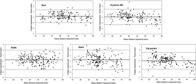
Bland–Altman plots comparing digital caliper and VECTRA H1 are shown in Figure 4. These plots illustrate the differences between measurement modalities and show the bias and the 95% limits of agreement between digital caliper and VECTRA H1 measurements for each facial movement tested. A bias of −0.62 mm and 95% confidence limit of −2.94 to 1.70 mm was shown under the rest condition (Fig. 4A). Results from the other facial movement conditions include a bias of −0.83 mm and the confidence limit of −3.60 to 1.94 mm under the eyebrow lift condition (Fig. 4B), a bias of −0.54 mm and the confidence limit of −3.43 to 2.35 mm under the smile condition (Fig. 4C), a bias of −0.51 and the confidence limit of −4.04 to 3.02 under the snarl condition (Fig. 4D), and a bias of −0.94 mm and the confidence limit of −3.93 to 2.05 under the lip pucker condition (Fig. 4E).
Fig. 4.
Bland–Altman plots showing the mean difference between caliper and VECTRA H1 measurements as well as the amount of scatter around the mean for rest (A), eyebrow lift (B), smile (C), snarl (D) and lip pucker (E) facial movements.
Intrarater Reliability
The same rater repeated image capture and facial distance measurements using the VECTRA H1 system for all 14 subjects to assess intrarater reliability. One subject was removed from analysis under the eyebrow lift condition due to a corrupted image that prevented successful 3D image merging. ICCs ranged from 0.936 (RAL-RCH) to 0.993 (REX-RCH) under the rest condition, 0.911 (TRI-N, RAL-RCH) to 0.988 (REX-RCH) under the eyebrow lift condition, 0.899 (RAL-RCH) to 0.991 (TRI-N) under the smile condition, 0.912 (RCH-LCH) to 0.986 (RCH-PG) under the snarl condition, and 0.876 (LAL-LCH) to 0.992 (TRI-N) under the lip pucker condition (Table 4). Median ICCs were “very good,” ranging from 0.946 (snarl) to 0.975 (smile) (Table 4). The error between the means of the two sets of measurements taken in millimeters and the Bland–Altman limits of agreement are displayed in Table 5 along with the mean absolute error between 3D image sets. The error between measurement sets was very small when calculated for each individual distance measured, with the greatest error being −0.77 mm under the smile condition (RCH-LCH). Mean absolute error was also less than 1 mm for all facial movements tested.
Table 4.
ICC and 95% CIs Comparing Two Sets of Measurements Taken by the Same Rater with the VECTRA H1 3D Imaging System
| Rest | Eyebrow Lift | Smile | Snarl | Lip Pucker | |
|---|---|---|---|---|---|
| Distance | ICC (95% CI) | ICC (95% CI) | ICC (95% CI) | ICC (95% CI) | ICC (95% CI) |
| 1. TRI-RSCI | 0.991 (0.973–0.997) | 0.960 (0.875–0.988) | 0.990 (0.971–0.997) | 0.936 (0.816–0.979) | 0.970 (0.882–0.991) |
| 2. TRI-LSCI | 0.991 (0.971–0.997) | 0.960 (0.875–0.987) | 0.984 (0.950–0.995) | 0.914 (0.761–0.971) | 0.984 (0.954–0.995) |
| 3. TRI-N | 0.990 (0.970–0.997) | 0.911 (0.734–0.972) | 0.991 (0.972–0.997) | 0.951 (0.857–0.984) | 0.992 (0.968–0.998) |
| 4. N-RAL | 0.989 (0.967–0.997) | 0.980 (0.939–0.994) | 0.984 (0.952–0.995) | 0.973 (0.917–0.991) | 0.981 (0.942–0.994) |
| 5. N-LAL | 0.988 (0.954–0.996) | 0.980 (0.935–0.994) | 0.974 (0.923–0.992) | 0.946 (0.714–0.985) | 0.948 (0.847–0.983) |
| 6. RCH-PG | 0.978 (0.935–0.993) | 0.983 (0.947–0.995) | 0.967 (0.896–0.989) | 0.986 (0.959–0.995) | 0.947 (0.844–0.983) |
| 7. LCH-PG | 0.981 (0.944–0.994) | 0.932 (0.747–0.980) | 0.906 (0.737–0.969) | 0.975 (0.925–0.992) | 0.956 (0.873–0.986) |
| 8. LS-LI | 0.940 (0.823–0.980) | 0.971 (0.909–0.991) | 0.956 (0.872–0.986) | 0.915 (0.714–0.973) | 0.972 (0.917–0.991) |
| 9. RCH-LCH | 0.969 (0.903–0.990) | 0.940 (0.817–0.981) | 0.975 (0.836–0.994) | 0.912 (0.747–0.971) | 0.945 (0.843–0.982) |
| 10. LEX-LCH | 0.984 (0.953–0.995) | 0.983 (0.944–0.995) | 0.976 (0.909–0.993) | 0.932 (0.806–0.978) | 0.988 (0.964–0.996) |
| 11. REX-RCH | 0.993 (0.979–0.998) | 0.988 (0.961–0.996) | 0.978 (0.936–0.993) | 0.974 (0.923–0.991) | 0.968 (0.906–0.989) |
| 12. RAL-RCH | 0.936 (0.818–0.979) | 0.911 (0.744–0.972) | 0.899 (0.720–0.966) | 0.915 (0.759–0.972) | 0.915 (0.756–0.972) |
| 13. LAL-LCH | 0.972 (0.918–0.991) | 0.950 (0.845–0.985) | 0.936 (0.813–0.979) | 0.963 (0.880–0.988) | 0.876 (0.668–0.958) |
| Median | 0.984 | 0.960 | 0.975 | 0.946 | 0.968 |
CI, confidence interval.
Table 5.
Difference between Means for Each Facial Distance Measured and Bland–Altman Lower and Upper Limits of Agreement Estimated by Mean Difference ± 1.96 SD Comparing 2 Sets of Measurements Taken by the Same Rater with the VECTRA H1 3D Imaging System
| Rest | Eyebrow Lift | Smile | Snarl | Lip Pucker | ||||||
|---|---|---|---|---|---|---|---|---|---|---|
| Distance | Error | Limits | Error | Limits | Error | Limits | Error | Limits | Error | Limits |
| 1. TRI-RSCI | 0.10 | −0.74, 0.95 | 0.02 | −1.63, 1.68 | −0.08 | −1.04, 0.88 | −0.45 | −2.73, 1.83 | −0.41 | −1.72, 0.89 |
| 2. TRI-LSCI | −0.01 | −0.72, 0.70 | −0.11 | −1.69, 1.48 | −0.04 | −1.01, 0.94 | −0.42 | −2.92, 2.08 | −0.14 | −0.98, 0.70 |
| 3. TRI-N | −0.05 | −1.23, 1.12 | 0.10 | −2.19, 2.39 | −0.19 | −1.34, 0.96 | −0.24 | −2.86, 2.38 | −0.27 | −1.11, 0.58 |
| 4. N-RAL | 0.06 | −0.93, 1.05 | 0.30 | −1.46, 2.06 | −0.03 | −1.39, 1.34 | 0.40 | −1.51, 2.30 | 0.08 | −1.60, 1.75 |
| 5. N-LAL | 0.27 | −0.62, 1.15 | −0.04 | −1.93, 1.86 | −0.09 | −1.68, 1.49 | 0.80 | −1.11, 2.72 | −0.16 | −2.77, 2.45 |
| 6. RCH-PG | 0.14 | −1.39, 1.66 | −0.08 | −1.61, 1.44 | −0.45 | −2.34, 1.45 | 0.20 | −1.42, 1.83 | 0.13 | −2.07, 2.32 |
| 7. LCH-PG | 0.19 | −1.18, 1.55 | 0.83 | −1.77, 3.43 | −0.32 | −3.80, 3.15 | −0.06 | −1.95, 1.83 | −0.22 | −2.42, 1.97 |
| 8. LS-LI | −0.01 | −1.34, 1.33 | 0.19 | −0.80, 1.18 | −0.17 | −2.12, 1.77 | 0.65 | −1.50, 2.80 | −0.22 | −2.90, 2.46 |
| 9. RCH-LCH | −0.30 | −1.65, 1.05 | −0.05 | −2.32, 2.21 | −0.77 | −2.49, 0.96 | −0.10 | −3.99, 3.79 | 0.51 | −2.67, 3.68 |
| 10. LEX-LCH | 0.11 | −1.40, 1.62 | −0.30 | −1.71, 1.11 | 0.60 | −1.38, 2.57 | 0.52 | −2.22, 3.25 | −0.11 | −1.77, 1.55 |
| 11. REX-RCH | −0.20 | −1.22, 0.83 | 0.20 | −1.22, 1.61 | 0.42 | −1.98, 2.81 | 0.28 | −1.64, 2.20 | −0.35 | −3.09, 2.38 |
| 12. RAL-RCH | −0.15 | −1.70, 1.40 | −0.24 | −1.92, 1.44 | −0.18 | −2.50, 2.14 | −0.60 | −3.49, 2.29 | 0.02 | −1.78, 1.82 |
| 13. LAL-LCH | −0.17 | −1.37, 1.03 | −0.01 | −1.63, 1.61 | 0.09 | −1.76, 1.93 | −0.48 | −2.31, 1.36 | 0.34 | −2.37, 3.04 |
| Mean (SD) | 0.48 (0.40) | 0.67 (0.63) | 0.71 (0.72) | 0.90 (0.88) | 0.76 (0.79) |
Mean absolute difference between both sets of measurements are also shown ( Mean ).
Interrater Reliability
Mean measurements obtained by each rater for the five facial movements are shown in Table 6. The mean error between reviewers was calculated based on these means, and very small differences between reviewers was noted, with the average error between reviewers being less than 0.5 mm. Group ICCs for each facial movement were very good, ranging from 0.997 (smile) to 0.999 (rest, eyebrow lift).
Table 6.
Mean Distances Measured by Each Reviewer, ICC between Rater Measurements for Each Facial Movement Tested and Mean Error in Measurements between Reviewers
| Rest | Eyebrow Lift | Smile | Snarl | Lip Pucker | |
|---|---|---|---|---|---|
| Raters | Mean (mm) | Mean (mm) | Mean (mm) | Mean (mm) | Mean (mm) |
| R1 | 46.43 | 46.21 | 48.75 | 42.80 | 47.42 |
| R2 | 46.36 | 46.50 | 48.07 | 43.00 | 47.86 |
| R3 | 46.51 | 46.46 | 48.76 | 43.34 | 48.48 |
| R4 | 46.38 | 46.42 | 48.65 | 43.43 | 48.87 |
| R5 | 46.48 | 46.40 | 48.52 | 43.41 | 48.58 |
| ICC (CI) | 0.999 (0.999–1.00) | 0.999 (0.998–1.00) | 0.997 (0.994–0.999) | 0.998 (0.996–0.999) | 0.998 (0.995–0.999) |
| Mean error (SD) | 0.08 (0.02) | 0.10 (0.10) | 0.28 (0.20) | 0.25 (0.23) | 0.43 (0.19) |
The mean error between reviewers was calculated by averaging the absolute value of the error between reviewers mean measurements ( Mean error ).
DISCUSSION
The results of the present study showed that the VECTRA H1 3D imaging system can accurately and reliably measure distances between fine facial landmarks while subjects perform facial movements common to facial nerve palsy assessments. A high level of agreement was found between VECTRA H1 and digital caliper measurements as median ICC coefficients ranged from 0.902 under the snarl condition to 0.921 under the smile condition. The mean absolute error between the VECTRA H1 and digital caliper measurements was under 2 mm under all facial movement conditions tested, ranging from 1.08 mm under the smile condition to 1.53 mm under the snarl condition, suggesting a clinically acceptable level of accuracy.17–19 Average TEM found in the present study (rest: 0.87 mm; eyebrow lift: 1.05 mm; smile: 1.05 mm; snarl: 1.25 mm; lip pucker: 1.14 mm) was comparable to previous studies that reported average TEM of 0.84 and 1.17 mm.11,20
Intrarater reliability of the VECTRA H1 was excellent, as median ICC coefficients ranged from 0.946 under the snarl condition to 0.975 under the smile condition. The mean absolute error between the researcher’s two sets of measurements was also small (<1 mm). Interrater reliability of the VECTRA H1 was excellent, as ICCs approached 1.00 (0.997–0.999) and mean absolute error between reviewers was less than 0.5 mm under all facial movement conditions tested.
Although 3D photogrammetry is an attractive technology for grading facial nerve function, many currently available 3D photogrammetry technologies have significant drawbacks that make them difficult to integrate into clinical practice. Examples include the requirement of bulky and expensive equipment,21,22 standardized lighting conditions,23,24 physical facial markers,22,25 or multiple cameras that require frequent calibration.26–29 The VECTRA H1 is a handheld device that is relatively inexpensive and does not require special lighting conditions or physical facial markers to be placed on the subject. A potential drawback of the VECTRA H1 system, however, is the requirement of a three-image capture procedure which makes image distortion and measurement error more probable if the patient cannot remain still or if images are not taken precisely.
Numerous validation studies of the VECTRA H1 have been conducted and have determined that the system meets clinically acceptable standards when imaging static, resting human faces, however none have investigated the system’s ability to image facial movement. Camison et al11 studied the accuracy and repeatability of the VECTRA H1 by comparing anthropometric measurements obtained with the VECTRA H1 to those of a previously validated imaging system, the 3dMDface system. The authors reported high levels of agreement and submillimeter error between the two systems when imaging 26 adult subjects. Repeatability of the system was also evaluated and deemed to be sufficient for clinical and research applications by comparing repeated 3D measurements on a mannequin head.
Gibelli et al,10 Kim et al,20 and Junqueira-Júnior et al30 used a similar approach to validation by choosing to compare facial measurements obtained with the portable VECTRA H1 to the static VECTRA M3, another Canfield Imaging product. Similar results were reported with the error both between and within imaging systems being less than or equal to 2 mm. Interrater reliability was also excellent, with Junqueira-Júnior et al30 reporting a mean absolute error between raters less than 0.39 mm and Kim et al20 reporting ICCs greater than 0.900. The methodology used by Kim et al20 was unique in that the researchers also compared 3D measurements to direct caliper measurements, making results more easily comparable to the present study. Interestingly, the authors found that the VECTRA H1 yielded larger measurements compared to the direct caliper with a mean difference of 1.74 and 0.94 mm depending on the rater. Mean absolute differences between measures were similar in our study (1.08 mm under the rest condition) and VECTRA H1 measurements were also consistently larger than those obtained using digital caliper (Table 3), suggesting that the VECTRA H1 may tend to overestimate facial distances.
An important study limitation is the use of healthy individuals as subjects. Results may differ in subjects diagnosed with facial nerve palsy or movement disorders given that they may experience more involuntary facial movements or have difficulty holding static facial expressions. Nonetheless, this study serves to ensure that the VECTRA H1 can accurately and reliably measure facial distances during facial movement before testing the imaging system on a clinical population.
CONCLUSIONS
Linear measurements of facial distances taken using the VECTRA H1 3D imaging system closely correlated with measurements obtained using a digital caliper when imaging subjects performing four common facial movements. Intrarater and interrater reliability of the VECTRA H1 when imaging facial movement was also excellent, evidenced by high ICCs and low absolute errors. The results of this study demonstrate that the VECTRA H1 is capable of accurately and reliably measuring facial distances even when subjects perform facial movement. The portability and relatively low cost of the VECTRA H1 make this system a feasible option for integration into clinical practice.
PATIENT CONSENT
The patient provided written consent for the use of his image.
Footnotes
Published online 23 February 2023.
Disclosure: The authors have no financial interest to declare in relation to the content of this article.
Related Digital Media are available in the full-text version of the article on www.PRSGlobalOpen.com.
REFERENCES
- 1.Pucciarelli V, Bertoli S, Codari M, et al. Facial evaluation in holoprosencephaly. J Craniofac Surg. 2017;28:e22–e28. [DOI] [PubMed] [Google Scholar]
- 2.Pucciarelli V, Bertoli S, Codari M, et al. The face of Glut1-DS patients: a 3D craniofacial morphometric analysis. Clin Anat. 2017;30:644–652. [DOI] [PubMed] [Google Scholar]
- 3.Alshammery FA. Three dimensional (3D) imaging techniques in orthodontics—an update. J Family Med Prim Care. 2020;9:2626–2630. [DOI] [PMC free article] [PubMed] [Google Scholar]
- 4.Kau CH, Richmond S, Incrapera A, et al. Three-dimensional surface acquisition systems for the study of facial morphology and their application to maxillofacial surgery. Int J Med Robot. 2007;3:97–110. [DOI] [PubMed] [Google Scholar]
- 5.Verhulst A, Hol M, Vreeken R, et al. Three-dimensional imaging of the face: a comparison between three different imaging modalities. Aesthet Surg J. 2018;38:579–585. [DOI] [PubMed] [Google Scholar]
- 6.Gross MM, Trotman CA, Moffatt KS. A comparison of three-dimensional and two-dimensional analyses of facial motion. Angle Orthod. 1996;66:189–194. [DOI] [PubMed] [Google Scholar]
- 7.Ghoddousi H, Edler R, Haers P, et al. Comparison of three methods of facial measurement. Int J Oral Maxillofac Surg. 2007;36:250–258. [DOI] [PubMed] [Google Scholar]
- 8.Savoldelli C, Benat G, Castillo L, et al. Accuracy, repeatability and reproducibility of a handheld three-dimensional facial imaging device: the Vectra H1. J Stomatol Oral Maxillofac Surg. 2019;120:289–296. [DOI] [PubMed] [Google Scholar]
- 9.Tzou CH, Artner NM, Pona I, et al. Comparison of three-dimensional surface-imaging systems. J Plast Reconstr Aesthet Surg. 2014;67:489–497. [DOI] [PubMed] [Google Scholar]
- 10.Gibelli D, Pucciarelli V, Cappella A, et al. Are portable stereophotogrammetric devices reliable in facial imaging? A validation study of VECTRA H1 device. J Oral Maxillofac Surg. 2018;76:1772–1784. [DOI] [PubMed] [Google Scholar]
- 11.Camison L, Bykowski M, Lee WW, et al. Validation of the Vectra H1 portable three-dimensional photogrammetry system for facial imaging. Int J Oral Maxillofac Surg. 2018;47:403–410. [DOI] [PMC free article] [PubMed] [Google Scholar]
- 12.Fattah AY, Gurusinghe ADR, Gavilan J, et al. ; Sir Charles Bell Society. Facial nerve grading instruments: systematic review of the literature and suggestion for uniformity. Plast Reconstr Surg. 2015;135:569–579. [DOI] [PubMed] [Google Scholar]
- 13.Farkas LG, Posnick JC, Hreczko TM. Anthropometric growth study of the head. Cleft Palate Craniofac J. 1992; 29:303–308.. [DOI] [PubMed] [Google Scholar]
- 14.Deutsch C.K., Shell A.R., Francis R.W., Bird B.D. (2012) The Farkas system of craniofacial anthropometry: methodology and normative databases. In: Preedy V. (eds) Handbook of Anthropometry. Springer; New York, N.Y. [Google Scholar]
- 15.Ross BG, Fradet G, Nedzelski JM. Development of a sensitive clinical facial grading system. Otolaryngol Head Neck Surg. 1996;114:380–386. [DOI] [PubMed] [Google Scholar]
- 16.Altman DG. Practical Statistics for Medical Research. London: Chapman and Hall; 1991. [Google Scholar]
- 17.Weinberg SM, Scott NM, Neiswanger K, et al. Digital three-dimensional photogrammetry: evaluation of anthropometric precision and accuracy using a Genex 3D camera system. Cleft Palate Craniofac J. 2004;41:507–518. [DOI] [PubMed] [Google Scholar]
- 18.Weinberg SM, Naidoo S, Govier DP, et al. Anthropometric precision and accuracy of digital three-dimensional photogrammetry: comparing the Genex and 3dMD imaging systems with one another and with direct anthropometry. J Craniofac Surg. 2006;17:477–483. [DOI] [PubMed] [Google Scholar]
- 19.Wong JY, Oh AK, Ohta E, et al. Validity and reliability of craniofacial anthropometric measurement of 3D digital photogrammetric images. Cleft Palate Craniofac J. 2008;45:232–239. [DOI] [PubMed] [Google Scholar]
- 20.Kim AJ, Gu D, Chandiramani R, Linjawi I, Deutsch ICK, Allareddy V, et al. Accuracy and reliability of digital craniofacial measurements using a small-format, handheld 3D camera. Orthod Craniofacial Res. 2018;21:132–139. [DOI] [PubMed] [Google Scholar]
- 21.Sawyer AR, See M, Nduka C. Quantitative analysis of normal smile with 3D stereophotogrammetry–an aid to facial reanimation. J Plast Reconstr Aesthet Surg. 2010;63:65–72. [DOI] [PubMed] [Google Scholar]
- 22.Frey M, Tzou CH, Michaelidou M, et al. 3D video analysis of facial movements. Facial Plast Surg Clin North Am. 2011;19:639–46, viii. [DOI] [PubMed] [Google Scholar]
- 23.Nakata S, Sato Y, Gunaratne P, Suzuki Y, Sugiura S, Nakashima T. Quantification of facial motion for objective evaluation using a high-speed three-dimensional face measurement system-A pilot study. Otolo Neurotol. 2006;27:1023–1029. [DOI] [PubMed] [Google Scholar]
- 24.Katsumi S, Esaki S, Hattori K, et al. Quantitative analysis of facial palsy using a three-dimensional facial motion measurement system. Auris Nasus Larynx. 2015;42:275–283. [DOI] [PubMed] [Google Scholar]
- 25.Sforza C, Guzzo M, Mapelli A, et al. Facial mimicry after conservative parotidectomy: a three-dimensional optoelectronic study. Int J Oral Maxillofac Surg. 2012;41:986–993. [DOI] [PubMed] [Google Scholar]
- 26.Jorge JJ, Jr, Pialarissi PR, Borges GC, et al. Objective computerized evaluation of normal patterns of facial muscles contraction. Braz J Otorhinolaryngol. 2012;78:41–51. [DOI] [PMC free article] [PubMed] [Google Scholar]
- 27.Lee JG, Jung SJ, Lee HJ, et al. Quantitative anatomical analysis of facial expression using a 3D motion capture system: application to cosmetic surgery and facial recognition technology. Clin Anat. 2015;28:735–744. [DOI] [PubMed] [Google Scholar]
- 28.Hontanilla B, Aubá C. Automatic three-dimensional quantitative analysis for evaluation of facial movement. J Plast Reconstr Aesthet Surg. 2008;61:18–30. [DOI] [PubMed] [Google Scholar]
- 29.Schimmel M, Leemann B, Christou P, et al. Quantitative assessment of facial muscle impairment in patients with hemispheric stroke. J Oral Rehabil. 2011;38:800–809. [DOI] [PubMed] [Google Scholar]
- 30.Junqueira-Júnior AA, Magri LV, Cazal MS, et al. Accuracy evaluation of tridimensional images performed by portable stereophotogrammetric system. Rev Odontol UNESP. 2019;48:1–15. [Google Scholar]


