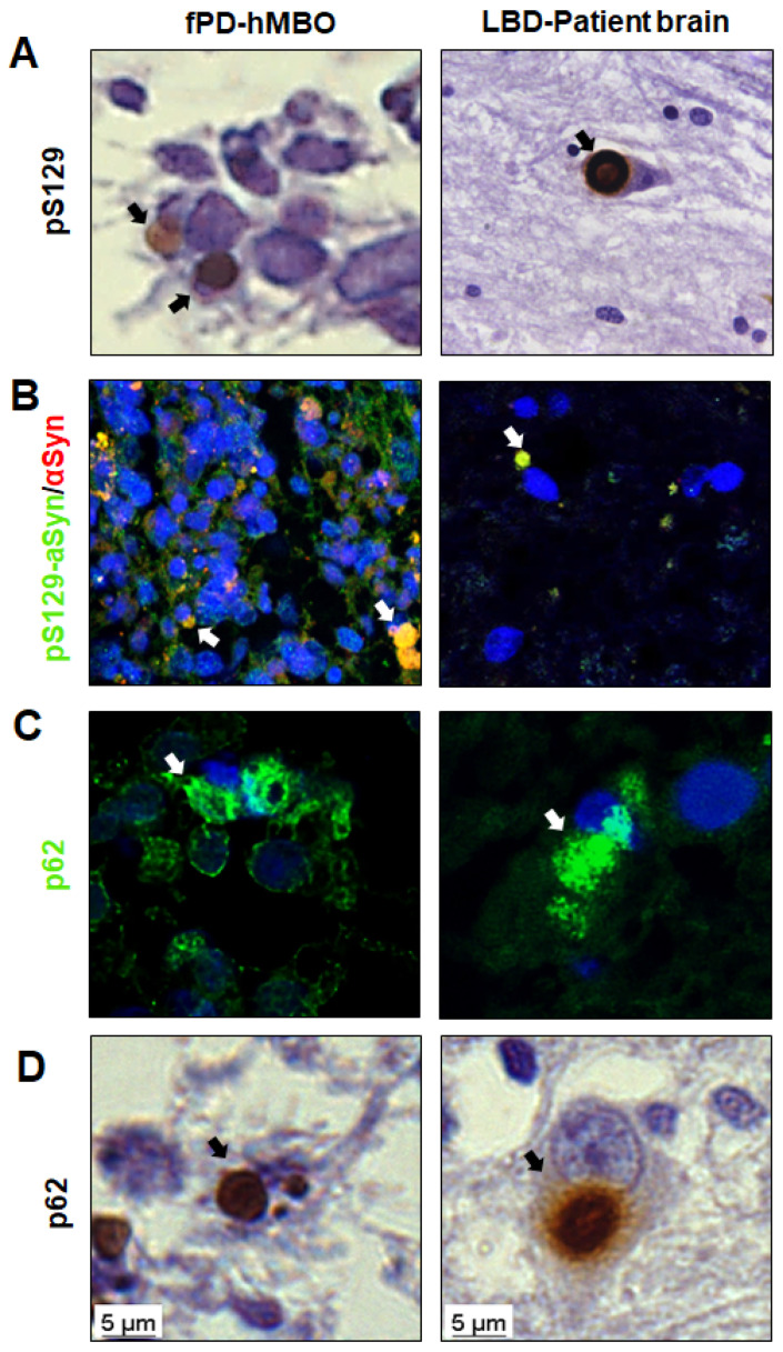
Figure 4.
Morphology and composition of LB-like αSyn accumulates in fPD-hMBOs. (A). Representative immunohistochemistry (IHC) images for pS129 counterstained with hematoxylin. (B). Representative IF images for pS129/αSyn and DAPI. (C). Representative IF images for p62 and DAPI. (D). Representative IHC images for p62 counterstained with hematoxylin. In all panels, the left shows the results with hMBOs, and the right shows the brain of patient affected by LBD as positive control. Arrows show the LB-like deposits in fPD-hMBOs and LBs in LBD brain sections. Scale bar 5 µm.

