Abstract
Biological aging is characterized by irreversible cell cycle blockade, a decreased capacity for tissue regeneration, and an increased risk of age-related diseases and mortality. A variety of genetic and epigenetic factors regulate aging, including the abnormal expression of aging-related genes, increased DNA methylation levels, altered histone modifications, and unbalanced protein translation homeostasis. The epitranscriptome is also closely associated with aging. Aging is regulated by both genetic and epigenetic factors, with significant variability, heterogeneity, and plasticity. Understanding the complex genetic and epigenetic mechanisms of aging will aid the identification of aging-related markers, which may in turn aid the development of effective interventions against this process. This review summarizes the latest research in the field of aging from a genetic and epigenetic perspective. We analyze the relationships between aging-related genes, examine the possibility of reversing the aging process by altering epigenetic age.
Keywords: healthy aging, senescence, genetics, epigenetics, epitranscriptome
1. Introduction
Throughout the centuries, there are countless examples of human beings attempting to escape the inevitable: the near-ubiquitous reality of aging and death. While such attempts have been in vain thus far, a number of theories regarding the occurrence of aging have been developed. Some believe that aging is determined primarily by genes [1], while others hypothesize that accumulated cellular damage is the main cause of systemic aging [2]. In fact, aging is a complex process resulting from multiple factors, including genetic and epigenetic molecular markers, such as telomere depletion, genomic instability, and epigenetic alterations [3]. With rapid advances being made in experimental techniques, an increasing body of evidence suggests that these genetic and epigenetic factors are not only individually associated with aging, but that they may work together to drive this process.
Cellular senescence is one of the important factors that trigger aging, and it is also the most widely studied target of aging intervention [4]. Hayflick first proposed the concept of cellular senescence, and he found that mammalian cell cultures divide to a certain stage, then appear senescent or die, which known as the Hayflick limit [5]. Cellular senescence is the process by which cellular functional aging leads to irreversible blockade of the cell cycle, and the two key signaling pathways that control the cell cycle are p53- cyclin-dependent kinase (CDK) inhibitor p21WAF1/CIP1-RB and p16INK4A–RB pathways [6]. Senescent cells are characterized by stagnation of DNA replication, increased expression of senescence-associated secretory phenotype (SASP), metabolic abnormalities of mitochondria, and lysosomes, changes in the nucleus, resistance to apoptosis, accumulation of DNA damage, epigenetic changes, etc [7]. Studies have found that the use of senolytic (“seno” is senescent, “lytic” meaning destroying) therapy to remove senescent cells can effectively improve aging [8].
Mutations in single genes have been known to significantly impact lifespan since the beginning of the 21st century. For example, Lamin A is one of the main components of the nuclear matrix, and mutations in exon 11 of the LMNA gene damage nuclear structure and function, which manifests as premature aging [9]. Termed progeria, this rare, genetic mutation-linked condition primarily drives aging via epigenetic changes, including altered histone H4 acetylation at lysine 16 (H4K16ac) [10], tri-methylation of H3 lysine 9 (H3H3K9me3) [11], and tri-methylation of lysine 27 on H3 (H3K27me3) [12] and heterochromatin protein 1 (HP1) [13]. Similar epigenetic alterations are also present in Werner syndrome (WS), which is characterized by premature aging and increased susceptibility to cancer [14,15]. Mutations in a single gene can cause drastic changes in lifespan, making us realize that the aging process is not disordered, but dynamically controllable. The mechanism of progeria and normal aging often have many similarities and studying progeria, or aging, can give us insight into the genetic mechanisms of aging and the complex network of factors. This article mainly summarizes the mechanism of aging and the effective intervention methods from the two aspects of the genetics and epigenetics of aging.
2. The Genetics of Aging
The lifespans of different biological species lie within a relatively stable range, and there are significant differences between species, which are indicated in the databases (Database of animal ageing and longevity) as summarized in (Table 1). Multiple factors contribute to this difference, including the ratio between body size and heart rate, environmental factors, energy uptake, and genetic factors [16]. In terms of genetic factors, whole genome sequencing has revealed that the mutation rate of non-germline somatic cells between species is an important factor affecting lifespan, with the somatic cell mutation rate having a strong inverse relationship with lifespan and no obvious correlation with body size [17]. It also supports Peto’s paradox, which is the correlation between body size, longevity, and cancer. Interestingly, the study found that not only is the size (number of cells) of individual animals independent of the relative lifespan of the species, but also that large species do not increase the chance of random mutation-induced cancer. [18]. Another important genetic factor is the telomere, which is a repeating double-stranded fragment located at the end of chromosomes in eukaryotic cells, where it maintains the integrity of chromosomes and contributes to controls cell division cycles [19]. Although the relationship between telomere length and species lifespan is somewhat controversial, increasing the length of telomeres in mice has been demonstrated to prolong their lifespan, and telomere shortening rate is an important factor affecting the lifespan of species [20]. The genetic basis of longevity is also closely associated with sex, age, and environmental factors, with the influence of genes on lifespan depending on sex, age, and genetic effects varying between males and females [21]. At present, research concerning genetics and aging mainly revolves around the discovery of gene inactivation and the extension of life expectancy by mutants overexpressing candidate genes. The analysis of large, genome-wide association studies (GWAS) has also resulted in the identification of potential biological markers and targets associated with aging: for example, 27 aging-related gene regions have been found, and a number of these lie close to the gene encoding apolipoprotein E (APOE). Recent studies have also demonstrated that APOE protein levels are upregulated in a variety of human stem cell aging models, driving cellular senescence by regulating the stability of heterochromatin [22,23]. The expression of apolipoprotein(a) (LPA) and cell adhesion molecule 1 (VCAM1) has been found to limit healthy lifespan, LPA performs a role in blood clotting and increases the risk of atherosclerosis. VCAM1 is mainly found on the surface of vascular endothelial cells, and high levels of VCAM1 can also lead to inflammation of blood vessels [23]. Screening genes related to aging through large-scale population data can help improve our understanding of the replication mechanism of aging and provide a good theoretical basis for improving healthy aging and aging-related diseases.
It is particularly noteworthy that sex and aging are closely related. Throughout nature, females generally live longer than males [24]. There are currently two main hypotheses that explain differences in lifespan between sexes: one is sex chromosome differences, and the other is mitochondrial DNA asymmetric inheritance [25]. Sex determination is when male and female sex is determined by different combinations of sex chromosomes [26]. Additionally, many studies have found that sex is profound in terms of longevity. Some aging interventions only work for males and not for females, and vice versa [27]. Between environmental conditions and sex-specific fertility costs and hormones are important causes of gender age differences [24].
Table 1.
Aging-related genetics and epigenetics databases, accessed on 20 December 2022.
| GenAge | The ageing gene database | https://genomics.senescence.info/genes/index.html | [28] |
| AnAge | Database of animal ageing and longevity | https://genomics.senescence.info/species/index.html | [28] |
| CellAge | Database of Cell Senescence Genes | https://genomics.senescence.info/cells/ | [29] |
| LongevityMap | Human longevity genetic variants | https://genomics.senescence.info/longevity/ | [30] |
| NIA Interventions Testing Program (ITP) Genetics | Conserved longevity gene prioritization | https://www.systems-genetics.org/itp-longevity | [21] |
| Aging Atlas | Transcriptomics Epigenomics Single-cell Transcriptomics Proteomics Pharmacogenomics Metabolomics |
https://ngdc.cncb.ac.cn/aging/index | [31] |
Endogenous and exogenous DNA damage can hinder cell function, and DNA repair mechanisms and specific gene mutations are key factors affecting cellular aging [32]. Previous studies have found that genetic mechanisms also underly the unusual longevity of certain groups. For example, some rare mutations carried by centenarians activate genes that inhibit cancer cell metastasis and promote DNA double-strand repair (DDR) [33]. Furthermore, a GWAS study found that APOE and G protein coupled receptor 78 (GPR78) variants are closely associated with human life expectancy [34]. Whole-exome sequencing (WES) also revealed that rare, longevity-associated coding variants are mainly concentrated in certain pathways of particular relevance to aging. For example, multiple rare variants in the Wnt pathway have been found to counteract the negative effects of APOE4 expression, improving longevity [35]. Aging-related genes and signaling pathways are the core genetic basis of the regulatory network of aging, and the mining of aging-related genes through bioinformatics and experimental exploration of their internal connections will help us unravel the mystery of aging.
2.1. Aging-Related Genes and Signaling Pathways
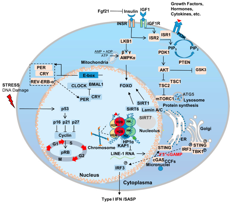
Aging is the most important risk factor for a broad array of diseases, including neurodegenerative diseases, cardiovascular disease, metabolic syndrome, chronic inflammation, and cancer. Additionally, genetic mutations that delay aging have been also found to delay the onset of age-related diseases [36,37]. Genes that regulate aging are relatively conserved among species and are enriched in certain signaling pathways [6] (Figure 1). The association between aging and disease makes fighting aging an even more attractive proposition; it is likely to fight the occurrence and progression of other diseases as well. In this section, we summarize the most important pathways and genes to the aging process.
Figure 1.
Genetic and signaling mechanisms underlying aging. Aging involves multiple genetic alterations in a range of pathways, including, but not limited to, nutrient sensing, sirtuins, nuclear skeleton proteins, immunity, inflammation and circadian rhythm. PI3K/AKT, AMPK, and mTORC1 serve as the core members of lipid, glucose, and amino acid sensing. Lamin A/C interacts with SIRT1, 6 and 7 to regulate chromatin and intracellular homeostasis. cGAS-STING responds to internal and external nuclear pressures and regulates senescence-associated secretory phenotype (SASP). The feedback regulation of circadian rhythm-associated genes is also affected by other aging-related genes. p16, cyclin-dependent kinase inhibitor 2A; p21, cyclin-dependent kinase inhibitor 1A; p27, cyclin-dependent kinase inhibitor 1B; Rb, retinoblastoma protein.
2.2. Nutrient Sensing
Cells rely on nutrient sensing for both the detection of stresses and, ultimately, their survival [38]. Nutrient availability and perception are important material basis for maintaining cell growth and normal function, and cellular metabolic homeostasis imbalance and cellular senescence complement each other [39]. For example, in Caenorhabditis elegans (C. elegans), mutations in the highly conserved daf-2 gene, which encodes an insulin-like receptor and regulates the insulin/insulin-like growth factor 1 (IGF-1) pathway, have been found to significantly prolong lifespan [40]. During aging, the mechanistic target of rapamycin (mTOR) signaling pathway is also important for perceiving stress signals and nutrient sensing, and protein translation [41,42]. Genetically inhibiting the insulin/IGF and mTOR pathways has also been demonstrated to extend mouse lifespan [43].
In 1939, researchers discovered that calorie restriction (CR) can ameliorate aging [44]. CR induces various metabolic changes in the body, and crosstalk between CR and proteins related to nutrient sensing-related pathways is an important reason for ameliorate aging [45]. To date, CR has been shown to extend the lifespan of Saccharomyces cerevisiae, C. elegans, normal and progeria mouse models, and non-human primate rhesus monkeys; at present, CR represents the most effective lifespan-extending intervention across species [46,47,48,49,50,51,52]. This is due to the fact that most molecular pathways involved in longevity are associated with increased stress resistance [53]. Compared with ad libitum access to food (AL), every-other-day feeding (EOD) increases the healthy lifespan of mice. Dietary restriction has also been found to limit the growth of various types of tumors [54]. The phosphatidylinositol-3-kinase (PI3K) pathway, which is a key insulin signaling component, is an important regulator of CR [50,55]. In addition, restricting the amount of branched-chain amino acids (BCAAs), such as leucine, in the diet has also been demonstrated to prolong the lifespan of LmnaG609G/G609G and Lmna–/– mice. In terms of physiological aging, a low-BCAA diet reduces weakness, but does not extend lifespan [56]. Thus, achieving CR via the regulation of metabolism and diet represents a promising anti-aging intervention.
2.3. Sirtuins
Sirtuins are another gene family that can extend the lifespan of C. elegans [57]. They are mainly responsible for regulating cell metabolism, genome stability, gene expression, signal transduction, and important for maintaining the health of the body [58]. There are seven sirtuins in mice and humans, and, under CR, SIRT1 expression is upregulated. This prolongs lifespan and is closely associated with the IGF signaling pathway [59]. Meanwhile, SIRT6 regulates the IGF1 levels and, thus, aging, with SIRT6 overexpression extending lifespan in male mice [27]. Recent studies have also confirmed that SIRT1 is a key protein in the regulation of endothelial cell aging; vascular endothelial cells are essential for maintaining the health and growth of blood vessels. Reducing the expression of SIRT1 in endothelial cells accelerates cellular aging and hinders the normal function of blood vessels [60]. Endothelial cell senescence performs a pivotal role in systemic aging, but the effects can be lessened via the overexpression of SIRT7 [61,62]. Furthermore, SIRT6 expression in endothelial cells has been shown to be important for maintaining heart function [63]. Taken together, these findings indicate that aging-related genes show tissue-dependent effects, and targeting specific types of senescent cells may represent an effective way to treat systemic aging.
2.4. Nuclear Skeleton-Associated Proteins
Intranuclear proteins, such as Lamins play an important role in regulating and maintaining the balance of aging and tumors. Mutations in the LMNA gene affect aging through a number of mechanisms. For example, Lamin A/C interacts with SIRT1, 6, and 7 and affects their intracellular activity and stability, thereby regulating aging [62,64,65]. Interactions between Lamin A/C and SIRT7 also inhibit the transcriptional activation of long interspersed elements-1 (LINE-1, L1) [66], which stabilizes heterochromatin structure. This inhibits the development of a SASP, such as a type I interferon response that triggers natural immune pathways, and can therefore delay the aging of human stem cells by reducing inflammation [67]. IGF-1/AKT signaling pathway protects cells from apoptosis [68]. Furthermore, recent studies have found that the abnormally processed progerin, which is classically located within the nucleus, is also localized outside it. Here, it interacts with IGF-1R and downregulates its expression, thereby impairing IGF-1/AKT signaling, inhibits cellular energy metabolism and accelerates cell aging [69]. Inhibiting isoprenylcysteine carboxylmethyltransferase (ICMT)-associated activation of AKT-mTOR signaling has been found to improve progeria symptoms [70]. Notably, an mTOR hypomorphic allele (MtorΔ/+) has also been found to improve aging characteristics and lifespan in LMNAG608G mice [71]. Taken together, these findings indicate that Lamin A and nutrient sensing share an intricate, important connection to the aging process.
Furthermore, another protein from the nuclear matrix, Lamin B1, is also closely related to aging. Cells respond to carcinogenic pressure by degrading Lamin B1 through autophagy, thus accelerating cell senescence [72]. Recent studies have found that intranuclear SIRT1 protein is the second major nuclear substrate for LC3-mediated selective autophagy, thus influencing cellular senescence through this degradation mechanism [73].
2.5. Immunity and Inflammation
Inflammaging is an important component of aging, which is a pathological phenomenon that brings together our knowledge of age-related chronic diseases, functional decline, and weakness [74]. In the process of aging, the innate and acquired immune system is remodeled, and the reliability and efficiency of the immune system decrease with age, which leads to the upregulation of inflammatory response and the occurrence of related degenerative diseases [75]. The drivers of the inflammatory response mainly include two parts: the degradation of immune receptors/immune sensors and the increase in stimuli that trigger inflammation [36,76]. Inflammation is also the result of lifelong exposure of the immune system to antigenic stimuli and complex genetic, environmental, and age-related mechanisms. Inflammation underlies aging and many age-related chronic diseases, which in turn increases the rate of aging [36]. Excess nutrients are an important factor in inflammation, diet performs an important role in the development and treatment of inflammation and related problems, and CR can slow inflammation and improve aging [77]. The activation of innate immune Toll-Like receptors perform an important role in the aging process, and when Toll-like receptors are knocked out, it can significantly ameliorate the aging of heart-related cells [78]. The Janus kinase/signal transducers and activators of transcription (JAK/STAT) signaling pathway plays an important role in regulating inflammatory response, and the inhibition of JAK/STAT signaling pathway can reduce age-related inflammatory response to a certain extent [79].
Innate immunity plays an important role in the aging process. The cytosolic cyclic GMP–AMP synthase (cGAS)-STING pathway is an important signaling pathway in cells whereby cytoplasmic sensory DNA activates immunity (Figure 1) [80]. During aging, cytoplasmic chromatin fragments (CCFs) leaked from the nucleus, and along with micronuclei or DNA that has escaped from the mitochondria, activate the cGAS-STING pathway, and thus facilitate SASP [81]. SASP promotes the senescence of adjacent or circulatory cells via paracrine signaling [82]. Recent studies have found that yes-associated protein 1 (YAP)/transcriptional coactivator with PDZ-binding motif (TAZ)-mediated control of cGAS-STING signaling is an important molecular mechanism in the regulation of aging in stromal cells and contractile cells. YAP/TAZ is also important for maintaining nuclear envelope stability via the modulation of Lamin B1 expression [83].
2.6. Circadian Rhythm
The production and maintenance of circadian rhythm is the result of positive and negative feedback loops regulated by a series of genes associated with the biological clock, including BMAL, CLOCK, PER, CRY, REV-ERB-α, ROR-β, etc. [84] (Figure 1). The circadian rhythm/clock genes are closely related to aging and two-way adjustment, with aging leading to the transcriptomic reprogramming of circadian genes. For example, the absence of the core clock transcription factor Bmal1 leads to multiple aging-like pathologies in mice [85]. Disturbances in the circadian rhythm accompany the occurrence of aging, and can contribute to the onset and progression of aging-related neurodegenerative diseases [86]. Notably, Salvador Aznar Benitah group and Sassone-Corsi group by comparing mice of different ages, revealed that a low-calorie diet can improve the circadian rhythm of somatic and stem cells, inhibiting the aging process [87,88].
3. The Epigenetics of Aging
Recent population studies have found that as aging progresses, genetics have a decreased influence on gene expression. Age-associated epigenetics perform a more important role than genetics in determining which genes in the body are expressed, and this affects susceptibility to disease [89,90]. The relationship between epigenetic modification and age has become increasingly apparent, and the impact of epigenetics on health, lifespan, and longevity has been widely studied. Epigenetic changes that occur during aging may not only serve as indicators of aging, but also drive age-associated transcriptional changes to directly affect the process [91]. Therefore, research thus far has primarily focused on the regulation of gene conditional expression.
3.1. Epigenetic Age
Epigenetic processes primarily regulate the aging process by regulated gene expression via dynamic changes in DNA methylation and histone modification, non-coding RNA, and chromatin remodeling [91]. Compared with euchromatin, heterochromatin is more involved in the maintenance of genome stability, which depends on specific heterochromatin-binding proteins, histone modifications, and DNA methylation [92]. Epigenetic changes are closely related to aging, and effect aging process are multifaceted, among which histone modifications and chromatin remodeling respond to epigenetic age from different levels.
3.1.1. Histone Modifications
There are numerous possible histone modifications, with chromatin conformation and gene expression being mainly determined by methylation, acetylation, phosphorylation, and ubiquitination [93]. Genome stability is necessary for maintaining normal physiological functions [94], and alterations to histone modifications occur in specific gene regions during cell aging [3]. During vascular aging, increased levels of histone H3 lysine 4 tri-methylation (H3K4me3) and H3K4 methyltransferase Smyd3 expression in endothelial cells results in the development of SASP [95]. Vascular stiffness increases with age, and in vivo studies in mice have found that H3K27me also significantly decreases during smooth muscle cell aging. H3K27me methyltransferase enhancer of zeste homolog 2 (EZH2) can be used as a new target to improve aging-induced vascular stiffness and fibrosis. [96]. Furthermore, recent studies have found that H3K27me3 is decreased and H3K27me1 is increased during healthy aging of human classical CD14+CD16− monocytes [97]. It can be seen that histone modifications in the aging process of different organs are closely related and heterogeneous.
3.1.2. Chromatin Remodeling
Chromatin remodeling and stability are closely related to aging. For example, in damaged mitochondria, acetyl-CoA acts as a signal to induce the accumulation of histone deacetylase complexes (NuRD), thereby mediating chromatin remodeling to regulate body aging [98]. Transcription factor activator protein 1 (AP-1) has also been found to drive SASP by reshaping the accessibility of specific chromatin regions [99]. Furthermore, zinc finger protein with KRAB and SCAN domains 3 (ZKSCAN3) can maintain heterochromatin stability and weaken cellular senescence by interacting with heterochromatin-related proteins [100].
Notably, the relationship between APOE protein and aging has also been revealed recently; APOE affects cellular senescence by regulating the stabilization of heterochromatin in stem cells [22]. HP1α is important for the maintenance of heterochromatin stability. During aging, the loss of HP1α is both an important cause of cellular aging and a biomarker of aging [14]. However, research on non-coding RNAs involved in structural changes in heterochromatin during cellular senescence is still limited, as is our understanding of the underlying mechanisms of action. Non-coding RNA is also involved in maintaining heterochromatin structure, and a recent study found that a combination of KCNQ1OT1 lncRNA with heterochromatin protein HP1α promoted genome-wide transposon repression. In this manner, the stability of heterochromatin structure was maintained, and cellular aging inhibited [101]. The association between chromatin remodeling and aging is more about homeostasis within the nucleus and the normal expression of genes, and the effects on other aspects of aging are gradually revealed.
3.2. Epigenetic Clock
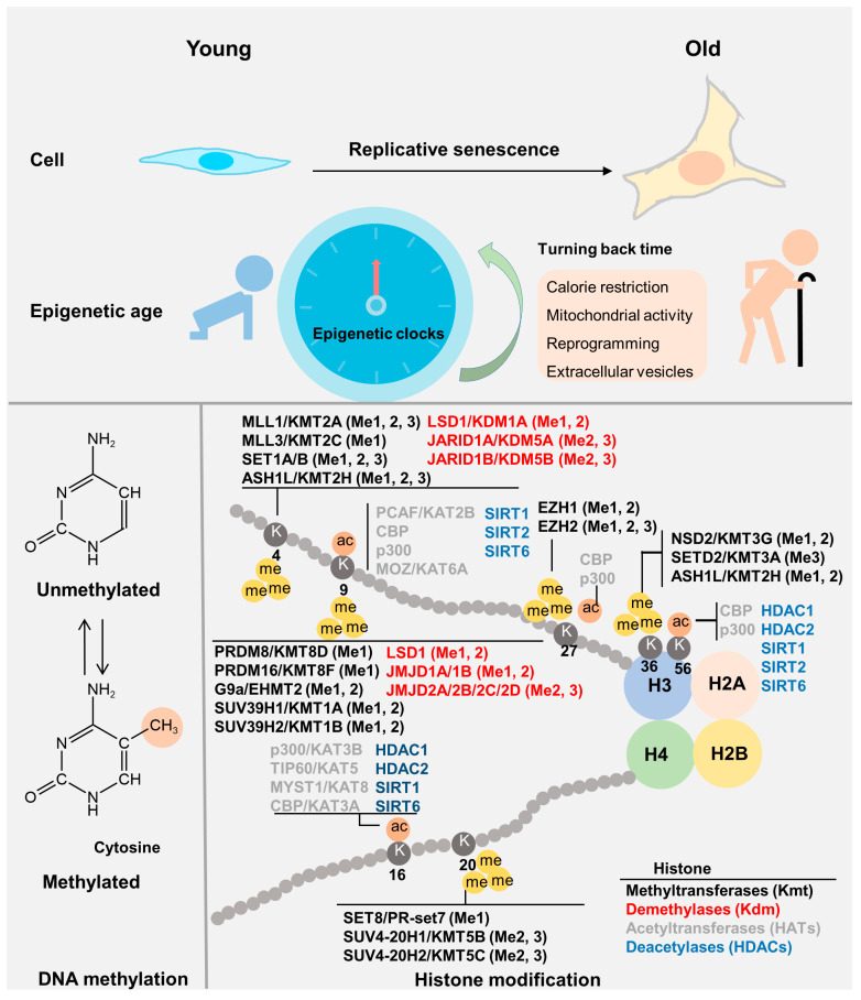
DNA methylation (5′ methylcytosine (5mC)) levels are clearly correlated with age and can be used to predict the chronological age of both blood and certain organs, including the kidneys and liver. Thus, this has been termed the “epigenetic clock” [102]. The epigenetic age of embryonic stem cells is almost zero. It is possible to use algorithms to calculate biological age based on how many sites in an individual’s genome bind to methyl groups (Figure 2). During the passage of mouse embryonic fibroblasts (MEFs) and the physiological aging of various tissues, DNA methylation levels are altered [103]. Radiation and oncogene expression can induce senescence in cells with epigenetic age (EpiAge) close to zero, replicative senescence exhibits increased EpiAge in human cells. Consistent results have also been found in murine cells [103,104]. Notably, fibroblasts taken from patients with Hutchinson Gilford Progeria Syndrome (HGPS) showed a weak correlation with EpiAge [105]. EpiAge has also been found to be closely related to nutrient sensing and the mitochondrial activity [104].
Figure 2.
Epigenetics of aging. The “epigenetic clock”, a biomedical measure of aging, primarily includes DNA methylation and histone modification status. During both cellular and systemic aging, epigenetic age increases. The epigenetic clock can be reversed by calorie restriction, mitochondrial activation, reprogramming, and extracellular vesicles. The section on histone modifications partially references the pathway and diagram of cell signaling technology (CST), and mainly lists histone modifications related to aging and regulatory kinases related to histone modifications. Ac, histone acetylation; me, histone methylation.
While technical noise is an important limiting factor in the reliability of aging biomarkers and the epigenetic clock [106], the reversibility of the epigenetic clock has become a favored topic of conversation in the field of aging research. Epigenetic reprogramming and the use of extracellular vesicles are two potential methods of reversing the epigenetic clock (Figure 2) and will be discussed below.
3.2.1. Epigenetic Reprogramming
Epigenetic remodeling is an important driver of aging [107]. Dialing back the epigenetic clock through epigenetic reprogramming seems to represent an effective way to reverse the aging process. For example, the partial reprogramming of Yamanaka factor (Oct4, Sox2, Klf4, and c-Myc (OSKM)) in progeroid mice has been shown to improve metabolic dysfunction and aging characteristics [108]. However, aging and reprogramming have a more complex relationship, and OSKM can also facilitate the reprogramming process by inducing cell damage and bringing cells into a state of senescent, organizational environment conducive to OSKM-driven reprogramming in neighboring cells [109]. Using adeno-associated virus (AAV) to introduce three transcription factors, Oct4, Sox2, and Klf4 (OSK) in aging mice, age-associated visual impairments were successfully reversed [110]. Recent studies have found that the in vivo reprogramming of normal aging mice restores youthful epigenetic characteristics in senescent cells, and significantly reduces the expression of inflammation/aging-related genes [111]. In addition, recent cocktail therapies utilizing reprogramming and senolytic strategies have successfully extended the lifespan of mice [112].
The results of these studies suggest that epigenetic reprogramming represents a highly promising and effective aging intervention. Compared with the introduction of genes to induce reprogramming, compound-induced reprogramming can effectively avoid safety problems associated with traditional transgenic operations [113]. Recently, human somatic cells have been successfully induced into pluripotent stem cells using only small chemical molecules, without relying on exogenous genes [114]. The application of compound-induced cellular reprogramming in aging is yet to be fully investigated, and may become an effective alternative to aging cocktail therapy in the future.
3.2.2. Extracellular Vesicles
Extracellular vehicles (EVs) mainly involved in the removal of excess or unnecessary substances from cells, the maintenance of homeostasis in the intracellular environment, and to serve as messengers during cell-to-cell communication [115]. Using EVs to deliver extracellular nicotinamide phosphoribosyltransferase (eNAMPT) to elderly mice has been found to alleviate age-related tissue function, and to significantly extend lifespan [116]. Recent studies have also found that injecting old mice with adipose mesenchymal stem cell extracellular vesicles (ADSC-sEVs) from young mice improves a number of aging problems in old mice; even reducing tissue epigenetic age [117]. As effective carriers of aging intervention mediator, such as proteins, nucleic acids, and lipids, exosomes have broad application prospects in the future, and will likely be of great research significance.
3.3. Epitranscriptomics
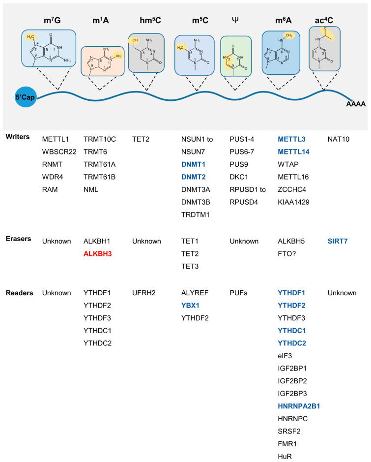
Epitranscriptomics focuses on the effects of RNA post-transcriptional modifications on the regulation of gene expression and has provided novel strategies to analyze the epigenetics of aging (Figure 3). RNAs can be covalently modified in a number of ways, including N6-methyladenosine (m6A), 5-methylcytidine (m5C), N1-methyladenosine (m1A), N4-acetylcytidine (ac4C), and N 7-methylguanosine (m7G), etc. [118]. Manipulating RNA modifications may represent a useful molecular mechanism through which longevity and stress tolerance might be improved, and we discuss some potential targets below.
Figure 3.
RNA modification and senescence. Seven types of RNA modifications have been studied thus far, including m7G, m1A, m6A, m5C hm5C, ac4C, and Ψ. A summary of the writer, eraser, and reader proteins of the related modifications is presented. Aging-related changes: red represents upregulation, while blue represents downregulation. M7G, 7-methylguanosine; m1A, N1-methyladenosine; m5C, 5-methylcytosine; hm5C, 5-hydroxymethylcytidine; m6A, N6-methyladenosine; Ψ, uridine-to-pseudouridine.
3.3.1. m6A
RNA m6A modification is dynamically regulated by methyltransferase-like (METTL) 3/14, fat mass and obesity-associated protein (FTO)/alkB homologue 5 (ALKBH5), and reader proteins, such as YTH domain-containing family (YTHDF)1/2/3 are involved in the functional outcome. m6A is involved in multiple aspects of RNA posttranscriptional processing, including RNA stability, translation, splicing, and export to the nucleus. A number of studies have highlighted the intrinsic link between RNA-modified enzymes and aging [119,120,121], but the relationship between m6A readers and aging needs to be studied in greater depth. YTHDF1/2, YTHDC1/2, heterogeneous nuclear ribonucleoprotein A2B1 (HNRNPA2B1), and heterogeneous nuclear ribonucleoprotein C (HNRNPC) protein expression levels have been shown to decrease during replicative senescence [122]. The loss of cell proliferation ability is a significant feature of cell senescence, and Forkhead box M1 (FOXM1) is a key proliferation-related transcription factor that regulates the expression G2/M phase transition genes. Notably, YTHDF1 regulates the translation of FOXM1 in an m6A-dependent manner [123], and FOXM1 overexpression has been shown to significantly improve physiological and pathological aging [124]. As aforementioned, Insulin/IGF-1 transmembrane receptor (IGFR) is particularly important for aging, and YTHDF3 expression patterns have been found to be consistent with insulin receptor (INSR) expression during aging [125]. YTHDF3 binds to m1A modifications on IGF1R mRNA to promote its degradation, thereby inhibiting IGF1R expression [126]. YTHDF3 also regulates the expression of longevity gene forkhead box O-3 (FOXO3) in both m6A-dependent and non-dependent ways, thereby regulating immune activation and autophagy [127,128].
3.3.2. ac4C
N-Acetyltransferase 10 (NAT10) is the key enzyme involved in the ac4C RNA acetylation modification and is essential for normal cell function [129]. Remodelin, an inhibitor of NAT10, improves the nuclear morphology of HGPS [130], and Nat10 heterozygosity has been shown to extend the lifespan of progeroid mice [131]. Recent studies have found that the ac4C modification can be removed by SIRT7 in vitro [132]. However, the dynamic changes of ac4C in vivo and its impact on aging remain to be fully characterized.
3.3.3. m1A and m5C
Other RNA modifications have also been found to be closely related to aging. Demethylase ALKBH3 of N1-methyladenosine (m1A) increases during aging and regulates hematopoietic system function [133]. The m5C modification is catalyzed by methyltransferase NSUN2/TRDMT1 and oxidized by dioxygenase ten-eleven translocation (TET) to form hm5C. NSUN2 can add the m5C modification to the p16 mRNA strand, which can inhibit the degradation of p16 mRNA and increase its stability [134].
4. Interventions for Aging
The rapid progress in the study of the molecular mechanism of aging has made us realize that the speed of aging and the length of life expectancy can be intervened by humans. To understand the genetic and epigenetic biological mechanisms behind aging, gain insight into the plasticity of aging, and, ultimately, achieve effective intervention in aging [135]. At present, the most effective aging intervention method is still CR, and the development of age-related genetic research has made us realize that aging-related pathways can also be used as targets for aging intervention, such as insulin-like signaling pathways, AMP-activated protein kinase (AMPK) signaling pathways, sirtuins, nicotinamide adenine dinucleotide (NAD+), circadian clock, chronic inflammation, cellular senescence, etc [136]. Increasing NAD+ levels can significantly ameliorate aging [137]. SASP is an important feature of cellular senescence, SASP can promote the aging of neighboring cells through autocrine or paracrine signaling pathways, and the accumulation of senescent cells is one of the important causes of aging with age. The intervention methods of senescent cells mainly include senolytics that eliminate senescent cells and senomophics that reduce SASP, Senomorphic compounds target pathological SASP signals, and senolytic eliminates potential senescent cells that release harmful SASP factors [138]. The use of senolytics to remove senescent cells and reduce the secretion of SASP can effectively delay tissue or overall aging. The use of drugs or other ways to reduce the expression of SASP can also significantly delay the aging of cells [8] Rapid development of biological theory and technology, gene therapy anti-aging has been widely studied and recognized. Gene therapy that delivers anti-aging genes through AAV introduction has great potential in treating multiple age-related diseases at once [139]. With the innovation of technology, gene editing and aging vaccines will also likely move towards anti-aging applications in the near future [140]. The cocktail of aging therapy combines different anti-aging methods to greatly enrich the anti-aging strategy [112].
5. Conclusions
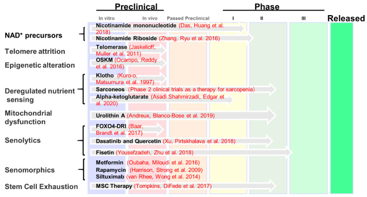
The study of genetic and epigenetic characteristics of aging is essential to the development of targeted treatments against aging. Still, our understanding of the genetics and epigenetics of aging is just the tip of the iceberg. Since the aging process is dynamic and complex, the use of more genomics and epitranscriptomics methods can effectively realize the real-time, dynamic, and multi-dimensional monitoring of the aging process. The genetic and epigenetic changes of aging were combined to select suitable target interventions. Then, a cocktail of therapy through a combination of multiple ways is possible to achieve a reversal of aging. With advances in scientific theories and technologies, personalized anti-aging treatments, improved health during aging, and increased longevity may not be far away. The US Food and Drug Administration (FDA) recently approved the use of the farnesyltransferase inhibitor Zokinvy (lonafarnib) to reduce and prevent the accumulation of defective presenile proteins, thereby reducing the risk of death caused by HGPS. This represents an important step from basic research to clinical application [141]. The Lifespan.io website (Lifespan.io is a nonprofit advocacy organization and news outlet covering aging and rejuvenation research) (Figure 4) provides us with current clinical application information for anti-aging medications or therapies. Notably, the Targeting Aging with Metformin (TAME) research plan involves the study of the anti-aging effects of metformin in a series of nationwide, six-year clinical trials at 14 leading research institutions, involving 3000 non-diabetic patients (65–79 years old) [142]. There is no shortage of exciting bonus scenes in aging research, in which a fancy study used deep neural networks to assess cellular senescence and found that nuclear morphology is an important biomarker of cellular senescence [143]. With the analysis of multiple big data related to the genetics and epigenetics of aging, the combination of artificial intelligence and aging research will greatly broaden people’s understanding of aging. It seems likely that in the near future, even more aging-related treatment modalities will be developed for clinical application. It is of great significance to understand the plasticity of aging process, the plasticity of aging biomarkers and drivers, and the plasticity of aging interventions from the perspectives of genetics and epigenetics, which is of great significance for improving healthy aging.
Figure 4.
Research and development of clinical anti-aging medication. The information presented in the figure is summarized at https://www.lifespan.io/road-maps/the-rejuvenation-roadmap/, accessed on 20 December 2022. A number of aging-related drugs are currently subject to preliminary exploratory investigations, while other disease-related or anti-cancer drugs have been gradually introduced into anti-aging research. At present, the most promising treatments include drugs targeting NAD+, telomere attrition, epigenetic alteration, deregulated nutrient sensing, mitochondrial dysfunction, senolytics, senomorphics, and stem cell exhaustion [60,108,144,145,146,147,148,149,150,151,152,153,154,155].
Author Contributions
J.Z. and S.W. searched data and references for the manuscript; J.Z. and B.L. wrote and edited the manuscript. All authors have read and agreed to the published version of the manuscript.
Institutional Review Board Statement
Not applicable.
Informed Consent Statement
Not applicable.
Data Availability Statement
Not applicable.
Conflicts of Interest
The authors have no conflict of interest to declare.
Funding Statement
This work was supported by grants from the Shenzhen Municipal Commission of Science and Technology Innovation (grant nos. ZDSYS20190902093401689, JCYJ20200814152850001 and JCYJ20180507182044945). The authors would like to thank Jessica Tamanini (Shenzhen University and ETediting) for editing the manuscript prior to submission.
Footnotes
Disclaimer/Publisher’s Note: The statements, opinions and data contained in all publications are solely those of the individual author(s) and contributor(s) and not of MDPI and/or the editor(s). MDPI and/or the editor(s) disclaim responsibility for any injury to people or property resulting from any ideas, methods, instructions or products referred to in the content.
References
- 1.Olovnikov A.M. Telomeres, telomerase, and aging: Origin of the theory. Exp. Gerontol. 1996;31:443–448. doi: 10.1016/0531-5565(96)00005-8. [DOI] [PubMed] [Google Scholar]
- 2.Gavrilov L.A., Gavrilova N.S. Evolutionary Theories of Aging and Longevity. Sci. World J. 2002;2:339–356. doi: 10.1100/tsw.2002.96. [DOI] [PMC free article] [PubMed] [Google Scholar]
- 3.Lopez-Otin C., Blasco M.A., Partridge L., Serrano M., Kroemer G. The hallmarks of aging. Cell. 2013;153:1194–1217. doi: 10.1016/j.cell.2013.05.039. [DOI] [PMC free article] [PubMed] [Google Scholar]
- 4.Childs B.G., Gluscevic M., Baker D.J., Laberge R.-M., Marquess D., Dananberg J., van Deursen J.M. Senescent cells: An emerging target for diseases of ageing. Nat. Rev. Drug Discov. 2017;16:718–735. doi: 10.1038/nrd.2017.116. [DOI] [PMC free article] [PubMed] [Google Scholar]
- 5.Hayflick L., Moorhead P.S. The serial cultivation of human diploid cell strains. Exp. Cell Res. 1961;25:585–621. doi: 10.1016/0014-4827(61)90192-6. [DOI] [PubMed] [Google Scholar]
- 6.Martínez-Zamudio R.I., Robinson L., Roux P.-F., Bischof O. SnapShot: Cellular Senescence Pathways. Cell. 2017;170:816. doi: 10.1016/j.cell.2017.07.049. [DOI] [PubMed] [Google Scholar]
- 7.Hernandez-Segura A., Nehme J., Demaria M. Hallmarks of Cellular Senescence. Trends Cell Biol. 2018;28:436–453. doi: 10.1016/j.tcb.2018.02.001. [DOI] [PubMed] [Google Scholar]
- 8.Gasek N.S., Kuchel G.A., Kirkland J.L., Xu M. Strategies for targeting senescent cells in human disease. Nat. Aging. 2021;1:870–879. doi: 10.1038/s43587-021-00121-8. [DOI] [PMC free article] [PubMed] [Google Scholar]
- 9.Eriksson M., Brown W.T., Gordon L.B., Glynn M.W., Singer J., Scott L., Erdos M.R., Robbins C.M., Moses T.Y., Berglund P., et al. Recurrent de novo point mutations in lamin A cause Hutchinson–Gilford progeria syndrome. Nature. 2003;423:293–298. doi: 10.1038/nature01629. [DOI] [PMC free article] [PubMed] [Google Scholar]
- 10.Krishnan V., Chow M.Z.Y., Wang Z., Zhang L., Liu B., Liu X., Zhou Z. Histone H4 lysine 16 hypoacetylation is associated with defective DNA repair and premature senescence in Zmpste24-deficient mice. Proc. Natl. Acad. Sci. USA. 2011;108:12325–12330. doi: 10.1073/pnas.1102789108. [DOI] [PMC free article] [PubMed] [Google Scholar]
- 11.Liu B., Wang Z., Zhang L., Ghosh S., Zheng H., Zhou Z. Depleting the methyltransferase Suv39h1 improves DNA repair and extends lifespan in a progeria mouse model. Nat. Commun. 2013;4:1868. doi: 10.1038/ncomms2885. [DOI] [PMC free article] [PubMed] [Google Scholar]
- 12.Shumaker D.K., Dechat T., Kohlmaier A., Adam S.A., Bozovsky M.R., Erdos M.R., Eriksson M., Goldman A.E., Khuon S., Collins F.S., et al. Mutant nuclear lamin A leads to progressive alterations of epigenetic control in premature aging. Proc. Natl. Acad. Sci. USA. 2006;103:8703–8708. doi: 10.1073/pnas.0602569103. [DOI] [PMC free article] [PubMed] [Google Scholar]
- 13.Scaffidi P., Misteli T. Lamin A-Dependent Nuclear Defects in Human Aging. Science. 2006;312:1059–1063. doi: 10.1126/science.1127168. [DOI] [PMC free article] [PubMed] [Google Scholar]
- 14.Zhang W., Li J., Suzuki K., Qu J., Wang P., Zhou J., Liu X., Ren R., Xu X., Ocampo A., et al. A Werner syndrome stem cell model unveils heterochromatin alterations as a driver of human aging. Science. 2015;348:1160–1163. doi: 10.1126/science.aaa1356. [DOI] [PMC free article] [PubMed] [Google Scholar]
- 15.Ozgenc A., Loeb L.A. Werner Syndrome, aging and cancer. Genome Dyn. 2006;1:206–217. doi: 10.1159/000092509. [DOI] [PubMed] [Google Scholar]
- 16.Singh P.P., Demmitt B.A., Nath R.D., Brunet A. The Genetics of Aging: A Vertebrate Perspective. Cell. 2019;177:200–220. doi: 10.1016/j.cell.2019.02.038. [DOI] [PMC free article] [PubMed] [Google Scholar]
- 17.Cagan A., Baez-Ortega A., Brzozowska N., Abascal F., Coorens T.H.H., Sanders M.A., Lawson A.R.J., Harvey L.M.R., Bhosle S., Jones D., et al. Somatic mutation rates scale with lifespan across mammals. Nature. 2022;604:517–524. doi: 10.1038/s41586-022-04618-z. [DOI] [PMC free article] [PubMed] [Google Scholar]
- 18.Vincze O., Colchero F., Lemaître J.-F., Conde D.A., Pavard S., Bieuville M., Urrutia A.O., Ujvari B., Boddy A.M., Maley C.C., et al. Cancer risk across mammals. Nature. 2021;601:263–267. doi: 10.1038/s41586-021-04224-5. [DOI] [PMC free article] [PubMed] [Google Scholar]
- 19.Gomes N.M.V., Ryder O.A., Houck M.L., Charter S.J., Walker W., Forsyth N.R., Austad S.N., Venditti C., Pagel M., Shay J.W., et al. Comparative biology of mammalian telomeres: Hypotheses on ancestral states and the roles of telomeres in longevity determination. Aging Cell. 2011;10:761–768. doi: 10.1111/j.1474-9726.2011.00718.x. [DOI] [PMC free article] [PubMed] [Google Scholar]
- 20.Muñoz-Lorente M.A., Cano-Martin A.C., Blasco M.A. Mice with hyper-long telomeres show less metabolic aging and longer lifespans. Nat. Commun. 2019;10:4723. doi: 10.1038/s41467-019-12664-x. [DOI] [PMC free article] [PubMed] [Google Scholar]
- 21.Sleiman M.B., Roy S., Gao A.W., Sadler M.C., von Alvensleben G.V.G., Li H., Sen S., Harrison D.E., Nelson J.F., Strong R., et al. Sex- and age-dependent genetics of longevity in a heterogeneous mouse population. Science. 2022;377:eabo3191. doi: 10.1126/science.abo3191. [DOI] [PMC free article] [PubMed] [Google Scholar]
- 22.Zhao H., Ji Q., Wu Z., Wang S., Ren J., Yan K., Wang Z., Hu J., Chu Q., Hu H., et al. Destabilizing heterochromatin by APOE mediates senescence. Nat. Aging. 2022;2:303–316. doi: 10.1038/s43587-022-00186-z. [DOI] [PubMed] [Google Scholar]
- 23.Timmers P.R.H.J., Tiys E.S., Sakaue S., Akiyama M., Kiiskinen T.T.J., Zhou W., Hwang S.-J., Yao C., Kamatani Y., Deelen J., et al. Mendelian randomization of genetically independent aging phenotypes identifies LPA and VCAM1 as biological targets for human aging. Nat. Aging. 2022;2:19–30. doi: 10.1038/s43587-021-00159-8. [DOI] [PubMed] [Google Scholar]
- 24.Lemaître J.-F., Ronget V., Tidière M., Allainé D., Berger V., Cohas A., Colchero F., Conde D.A., Garratt M., Liker A., et al. Sex differences in adult lifespan and aging rates of mortality across wild mammals. Proc. Natl. Acad. Sci. USA. 2020;117:8546–8553. doi: 10.1073/pnas.1911999117. [DOI] [PMC free article] [PubMed] [Google Scholar]
- 25.Trifunovic A. Mitochondrial DNA and ageing. Biochim. Biophys. Acta. 2006;1757:611–617. doi: 10.1016/j.bbabio.2006.03.003. [DOI] [PubMed] [Google Scholar]
- 26.Ellegren H. Sex-chromosome evolution: Recent progress and the influence of male and female heterogamety. Nat. Rev. Genet. 2011;12:157–166. doi: 10.1038/nrg2948. [DOI] [PubMed] [Google Scholar]
- 27.Kanfi Y., Naiman S., Amir G., Peshti V., Zinman G., Nahum L., Bar-Joseph Z., Cohen H.Y. The sirtuin SIRT6 regulates lifespan in male mice. Nature. 2012;483:218–221. doi: 10.1038/nature10815. [DOI] [PubMed] [Google Scholar]
- 28.Tacutu R., Thornton D., Johnson E., Budovsky A., Barardo D., Craig T., Diana E., Lehmann G., Toren D., Wang J., et al. Human Ageing Genomic Resources: New and updated databases. Nucleic Acids Res. 2017;46:D1083–D1090. doi: 10.1093/nar/gkx1042. [DOI] [PMC free article] [PubMed] [Google Scholar]
- 29.Avelar R.A., Ortega J.G., Tacutu R., Tyler E.J., Bennett D., Binetti P., Budovsky A., Chatsirisupachai K., Johnson E., Murray A., et al. A multidimensional systems biology analysis of cellular senescence in aging and disease. Genome Biol. 2020;21:91. doi: 10.1186/s13059-020-01990-9. [DOI] [PMC free article] [PubMed] [Google Scholar]
- 30.Budovsky A., Craig T., Wang J., Tacutu R., Csordas A., Lourenço J., Fraifeld V.E., de Magalhães J.P. LongevityMap: A database of human genetic variants associated with longevity. Trends Genet. 2013;29:559–560. doi: 10.1016/j.tig.2013.08.003. [DOI] [PubMed] [Google Scholar]
- 31.Aging Atlas C. Aging Atlas: A multi-omics database for aging biology. Nucleic Acids Res. 2021;49:D825–D830. doi: 10.1093/nar/gkaa894. [DOI] [PMC free article] [PubMed] [Google Scholar]
- 32.Liu B., Wang J., Chan K.M., Tjia W.M., Deng W., Guan X., Huang J.-D., Li K.M., Chau P.Y., Chen D.J., et al. Genomic instability in laminopathy-based premature aging. Nat. Med. 2005;11:780–785. doi: 10.1038/nm1266. [DOI] [PubMed] [Google Scholar]
- 33.Garagnani P., Marquis J., Delledonne M., Pirazzini C., Marasco E., Kwiatkowska K.M., Iannuzzi V., Bacalini M.G., Valsesia A., Carayol J., et al. Whole-genome sequencing analysis of semi-supercentenarians. Elife. 2021;10:e57849. doi: 10.7554/eLife.57849. [DOI] [PMC free article] [PubMed] [Google Scholar]
- 34.Deelen J., Evans D.S., Arking D.E., Tesi N., Nygaard M., Liu X., Wojczynski M.K., Biggs M.L., van der Spek A., Atzmon G., et al. A meta-analysis of genome-wide association studies identifies multiple longevity genes. Nat. Commun. 2019;10:3669. doi: 10.1038/s41467-019-11558-2. [DOI] [PMC free article] [PubMed] [Google Scholar]
- 35.Lin J.-R., Sin-Chan P., Napolioni V., Torres G.G., Mitra J., Zhang Q., Jabalameli M.R., Wang Z., Nguyen N., Gao T., et al. Rare genetic coding variants associated with human longevity and protection against age-related diseases. Nat. Aging. 2021;1:783–794. doi: 10.1038/s43587-021-00108-5. [DOI] [PubMed] [Google Scholar]
- 36.Franceschi C., Garagnani P., Parini P., Giuliani C., Santoro A. Inflammaging: A new immune–metabolic viewpoint for age-related diseases. Nat. Rev. Endocrinol. 2018;14:576–590. doi: 10.1038/s41574-018-0059-4. [DOI] [PubMed] [Google Scholar]
- 37.Amorim J.A., Coppotelli G., Rolo A.P., Palmeira C.M., Ross J.M., Sinclair D.A. Mitochondrial and metabolic dysfunction in ageing and age-related diseases. Nat. Rev. Endocrinol. 2022;18:243–258. doi: 10.1038/s41574-021-00626-7. [DOI] [PMC free article] [PubMed] [Google Scholar]
- 38.Efeyan A., Comb W.C., Sabatini D.M. Nutrient-sensing mechanisms and pathways. Nature. 2015;517:302–310. doi: 10.1038/nature14190. [DOI] [PMC free article] [PubMed] [Google Scholar]
- 39.Kulkarni A.S., Gubbi S., Barzilai N. Benefits of Metformin in Attenuating the Hallmarks of Aging. Cell Metab. 2020;32:15–30. doi: 10.1016/j.cmet.2020.04.001. [DOI] [PMC free article] [PubMed] [Google Scholar]
- 40.Kenyon C., Chang J., Gensch E., Rudner A., Tabtiang R. A C. elegans mutant that lives twice as long as wild type. Nature. 1993;366:461–464. doi: 10.1038/366461a0. [DOI] [PubMed] [Google Scholar]
- 41.Johnson S.C., Rabinovitch P.S., Kaeberlein M. mTOR is a key modulator of ageing and age-related disease. Nature. 2013;493:338–345. doi: 10.1038/nature11861. [DOI] [PMC free article] [PubMed] [Google Scholar]
- 42.Saxton R.A., Sabatini D.M. mTOR Signaling in Growth, Metabolism, and Disease. Cell. 2017;169:361–371. doi: 10.1016/j.cell.2017.03.035. [DOI] [PubMed] [Google Scholar]
- 43.Unnikrishnan A., Deepa S.S., Herd H.R., Richardson A. Chapter 19—Extension of Life Span in Laboratory Mice. In: Ram J.L., Conn P.M., editors. Conn’s Handbook of Models for Human Aging. 2nd ed. Academic Press; Cambridge, MA, USA: 2018. pp. 245–270. [Google Scholar]
- 44.McCay C.M., Maynard L.A., Sperling G., Barnes L.L. Retarded growth, life span, ultimate body size and age changes in the albino rat after feeding diets restricted in calories: Four figures. J. Nutr. 1939;18:1–13. doi: 10.1093/jn/18.1.1. [DOI] [PubMed] [Google Scholar]
- 45.Fontana L., Nehme J., Demaria M. Caloric restriction and cellular senescence. Mech. Ageing Dev. 2018;176:19–23. doi: 10.1016/j.mad.2018.10.005. [DOI] [PubMed] [Google Scholar]
- 46.Mattison J.A., Colman R.J., Beasley T.M., Allison D.B., Kemnitz J.W., Roth G.S., Ingram D.K., Weindruch R., de Cabo R., Anderson R.M. Caloric restriction improves health and survival of rhesus monkeys. Nat. Commun. 2017;8:14063. doi: 10.1038/ncomms14063. [DOI] [PMC free article] [PubMed] [Google Scholar]
- 47.Anderson R.M., Bitterman K.J., Wood J.G., Medvedik O., Sinclair D.A. Nicotinamide and PNC1 govern lifespan extension by calorie restriction in Saccharomyces cerevisiae. Nature. 2003;423:181–185. doi: 10.1038/nature01578. [DOI] [PMC free article] [PubMed] [Google Scholar]
- 48.Panowski S.H., Wolff S., Aguilaniu H., Durieux J., Dillin A. PHA-4/Foxa mediates diet-restriction-induced longevity of C. elegans. Nature. 2007;447:550–555. doi: 10.1038/nature05837. [DOI] [PubMed] [Google Scholar]
- 49.Vermeij W.P., Dollé M.E.T., Reiling E., Jaarsma D., Payan-Gomez C., Bombardieri C.R., Wu H., Roks A.J.M., Botter S.M., Van Der Eerden B.C., et al. Restricted diet delays accelerated ageing and genomic stress in DNA-repair-deficient mice. Nature. 2016;537:427–431. doi: 10.1038/nature19329. [DOI] [PMC free article] [PubMed] [Google Scholar]
- 50.Xie K., Neff F., Markert A., Rozman J., Aguilar-Pimentel J.A., Amarie O.V., Becker L., Brommage R., Garrett L., Henzel K.S., et al. Every-other-day feeding extends lifespan but fails to delay many symptoms of aging in mice. Nat. Commun. 2017;8:155. doi: 10.1038/s41467-017-00178-3. [DOI] [PMC free article] [PubMed] [Google Scholar]
- 51.Mattison J.A., Roth G.S., Beasley T.M., Tilmont E.M., Handy A.M., Herbert R.L., Longo D.L., Allison D.B., Young J.E., Bryant M., et al. Impact of caloric restriction on health and survival in rhesus monkeys from the NIA study. Nature. 2012;489:318–321. doi: 10.1038/nature11432. [DOI] [PMC free article] [PubMed] [Google Scholar]
- 52.Colman R.J., Anderson R.M., Johnson S.C., Kastman E.K., Kosmatka K.J., Beasley T.M., Allison D.B., Cruzen C., Simmons H.A., Kemnitz J.W., et al. Caloric Restriction Delays Disease Onset and Mortality in Rhesus Monkeys. Science. 2009;325:201–204. doi: 10.1126/science.1173635. [DOI] [PMC free article] [PubMed] [Google Scholar]
- 53.Parsons P.A. The limit to human longevity: An approach through a stress theory of ageing. Mech. Ageing Dev. 1996;87:211–218. doi: 10.1016/0047-6374(96)01710-1. [DOI] [PubMed] [Google Scholar]
- 54.Kanarek N., Petrova B., Sabatini D.M. Dietary modifications for enhanced cancer therapy. Nature. 2020;579:507–517. doi: 10.1038/s41586-020-2124-0. [DOI] [PubMed] [Google Scholar]
- 55.Kalaany N.Y., Sabatini D.M. Tumours with PI3K activation are resistant to dietary restriction. Nature. 2009;458:725–731. doi: 10.1038/nature07782. [DOI] [PMC free article] [PubMed] [Google Scholar]
- 56.Richardson N.E., Konon E.N., Schuster H.S., Mitchell A.T., Boyle C., Rodgers A.C., Finke M., Haider L.R., Yu D., Flores V., et al. Lifelong restriction of dietary branched-chain amino acids has sex-specific benefits for frailty and life span in mice. Nat. Aging. 2021;1:73–86. doi: 10.1038/s43587-020-00006-2. [DOI] [PMC free article] [PubMed] [Google Scholar]
- 57.Kennedy B.K., Austriaco N.R., Jr., Zhang J., Guarente L. Mutation in the silencing gene SIR4 can delay aging in S. cerevisiae. Cell. 1995;80:485–496. doi: 10.1016/0092-8674(95)90499-9. [DOI] [PubMed] [Google Scholar]
- 58.Wang T., Wang Y., Liu L., Jiang Z., Li X., Tong R., He J., Shi J. Research progress on sirtuins family members and cell senescence. Eur. J. Med. Chem. 2020;193:112207. doi: 10.1016/j.ejmech.2020.112207. [DOI] [PubMed] [Google Scholar]
- 59.Cohen H.Y., Miller C., Bitterman K.J., Wall N.R., Hekking B., Kessler B., Howitz K.T., Gorospe M., de Cabo R., Sinclair D.A. Calorie Restriction Promotes Mammalian Cell Survival by Inducing the SIRT1 Deacetylase. Science. 2004;305:390–392. doi: 10.1126/science.1099196. [DOI] [PubMed] [Google Scholar]
- 60.Das A., Huang G.X., Bonkowski M.S., Longchamp A., Li C., Schultz M.B., Kim L.-J., Osborne B., Joshi S., Lu Y., et al. Impairment of an Endothelial NAD+-H2S Signaling Network Is a Reversible Cause of Vascular Aging. Cell. 2019;176:944–945. doi: 10.1016/j.cell.2019.01.026. [DOI] [PMC free article] [PubMed] [Google Scholar]
- 61.Osmanagic-Myers S., Kiss A., Manakanatas C., Hamza O., Sedlmayer F., Szabo P.L., Fischer I., Fichtinger P., Podesser B.K., Eriksson M., et al. Endothelial progerin expression causes cardiovascular pathology through an impaired mechanoresponse. J. Clin. Investig. 2019;129:531–545. doi: 10.1172/JCI121297. [DOI] [PMC free article] [PubMed] [Google Scholar]
- 62.Sun S., Qin W., Tang X., Meng Y., Hu W., Zhang S., Qian M., Liu Z., Cao X., Pang Q., et al. Vascular endothelium–targeted Sirt7 gene therapy rejuvenates blood vessels and extends life span in a Hutchinson-Gilford progeria model. Sci. Adv. 2020;6:eaay5556. doi: 10.1126/sciadv.aay5556. [DOI] [PMC free article] [PubMed] [Google Scholar]
- 63.Wu X., Liu H., Brooks A., Xu S., Luo J., Steiner R., Mickelsen D.M., Moravec C.S., Jeffrey A.D., Small E.M., et al. SIRT6 Mitigates Heart Failure With Preserved Ejection Fraction in Diabetes. Circ. Res. 2022;131:926–943. doi: 10.1161/CIRCRESAHA.121.318988. [DOI] [PMC free article] [PubMed] [Google Scholar]
- 64.Liu B., Ghosh S., Yang X., Zheng H., Liu X., Wang Z., Jin G., Zheng B., Kennedy B.K., Suh Y., et al. Resveratrol rescues SIRT1-dependent adult stem cell decline and alleviates progeroid features in laminopathy-based progeria. Cell Metab. 2012;16:738–750. doi: 10.1016/j.cmet.2012.11.007. [DOI] [PubMed] [Google Scholar]
- 65.Ghosh S., Liu B., Wang Y., Hao Q., Zhou Z. Lamin A Is an Endogenous SIRT6 Activator and Promotes SIRT6-Mediated DNA Repair. Cell Rep. 2015;13:1396–1406. doi: 10.1016/j.celrep.2015.10.006. [DOI] [PubMed] [Google Scholar]
- 66.Vazquez B.N., Thackray J.K., Simonet N.G., Chahar S., Kane-Goldsmith N., Newkirk S.J., Lee S., Xing J., Verzi M.P., An W., et al. SIRT7 mediates L1 elements transcriptional repression and their association with the nuclear lamina. Nucleic Acids Res. 2019;47:7870–7885. doi: 10.1093/nar/gkz519. [DOI] [PMC free article] [PubMed] [Google Scholar]
- 67.Bi S., Liu Z., Wu Z., Wang Z., Liu X., Wang S., Ren J., Yao Y., Zhang W., Song M., et al. SIRT7 antagonizes human stem cell aging as a heterochromatin stabilizer. Protein Cell. 2020;11:483–504. doi: 10.1007/s13238-020-00728-4. [DOI] [PMC free article] [PubMed] [Google Scholar]
- 68.Kulik G., Weber M.J. Akt-Dependent and -Independent Survival Signaling Pathways Utilized by Insulin-Like Growth Factor I. Mol. Cell Biol. 1998;18:6711–6718. doi: 10.1128/MCB.18.11.6711. [DOI] [PMC free article] [PubMed] [Google Scholar]
- 69.Jiang B., Wu X., Meng F., Si L., Cao S., Dong Y., Sun H., Lv M., Xu H., Bai N., et al. Progerin modulates the IGF-1R/Akt signaling involved in aging. Sci. Adv. 2022;8:eabo0322. doi: 10.1126/sciadv.abo0322. [DOI] [PMC free article] [PubMed] [Google Scholar]
- 70.Ibrahim M.X., Sayin V.I., Akula M.K., Liu M., Fong L.G., Young S.G., Bergo M.O. Targeting Isoprenylcysteine Methylation Ameliorates Disease in a Mouse Model of Progeria. Science. 2013;340:1330–1333. doi: 10.1126/science.1238880. [DOI] [PMC free article] [PubMed] [Google Scholar]
- 71.Cabral W.A., Tavarez U.L., Beeram I., Yeritsyan D., Boku Y.D., Eckhaus M.A., Nazarian A., Erdos M.R., Collins F.S. Genetic reduction of mTOR extends lifespan in a mouse model of Hutchinson-Gilford Progeria syndrome. Aging Cell. 2021;20:e13457. doi: 10.1111/acel.13457. [DOI] [PMC free article] [PubMed] [Google Scholar]
- 72.Dou Z., Xu C., Donahue G., Shimi T., Pan J.-A., Zhu J., Ivanov A., Capell B.C., Drake A.M., Shah P.P., et al. Autophagy mediates degradation of nuclear lamina. Nature. 2015;527:105–109. doi: 10.1038/nature15548. [DOI] [PMC free article] [PubMed] [Google Scholar]
- 73.Xu C., Wang L., Fozouni P., Evjen G., Chandra V., Jiang J., Lu C., Nicastri M., Bretz C., Winkler J.D., et al. SIRT1 is downregulated by autophagy in senescence and ageing. Nature. 2020;22:1170–1179. doi: 10.1038/s41556-020-00579-5. [DOI] [PMC free article] [PubMed] [Google Scholar]
- 74.Franceschi C., Bonafe M., Valensin S., Olivieri F., De Luca M., Ottaviani E., De Benedictis G. Inflamm-aging: An evolutionary perspective on immunosenescence. Ann. N. Y. Acad. Sci. 2000;908:244–254. doi: 10.1111/j.1749-6632.2000.tb06651.x. [DOI] [PubMed] [Google Scholar]
- 75.Baylis D., Bartlett D.B., Patel H.P., Roberts H.C. Understanding how we age: Insights into inflammaging. Longev. Health. 2013;2:8. doi: 10.1186/2046-2395-2-8. [DOI] [PMC free article] [PubMed] [Google Scholar]
- 76.Hopfner K.P., Hornung V. Molecular mechanisms and cellular functions of cGAS-STING signalling. Nat. Rev. Mol. Cell Biol. 2020;21:501–521. doi: 10.1038/s41580-020-0244-x. [DOI] [PubMed] [Google Scholar]
- 77.Kim D.H., Bang E., Jung H.J., Noh S.G., Yu B.P., Choi Y.J., Chung H.Y. Anti-Aging Effects of Calorie Restriction (CR) and CR Mimetics Based on the Senoinflammation Concept. Nutrients. 2020;12:422. doi: 10.3390/nu12020422. [DOI] [PMC free article] [PubMed] [Google Scholar]
- 78.Wang S., Ge W., Harns C., Meng X., Zhang Y., Ren J. Ablation of toll-like receptor 4 attenuates aging-induced myocardial remodeling and contractile dysfunction through NCoRI-HDAC1-mediated regulation of autophagy. J. Mol. Cell Cardiol. 2018;119:40–50. doi: 10.1016/j.yjmcc.2018.04.009. [DOI] [PubMed] [Google Scholar]
- 79.Xu M., Tchkonia T., Ding H., Ogrodnik M., Lubbers E.R., Pirtskhalava T., White T.A., Johnson K.O., Stout M.B., Mezera V., et al. JAK inhibition alleviates the cellular senescence-associated secretory phenotype and frailty in old age. Proc. Natl. Acad. Sci. USA. 2015;112:E6301–E6310. doi: 10.1073/pnas.1515386112. [DOI] [PMC free article] [PubMed] [Google Scholar]
- 80.Ablasser A., Goldeck M., Cavlar T., Deimling T., Witte G., Röhl I., Hopfner K.-P., Ludwig J., Hornung V. cGAS produces a 2′-5′-linked cyclic dinucleotide second messenger that activates STING. Nature. 2013;498:380–384. doi: 10.1038/nature12306. [DOI] [PMC free article] [PubMed] [Google Scholar]
- 81.Dou Z., Ghosh K., Vizioli M.G., Zhu J., Sen P., Wangensteen K.J., Simithy J., Lan Y., Lin Y., Zhou Z., et al. Cytoplasmic chromatin triggers inflammation in senescence and cancer. Nature. 2017;550:402–406. doi: 10.1038/nature24050. [DOI] [PMC free article] [PubMed] [Google Scholar]
- 82.Glück S., Guey B., Gulen M.F., Wolter K., Kang T.-W., Schmacke N.A., Bridgeman A., Rehwinkel J., Zender L., Ablasser A. Innate immune sensing of cytosolic chromatin fragments through cGAS promotes senescence. Nature. 2017;19:1061–1070. doi: 10.1038/ncb3586. [DOI] [PMC free article] [PubMed] [Google Scholar]
- 83.Sladitschek-Martens H.L., Guarnieri A., Brumana G., Zanconato F., Battilana G., Xiccato R.L., Panciera T., Forcato M., Bicciato S., Guzzardo V., et al. AP/TAZ activity in stromal cells prevents ageing by controlling cGAS-STING. Nature. 2022;607:790–798. doi: 10.1038/s41586-022-04924-6. [DOI] [PMC free article] [PubMed] [Google Scholar]
- 84.Patke A., Young M.W., Axelrod S. Molecular mechanisms and physiological importance of circadian rhythms. Nat. Rev. Mol. Cell Biol. 2020;21:67–84. doi: 10.1038/s41580-019-0179-2. [DOI] [PubMed] [Google Scholar]
- 85.Janich P., Meng Q.-J., Benitah S.A. Circadian control of tissue homeostasis and adult stem cells. Curr. Opin. Cell Biol. 2014;31:8–15. doi: 10.1016/j.ceb.2014.06.010. [DOI] [PubMed] [Google Scholar]
- 86.Musiek E.S., Holtzman D.M. Mechanisms linking circadian clocks, sleep, and neurodegeneration. Science. 2016;354:1004–1008. doi: 10.1126/science.aah4968. [DOI] [PMC free article] [PubMed] [Google Scholar]
- 87.Sato S., Solanas G., Peixoto F.O., Bee L., Symeonidi A., Schmidt M.S., Brenner C., Masri S., Benitah S.A., Sassone-Corsi P. Circadian Reprogramming in the Liver Identifies Metabolic Pathways of Aging. Cell. 2017;170:664–677.e11. doi: 10.1016/j.cell.2017.07.042. [DOI] [PMC free article] [PubMed] [Google Scholar]
- 88.Solanas G., Peixoto F.O., Perdiguero E., Jardí M., Ruiz-Bonilla V., Datta D., Symeonidi A., Castellanos A., Welz P.-S., Caballero J.M., et al. Aged Stem Cells Reprogram Their Daily Rhythmic Functions to Adapt to Stress. Cell. 2017;170:678–692.e20. doi: 10.1016/j.cell.2017.07.035. [DOI] [PubMed] [Google Scholar]
- 89.Yamamoto R., Chung R., Vazquez J.M., Sheng H., Steinberg P.L., Ioannidis N.M., Sudmant P.H. Tissue-specific impacts of aging and genetics on gene expression patterns in humans. Nat. Commun. 2022;13:5803. doi: 10.1038/s41467-022-33509-0. [DOI] [PMC free article] [PubMed] [Google Scholar]
- 90.Kaplanis J., Gordon A., Shor T., Weissbrod O., Geiger D., Wahl M., Gershovits M., Markus B., Sheikh M., Gymrek M., et al. Quantitative analysis of population-scale family trees with millions of relatives. Science. 2018;360:171–175. doi: 10.1126/science.aam9309. [DOI] [PMC free article] [PubMed] [Google Scholar]
- 91.Zhang W., Qu J., Liu G.-H., Belmonte J.C.I. The ageing epigenome and its rejuvenation. Nat. Rev. Mol. Cell Biol. 2020;21:137–150. doi: 10.1038/s41580-019-0204-5. [DOI] [PubMed] [Google Scholar]
- 92.Grewal S.I.S., Jia S. Heterochromatin revisited. Nat. Rev. Genet. 2007;8:35–46. doi: 10.1038/nrg2008. [DOI] [PubMed] [Google Scholar]
- 93.Tessarz P., Kouzarides T. Histone core modifications regulating nucleosome structure and dynamics. Nat. Rev. Mol. Cell Biol. 2014;15:703–708. doi: 10.1038/nrm3890. [DOI] [PubMed] [Google Scholar]
- 94.Millán-Zambrano G., Burton A., Bannister A.J., Schneider R. Histone post-translational modifications—cause and consequence of genome function. Nat. Rev. Genet. 2022;23:563–580. doi: 10.1038/s41576-022-00468-7. [DOI] [PubMed] [Google Scholar]
- 95.Yang D., Wei G., Long F., Nie H., Tian X., Qu L., Wang S., Li P., Qiu Y., Wang Y., et al. Histone methyltransferase Smyd3 is a new regulator for vascular senescence. Aging Cell. 2020;19:e13212. doi: 10.1111/acel.13212. [DOI] [PMC free article] [PubMed] [Google Scholar]
- 96.Ibarrola J., Kim S.K., Lu Q., DuPont J.J., Creech A., Sun Z., A Hill M., Jaffe J.D., Jaffe I.Z. Smooth muscle mineralocorticoid receptor as an epigenetic regulator of vascular ageing. Cardiovasc. Res. 2022;118:3386–3400. doi: 10.1093/cvr/cvac007. [DOI] [PMC free article] [PubMed] [Google Scholar]
- 97.Shchukina I., Bagaitkar J., Shpynov O., Loginicheva E., Porter S., Mogilenko D.A., Wolin E., Collins P., Demidov G., Artomov M., et al. Enhanced epigenetic profiling of classical human monocytes reveals a specific signature of healthy aging in the DNA methylome. Nat. Aging. 2021;1:124–141. doi: 10.1038/s43587-020-00002-6. [DOI] [PMC free article] [PubMed] [Google Scholar]
- 98.Zhu D., Wu X., Zhou J., Li X., Huang X., Li J., Wu J., Bian Q., Wang Y., Tian Y. NuRD mediates mitochondrial stress-induced longevity via chromatin remodeling in response to acetyl-CoA level. Sci. Adv. 2020;6:eabb2529. doi: 10.1126/sciadv.abb2529. [DOI] [PMC free article] [PubMed] [Google Scholar]
- 99.Zhang C., Zhang X., Huang L., Guan Y., Huang X., Tian X., Zhang L., Tao W. ATF3 drives senescence by reconstructing accessible chromatin profiles. Aging Cell. 2021;20:e13315. doi: 10.1111/acel.13315. [DOI] [PMC free article] [PubMed] [Google Scholar]
- 100.Hu H., Ji Q., Song M., Ren J., Liu Z., Wang Z., Liu X., Yan K., Hu J., Jing Y., et al. ZKSCAN3 counteracts cellular senescence by stabilizing heterochromatin. Nucleic Acids Res. 2020;48:6001–6018. doi: 10.1093/nar/gkaa425. [DOI] [PMC free article] [PubMed] [Google Scholar]
- 101.Zhang X., Jiang Q., Li J., Zhang S., Cao Y., Xia X., Cai D., Tan J., Chen J., Han J.-D.J. KCNQ1OT1 promotes genome-wide transposon repression by guiding RNA–DNA triplexes and HP1 binding. Nature. 2022;24:1617–1629. doi: 10.1038/s41556-022-01008-5. [DOI] [PubMed] [Google Scholar]
- 102.Horvath S., Raj K. DNA methylation-based biomarkers and the epigenetic clock theory of ageing. Nat. Rev. Genet. 2018;19:371–384. doi: 10.1038/s41576-018-0004-3. [DOI] [PubMed] [Google Scholar]
- 103.Minteer C., Morselli M., Meer M., Cao J., Higgins-Chen A., Lang S.M., Pellegrini M., Yan Q., Levine M.E. Tick tock, tick tock: Mouse culture and tissue aging captured by an epigenetic clock. Aging Cell. 2022;21:e13553. doi: 10.1111/acel.13553. [DOI] [PMC free article] [PubMed] [Google Scholar]
- 104.Kabacik S., Lowe D., Fransen L., Leonard M., Ang S.-L., Whiteman C., Corsi S., Cohen H., Felton S., Bali R., et al. The relationship between epigenetic age and the hallmarks of aging in human cells. Nat. Aging. 2022;2:484–493. doi: 10.1038/s43587-022-00220-0. [DOI] [PMC free article] [PubMed] [Google Scholar]
- 105.Horvath S., Oshima J., Martin G.M., Lu A.T., Quach A., Cohen H., Felton S., Matsuyama M., Lowe D., Kabacik S., et al. Epigenetic clock for skin and blood cells applied to Hutchinson Gilford Progeria Syndrome and ex vivo studies. Aging. 2018;10:1758–1775. doi: 10.18632/aging.101508. [DOI] [PMC free article] [PubMed] [Google Scholar]
- 106.Higgins-Chen A.T., Thrush K.L., Wang Y., Minteer C.J., Kuo P.-L., Wang M., Niimi P., Sturm G., Lin J., Moore A.Z., et al. A computational solution for bolstering reliability of epigenetic clocks: Implications for clinical trials and longitudinal tracking. Nat. Aging. 2022;2:644–661. doi: 10.1038/s43587-022-00248-2. [DOI] [PMC free article] [PubMed] [Google Scholar]
- 107.Benayoun B., Pollina E.A., Brunet A. Epigenetic regulation of ageing: Linking environmental inputs to genomic stability. Nat. Rev. Mol. Cell Biol. 2015;16:593–610. doi: 10.1038/nrm4048. [DOI] [PMC free article] [PubMed] [Google Scholar]
- 108.Ocampo A., Reddy P., Martinez-Redondo P., Platero-Luengo A., Hatanaka F., Hishida T., Li M., Lam D., Kurita M., Beyret E., et al. In Vivo Amelioration of Age-Associated Hallmarks by Partial Reprogramming. Cell. 2016;167:1719–1733.e12. doi: 10.1016/j.cell.2016.11.052. [DOI] [PMC free article] [PubMed] [Google Scholar]
- 109.Mosteiro L., Pantoja C., Alcazar N., Marión R.M., Chondronasiou D., Rovira M., Fernandez-Marcos P.J., Muñoz-Martin M., Blanco-Aparicio C., Pastor J., et al. Tissue damage and senescence provide critical signals for cellular reprogramming in vivo. Science. 2016;354:aaf4445. doi: 10.1126/science.aaf4445. [DOI] [PubMed] [Google Scholar]
- 110.Lu Y., Brommer B., Tian X., Krishnan A., Meer M., Wang C., Vera D.L., Zeng Q., Yu D., Bonkowski M.S., et al. Reprogramming to recover youthful epigenetic information and restore vision. Nature. 2020;588:124–129. doi: 10.1038/s41586-020-2975-4. [DOI] [PMC free article] [PubMed] [Google Scholar]
- 111.Browder K.C., Reddy P., Yamamoto M., Haghani A., Guillen I.G., Sahu S., Wang C., Luque Y., Prieto J., Shi L., et al. In vivo partial reprogramming alters age-associated molecular changes during physiological aging in mice. Nat. Aging. 2022;2:243–253. doi: 10.1038/s43587-022-00183-2. [DOI] [PubMed] [Google Scholar]
- 112.Kaur P., Otgonbaatar A., Ramamoorthy A., Chua E.H.Z., Harmston N., Gruber J., Tolwinski N.S. Combining stem cell rejuvenation and senescence targeting to synergistically extend lifespan. Aging. 2022;14:8270–8291. doi: 10.18632/aging.204347. [DOI] [PMC free article] [PubMed] [Google Scholar]
- 113.Hou P., Li Y., Zhang X., Liu C., Guan J., Li H., Zhao T., Ye J., Yang W., Liu K., et al. Pluripotent Stem Cells Induced from Mouse Somatic Cells by Small-Molecule Compounds. Science. 2013;341:651–654. doi: 10.1126/science.1239278. [DOI] [PubMed] [Google Scholar]
- 114.Guan J., Wang G., Wang J., Zhang Z., Fu Y., Cheng L., Meng G., Lyu Y., Zhu J., Li Y., et al. Chemical reprogramming of human somatic cells to pluripotent stem cells. Nature. 2022;605:325–331. doi: 10.1038/s41586-022-04593-5. [DOI] [PubMed] [Google Scholar]
- 115.Kalluri R., LeBleu V.S. The biology, function, and biomedical applications of exosomes. Science. 2020;367:eaau6977. doi: 10.1126/science.aau6977. [DOI] [PMC free article] [PubMed] [Google Scholar]
- 116.Yoshida M., Satoh A., Lin J.B., Mills K.F., Sasaki Y., Rensing N., Wong M., Apte R.S., Imai S.-I. Extracellular Vesicle-Contained eNAMPT Delays Aging and Extends Lifespan in Mice. Cell Metab. 2019;30:329–342.e5. doi: 10.1016/j.cmet.2019.05.015. [DOI] [PMC free article] [PubMed] [Google Scholar]
- 117.Sanz-Ros J., Romero-García N., Mas-Bargues C., Monleón D., Gordevicius J., Brooke R.T., Dromant M., Díaz A., Derevyanko A., Guío-Carrión A., et al. Small extracellular vesicles from young adipose-derived stem cells prevent frailty, improve health span, and decrease epigenetic age in old mice. Sci. Adv. 2022;8:eabq2226. doi: 10.1126/sciadv.abq2226. [DOI] [PMC free article] [PubMed] [Google Scholar]
- 118.Wiener D., Schwartz S. The epitranscriptome beyond m6A. Nat. Rev. Genet. 2021;22:119–131. doi: 10.1038/s41576-020-00295-8. [DOI] [PubMed] [Google Scholar]
- 119.Zhang J., Ao Y., Zhang Z., Mo Y., Peng L., Jiang Y., Wang Z., Liu B. Lamin A safeguards the m6A methylase METTL14 nuclear speckle reservoir to prevent cellular senescence. Aging Cell. 2020;19:e13215. doi: 10.1111/acel.13215. [DOI] [PMC free article] [PubMed] [Google Scholar]
- 120.Min K.-W., Zealy R.W., Davila S., Fomin M., Cummings J.C., Makowsky D., Mcdowell C.H., Thigpen H., Hafner M., Kwon S.-H., et al. Profiling of m6A RNA modifications identified an age-associated regulation of AGO2 mRNA stability. Aging Cell. 2018;17:e12753. doi: 10.1111/acel.12753. [DOI] [PMC free article] [PubMed] [Google Scholar]
- 121.Wu Z., Shi Y., Lu M., Song M., Yu Z., Wang J., Wang S., Ren J., Yang Y.-G., Liu G.-H., et al. METTL3 counteracts premature aging via m6A-dependent stabilization of MIS12 mRNA. Nucleic Acids Res. 2020;48:11083–11096. doi: 10.1093/nar/gkaa816. [DOI] [PMC free article] [PubMed] [Google Scholar]
- 122.Wu F., Zhang L., Lai C., Peng X., Yu S., Zhou C., Zhang B., Zhang W. Dynamic Alteration Profile and New Role of RNA m6A Methylation in Replicative and H2O2-Induced Premature Senescence of Human Embryonic Lung Fibroblasts. Int. J. Mol. Sci. 2022;23:9271. doi: 10.3390/ijms23169271. [DOI] [PMC free article] [PubMed] [Google Scholar]
- 123.Chen H., Yu Y., Yang M., Huang H., Ma S., Hu J., Xi Z., Guo H., Yao G., Yang L., et al. YTHDF1 promotes breast cancer progression by facilitating FOXM1 translation in an m6A-dependent manner. Cell Biosci. 2022;12:19. doi: 10.1186/s13578-022-00759-w. [DOI] [PMC free article] [PubMed] [Google Scholar]
- 124.Ribeiro R., Macedo J.C., Costa M., Ustiyan V., Shindyapina A.V., Tyshkovskiy A., Gomes R.N., Castro J.P., Kalin T.V., Vasques-Nóvoa F., et al. In vivo cyclic induction of the FOXM1 transcription factor delays natural and progeroid aging phenotypes and extends healthspan. Nat. Aging. 2022;2:397–411. doi: 10.1038/s43587-022-00209-9. [DOI] [PubMed] [Google Scholar]
- 125.Lu Y., He X., Zhong S. Cross-species microarray analysis with the OSCAR system suggests an INSR->Pax6->NQO1 neuro-protective pathway in aging and Alzheimer’s disease. Nucleic Acids Res. 2007;35:W105–W114. doi: 10.1093/nar/gkm408. [DOI] [PMC free article] [PubMed] [Google Scholar]
- 126.Zheng Q., Gan H., Yang F., Yao Y., Hao F., Hong L., Jin L. Cytoplasmic m1A reader YTHDF3 inhibits trophoblast invasion by downregulation of m1A-methylated IGF1R. Cell Discov. 2020;6:12. doi: 10.1038/s41421-020-0144-4. [DOI] [PMC free article] [PubMed] [Google Scholar]
- 127.Zhang Y., Wang X., Zhang X., Wang J., Ma Y., Zhang L., Cao X. RNA-binding protein YTHDF3 suppresses interferon-dependent antiviral responses by promoting FOXO3 translation. Proc. Natl. Acad. Sci. USA. 2019;116:976–981. doi: 10.1073/pnas.1812536116. [DOI] [PMC free article] [PubMed] [Google Scholar]
- 128.Hao W., Dian M., Zhou Y., Zhong Q., Pang W., Li Z., Zhao Y., Ma J., Lin X., Luo R., et al. Autophagy induction promoted by m6A reader YTHDF3 through translation upregulation of FOXO3 mRNA. Nat. Commun. 2022;13:5845. doi: 10.1038/s41467-022-32963-0. [DOI] [PMC free article] [PubMed] [Google Scholar]
- 129.Arango D., Sturgill D., Alhusaini N., Dillman A.A., Sweet T.J., Hanson G., Hosogane M., Sinclair W.R., Nanan K.K., Mandler M.D., et al. Acetylation of Cytidine in mRNA Promotes Translation Efficiency. Cell. 2018;175:1872–1886.e24. doi: 10.1016/j.cell.2018.10.030. [DOI] [PMC free article] [PubMed] [Google Scholar]
- 130.Larrieu D., Britton S., Demir M., Rodriguez R., Jackson S.P. Chemical Inhibition of NAT10 Corrects Defects of Laminopathic Cells. Science. 2014;344:527–532. doi: 10.1126/science.1252651. [DOI] [PMC free article] [PubMed] [Google Scholar]
- 131.Balmus G., Larrieu D., Barros A.C., Collins C., Abrudan M., Demir M., Geisler N.J., Lelliott C.J., White J.K., Karp N.A., et al. Targeting of NAT10 enhances healthspan in a mouse model of human accelerated aging syndrome. Nat. Commun. 2018;9:1700. doi: 10.1038/s41467-018-03770-3. [DOI] [PMC free article] [PubMed] [Google Scholar]
- 132.Xu C., Zhang J., Zhang J., Liu B. SIRT7 is a deacetylase of N4-acetylcytidine on ribosomal RNA. Genome Instab. Dis. 2021;2:253–260. doi: 10.1007/s42764-021-00046-x. [DOI] [Google Scholar]
- 133.He H., Wang Y., Wang J. ALKBH3 is dispensable in maintaining hematopoietic stem cells but forced ALKBH3 rectified the differentiation skewing of aged hematopoietic stem cells. Blood Sci. 2020;2:137–143. doi: 10.1097/BS9.0000000000000057. [DOI] [PMC free article] [PubMed] [Google Scholar]
- 134.Zhang X., Liu Z., Yi J., Tang H., Xing J., Yu M., Tong T., Shang Y., Gorospe M., Wang W. The tRNA methyltransferase NSun2 stabilizes p16INK4 mRNA by methylating the 3′-untranslated region of p16. Nat. Commun. 2012;3:712. doi: 10.1038/ncomms1692. [DOI] [PMC free article] [PubMed] [Google Scholar]
- 135.Morris B.J., Willcox B.J., Donlon T.A. Genetic And epigenetic regulation of human aging and longevity. Biochim. Biophys. Acta. Mol. Basis Dis. 2019;1865:1718–1744. doi: 10.1016/j.bbadis.2018.08.039. [DOI] [PMC free article] [PubMed] [Google Scholar]
- 136.Campisi J., Kapahi P., Lithgow G.J., Melov S., Newman J.C., Verdin E. From discoveries in ageing research to therapeutics for healthy ageing. Nature. 2019;571:183–192. doi: 10.1038/s41586-019-1365-2. [DOI] [PMC free article] [PubMed] [Google Scholar]
- 137.Covarrubias A.J., Perrone R., Grozio A., Verdin E. NAD+ metabolism and its roles in cellular processes during ageing. Nat. Rev. Mol. Cell Biol. 2021;22:119–141. doi: 10.1038/s41580-020-00313-x. [DOI] [PMC free article] [PubMed] [Google Scholar]
- 138.Lagoumtzi S.M., Chondrogianni N. Senolytics and senomorphics: Natural and synthetic therapeutics in the treatment of aging and chronic diseases. Free. Radic. Biol. Med. 2021;171:169–190. doi: 10.1016/j.freeradbiomed.2021.05.003. [DOI] [PubMed] [Google Scholar]
- 139.Davidsohn N., Pezzone M., Vernet A., Graveline A., Oliver D., Slomovic S., Punthambaker S., Sun X., Liao R., Bonventre J.V., et al. A single combination gene therapy treats multiple age-related diseases. Proc. Natl. Acad. Sci. USA. 2019;116:23505–23511. doi: 10.1073/pnas.1910073116. [DOI] [PMC free article] [PubMed] [Google Scholar]
- 140.Suda M., Shimizu I., Katsuumi G., Yoshida Y., Hayashi Y., Ikegami R., Matsumoto N., Yoshida Y., Mikawa R., Katayama A., et al. Senolytic vaccination improves normal and pathological age-related phenotypes and increases lifespan in progeroid mice. Nat. Aging. 2021;1:1117–1126. doi: 10.1038/s43587-021-00151-2. [DOI] [PubMed] [Google Scholar]
- 141.Misteli T. Farnesyltransferase inhibition in HGPS. Cell. 2021;184:293. doi: 10.1016/j.cell.2020.12.029. [DOI] [PubMed] [Google Scholar]
- 142.Partridge L., Fuentealba M., Kennedy B.K. The quest to slow ageing through drug discovery. Nat. Rev. Drug Discov. 2020;19:513–532. doi: 10.1038/s41573-020-0067-7. [DOI] [PubMed] [Google Scholar]
- 143.Heckenbach I., Mkrtchyan G.V., Ezra M.B., Bakula D., Madsen J.S., Nielsen M.H., Oró D., Osborne B., Covarrubias A.J., Idda M.L., et al. Nuclear morphology is a deep learning biomarker of cellular senescence. Nat. Aging. 2022;2:742–755. doi: 10.1038/s43587-022-00263-3. [DOI] [PMC free article] [PubMed] [Google Scholar]
- 144.Ryu D., Zhang H., Ropelle E.R., Sorrentino V., Mazala D.A., Mouchiroud L., Marshall P.L., Campbell M.D., Ali A.S., Knowels G.M., et al. NAD+ repletion improves muscle function in muscular dystrophy and counters global PARylation. Sci. Transl. Med. 2016;8:361ra139. doi: 10.1126/scitranslmed.aaf5504. [DOI] [PMC free article] [PubMed] [Google Scholar]
- 145.Jaskelioff M., Muller F.L., Paik J.H., Thomas E., Jiang S., Adams A.C., Sahin E., Kost-Alimova M., Protopopov A., Cadinanos J., et al. Telomerase reactivation reverses tissue degeneration in aged telomerase-deficient mice. Nature. 2011;469:102–106. doi: 10.1038/nature09603. [DOI] [PMC free article] [PubMed] [Google Scholar]
- 146.Kuro-o M., Matsumura Y., Aizawa H., Kawaguchi H., Suga T., Utsugi T., Ohyama Y., Kurabayashi M., Kaname T., Kume E., et al. Mutation of the mouse klotho gene leads to a syndrome resembling ageing. Nature. 1997;390:45–51. doi: 10.1038/36285. [DOI] [PubMed] [Google Scholar]
- 147.Asadi Shahmirzadi A., Edgar D., Liao C.Y., Hsu Y.M., Lucanic M., Asadi Shahmirzadi A., Wiley C.D., Gan G., Kim D.E., Kasler H.G., et al. Alpha-Ketoglutarate, an Endogenous Metabolite, Extends Lifespan and Compresses Morbidity in Aging Mice. Cell Metab. 2020;32:447–456. doi: 10.1016/j.cmet.2020.08.004. [DOI] [PMC free article] [PubMed] [Google Scholar]
- 148.Andreux P.A., Blanco-Bose W., Ryu D., Burdet F., Ibberson M., Aebischer P., Auwerx J., Singh A., Rinsch C. The mitophagy activator urolithin A is safe and induces a molecular signature of improved mitochondrial and cellular health in humans. Nat. Metab. 2019;1:595–603. doi: 10.1038/s42255-019-0073-4. [DOI] [PubMed] [Google Scholar]
- 149.Baar M.P., Brandt R.M.C., Putavet D.A., Klein J.D.D., Derks K.W.J., Bourgeois B.R.M., Stryeck S., Rijksen Y., van Willigenburg H., Feijtel D.A., et al. Targeted Apoptosis of Senescent Cells Restores Tissue Homeostasis in Response to Chemotoxicity and Aging. Cell. 2017;169:132–147. doi: 10.1016/j.cell.2017.02.031. [DOI] [PMC free article] [PubMed] [Google Scholar]
- 150.Xu M., Pirtskhalava T., Farr J.N., Weigand B.M., Palmer A.K., Weivoda M.M., Inman C.L., Ogrodnik M.B., Hachfeld C.M., Fraser D.G., et al. Senolytics improve physical function and increase lifespan in old age. Nat. Med. 2018;24:1246–1256. doi: 10.1038/s41591-018-0092-9. [DOI] [PMC free article] [PubMed] [Google Scholar]
- 151.Yousefzadeh M.J., Zhu Y., McGowan S.J., Angelini L., Fuhrmann-Stroissnigg H., Xu M., Ling Y.Y., Melos K.I., Pirtskhalava T., Inman C.L., et al. Fisetin is a senotherapeutic that extends health and lifespan. EBioMedicine. 2018;36:18–28. doi: 10.1016/j.ebiom.2018.09.015. [DOI] [PMC free article] [PubMed] [Google Scholar]
- 152.Oubaha M., Miloudi K., Dejda A., Guber V., Mawambo G., Germain M.A., Bourdel G., Popovic N., Rezende F.A., Kaufman R.J., et al. Senescence-associated secretory phenotype contributes to pathological angiogenesis in retinopathy. Sci. Transl. Med. 2016;8:362ra144. doi: 10.1126/scitranslmed.aaf9440. [DOI] [PubMed] [Google Scholar]
- 153.Harrison D.E., Strong R., Sharp Z.D., Nelson J.F., Astle C.M., Flurkey K., Nadon N.L., Wilkinson J.E., Frenkel K., Carter C.S., et al. Rapamycin fed late in life extends lifespan in genetically heterogeneous mice. Nature. 2009;460:392–395. doi: 10.1038/nature08221. [DOI] [PMC free article] [PubMed] [Google Scholar]
- 154.van Rhee F., Wong R.S., Munshi N., Rossi J.F., Ke X.Y., Fossa A., Simpson D., Capra M., Liu T., Hsieh R.K., et al. Siltuximab for multicentric Castleman’s disease: A randomised, double-blind, placebo-controlled trial. Lancet Oncol. 2014;15:966–974. doi: 10.1016/S1470-2045(14)70319-5. [DOI] [PubMed] [Google Scholar]
- 155.Tompkins B.A., DiFede D.L., Khan A., Landin A.M., Schulman I.H., Pujol M.V., Heldman A.W., Miki R., Goldschmidt-Clermont P.J., Goldstein B.J., et al. Allogeneic Mesenchymal Stem Cells Ameliorate Aging Frailty: A Phase II Randomized, Double-Blind, Placebo-Controlled Clinical Trial. J. Gerontol. A Biol. Sci. Med. Sci. 2017;72:1513–1522. doi: 10.1093/gerona/glx137. [DOI] [PMC free article] [PubMed] [Google Scholar]
Associated Data
This section collects any data citations, data availability statements, or supplementary materials included in this article.
Data Availability Statement
Not applicable.


