Abstract
The severity of the symptoms associated with COVID-19 is highly variable, and has been associated with circulating amino acids as a group of analytes in metabolomic studies. However, for each individual amino acid, there are discordant results among studies. The aims of the present study were: (i) to investigate the association between COVID-19-symptom severity and circulating amino-acid concentrations; and (ii) to assess the ability of circulating amino-acid levels to predict adverse outcomes (intensive-care-unit admission or hospital death). We studied a sample of 736 participants from the Biobanque Québécoise COVID-19. All participants tested positive for COVID-19, and the severity of symptoms was determined using the World-Health-Organization criteria. Circulating amino acids were measured by HPLC-MS/MS. We used logistic models to assess the association between circulating amino acids concentrations and the odds of presenting mild vs. severe or mild vs. moderate symptoms, as well as their accuracy in predicting adverse outcomes. Patients with severe COVID-19 symptoms were older on average, and they had a higher prevalence of obesity and type 2 diabetes. Out of 20 amino acids tested, 16 were significantly associated with disease severity, with phenylalanine (positively) and cysteine (inversely) showing the strongest associations. These associations remained significant after adjustment for age, sex and body mass index. Phenylalanine had a fair ability to predict the occurrence of adverse outcomes, similar to traditionally measured laboratory variables. A multivariate model including both circulating amino acids and clinical variables had a 90% accuracy at predicting adverse outcomes in this sample. In conclusion, patients presenting severe COVID-19 symptoms have an altered amino-acid profile, compared to those with mild or moderate symptoms.
Keywords: BQC19, COVID-19, amino acids, obesity
1. Introduction
The symptoms associated with coronavirus disease 2019 (COVID-19) are very heterogenous. Around 80% of people infected are asymptomatic or present mild symptoms and do not require hospitalization [1]. This leaves a significant percentage (20%) of patients needing medical care, a quarter of whom require extensive treatments such as assisted ventilation [1]. In order to provide the best care for patients and to organize health care efficiently, it is important to understand the pathophysiology of the disease and to find relevant predictive markers of severe symptoms.
Since the beginning of the pandemic, an impressive number of studies investigating metabolomics in relation to COVID-19 have been published. Most of the studies aimed to either define the unique metabolic signature of COVID-19 or to predict the severity of and/or mortality from the disease. Reviews of this literature are now available [2,3,4] and they highlight the fact that one of the groups of metabolites most often altered in the context of severe COVID-19 symptoms is circulating amino acids. However, the results for individual amino acids are sometimes discordant. For example, four studies have reported that phenylalanine level was positively associated with symptom severity [5,6,7,8], whereas one study reported an inverse association [9], and four studies reported no association [10,11,12,13]. Moreover, very few studies have evaluated the potential of using circulating amino acids to identify patients who will develop life-threatening symptoms.
Studies have demonstrated that patients living with obesity are at greater risk of presenting severe symptoms when infected with SARS-CoV-2, compared to those without obesity [14]. Moreover, visceral adiposity appears to be a better predictor of disease severity compared to subcutaneous adiposity [15]. It has been suggested that the low-grade chronic inflammation associated with visceral obesity could exacerbate the inflammation storm observed in COVID-19 [14]. Circulating levels of some amino acids have been shown to be positively associated with overall adiposity, the most studied being branched-chain amino acids (BCAAs, namely leucine, isoleucine and valine) [16]. We have previously demonstrated that circulating glutamate level was significantly associated with waist circumference and visceral adiposity, more strongly than any other amino acid [17,18,19]. The aims of the present study were: i) to determine which circulating amino acids are associated with COVID-19-symptom severity; and ii) to determine whether they could be relevant predictors of adverse outcomes. We hypothesized that amino acids previously shown to be associated with adiposity indices (leucine, isoleucine, valine and glutamate) were the most strongly associated with symptom severity and the best predictors of adverse outcomes. This study is targeted on amino acids and does not aim to find the metabolite (or group of metabolites) most strongly associated with COVID-19 severity.
2. Materials and Methods
2.1. Study Population and End-Points
We studied a sample of the Biobanque Québécoise COVID-19 (BQC19) [20], a biobanking infrastructure in the province of Quebec that aims to provide researchers with data and biological samples to perform studies on COVID-19. It was initiated in the spring of 2020, and recruitment of patients took place in 11 different hospitals across the province. The BQC19 inclusion criteria were: i) having a polymerase-chain-reaction (PCR)-diagnostic test for COVID-19 (be it positive or negative); and ii) being able to provide informed consent. The BQC19 is an ongoing organization, and the last update available for analyses spans from February 2020 to August 2022. The following variants were present in the province of Quebec during this period: delta (B.1.617.2), omicron (B.1.1.529), alpha (B.1.1.7), beta (B.1.351) and gamma (P.1) [21].
COVID-19 severity was determined at inclusion based on the World Health Organization (WHO) Clinical Progression Scale as mild, moderate, severe or deceased [22]. There were very few deceased patients at inclusion; therefore, we combined the severe and deceased categories for all analyses (subsequently referred to as “severe”).
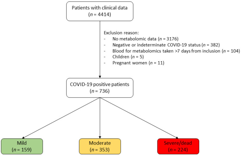
For the present study, we excluded: (i) patients without metabolomic measurement from blood taken within 7 days of inclusion; (ii) patients who tested negative for COVID-19; (iii) patients without a disease-severity score at inclusion; and (iv) pregnant women and children. This resulted in a sample of 736 individuals. A flowchart of patient selection is presented in Figure 1.
Figure 1.
Patient-selection flowchart. PCR tests were used to assess COVID-19 status. Symptom severity was established at inclusion, using the World Health Organization (WHO) criteria [22].
To determine the ability of amino acids to predict adverse outcomes, we considered a subsample of 476 hospitalized patients with data on ICU admission and/or vital status at discharge, and for whom metabolomic measurements were carried out using plasma collected before their ICU admission or death. We defined adverse outcomes as admission to the intensive care unit (ICU) or hospital death.
2.2. Metabolomics Measurements
Plasma samples were analyzed by Metabolon Inc. [23]. Briefly, samples were mixed with methanol, centrifuged and analyzed using ultra-performance liquid chromatography and tandem mass spectrometry (UPLC-MS/MS) using both reverse-phase and hydrophilic-interaction liquid chromatography and both positive and negative ionization. Pooled samples and blanks were used to measure extraction and injection variability. Overall, 1438 metabolites were measured, including 1155 identified compounds and 280 unknown compounds. For more details about the protocol, see [23].
We focus on circulating amino acids for two reasons. First, abdominal obesity has previously been associated with COVID-19 severity, and we have previously demonstrated that circulating concentrations of some amino acids are correlated with central fat accumulation. Second, the existing literature shows that amino acids, as a group, are almost invariably altered in the context of severe COVID-19, although results for specific amino acids are heterogeneous. When more than one metabolomic measurement was available in our 7-day timeframe, we used the closest to study inclusion.
2.3. Statistical Analyses
We compared patient characteristics and comorbidities between severity groups, using Krustal–Wallis and chi-squared tests. To examine the association between disease severity and circulating-amino-acid levels, we used univariate and adjusted logistic-regression models comparing mild patients to moderate and severe patients. We reported the results as odd ratios (OR) for each 1 SD increment in circulating amino acids. We compared the level of metabolites annotated to the TCA and urea cycle between severity groups using the Krustal–Wallis test.
To determine the ability of individual amino-acid levels to predict adverse outcomes, we performed univariate logistic-regression models and computed receiving-operator-characteristic (ROC) curves. The latter is a graphical tool that allows the visualization of the trade-off between sensitivity and specificity of a given predictor. It also allows the calculation of the area under the ROC curve (ROC_AUC), which is a metric of the ability of the predictor to discriminate between two groups. For example, a ROC_AUC of 0.70 indicates that the predictor has 70% accuracy at classifying observations in the correct group. A predictor is considered good when it has a ROC_AUC above 0.8, and excellent above 0.9 [24]. We did not identify optimal thresholds from the ROC curves because the amino-acid levels were expressed as relative abundance, and therefore thresholds would have been of limited use.
We also ran univariate logistic analysis on clinical variables for comparison purposes. Additionally, we built a multivariate logistic-regression model using circulating-amino-acid levels and the clinical variables significantly associated with the outcome in univariate analysis as candidate predictors. Missing values were imputed using multivariate imputation by chained equations using predictive mean matching for continuous variables and logistic regression for ordinal variables. Predictive variables were scaled, and those with ≥50% missing values were discarded. Fifty imputations were run, and forward as well as backward logistic-models were fit with the candidate variables on each imputation. The variables used in the majority (≥50%) of models were selected for the final model. The final model was run on the imputed data, and the results were pooled.
A p-value below 0.05 was considered significant, except for post hoc analyses, where the Bonferroni correction was applied.
3. Results
Overall, 736 individuals were included for analysis (Figure 1). According to WHO criterion for COVID-19 disease severity [22], 159 had mild symptoms, 353 moderate symptoms and 224 severe symptoms.
Sample characteristics by severity are presented in Table 1. Patients with moderate and severe symptoms were significantly older than those with mild symptoms. Sex was also associated with severity, with the severe subgroup counting more males than the other two groups. Glucose level was significantly higher in patients with severe symptoms. C-reactive protein (CRP), creatinine, urea, and white-blood-cell count were significantly higher, whereas albumin and hemoglobin were lower in severe vs. mild and moderate patients. Although the percentage of participants vaccinated was not different across groups, the number of doses received differed significantly. Indeed, there was a greater prevalence of participants with three vaccine doses in the mild group (19%) compared to the moderate and severe groups (1%).
Table 1.
Sample characteristics by COVID-19 severity.
| Variables (Unit) | n= | Mild (n = 159) | Moderate (n = 353) | Severe (n = 224) | p-Value | ||
|---|---|---|---|---|---|---|---|
| Age (years) | 736 | 55.7 (44.2–67.2) | 67.3 (51.8–81.9) | # | 66.0 (54.8–73.6) | # | 2.4 × 10⁻¹⁰ |
| Sex (female) | 736 | 86 (54%)86 (54%) | 168 (48%) | 66 (29%) | #& | 1.0 × 10⁻⁶ | |
| BMI (kg/m2) | 372 | 28.1 (24.7–31.7) | 26.7 (23.6–30.4) | 28.7 (25.6–33.9) | & | 0.0183 | |
| Vaccinated (yes) | 321 | 91 (90%) | 104 (84%) | 83 (86%) | 0.3938 | ||
| Number of vaccine doses (%, 1/2/3 doses) | 277 | 23%/58%/19% | 41%/58%/1% | # | 37%/61%/1% | # | 1.5 × 10⁻⁶ |
| Outpatient (yes) | 736 | 80 (50%) | 3 (1%) | # | 1 (0%) | # | 1.2 × 10−66 |
| CRP (mg/L) * | 391 | 16.4 (1.9–56.2) | 42.3 (18.5–89.1) | # | 96.1 (45.4–173.2) | #& | 1.2 × 10⁻¹² |
| ALT (U/L) | 391 | 25.5 (18.8–48.2) | 27.0 (17.0–51.0) | 35.5 (22.2–69.0) | & | 0.0061 | |
| Glucose (mmol/L) | 451 | 5.65 (5.23–6.57) | 5.90 (5.10–7.68) | 7.40 (6.10–9.90) | #& | 2.9 × 10⁻⁹ | |
| Creatinine (mmol/L) | 519 | 72.0 (60.0–87.5) | 74.0 (57.0–94.0) | 84.0 (63.5–125.0) | #& | 0.0011 | |
| Haemoglobin (g/L) | 508 | 137 (123–151) | 125 (112–137) | # | 120 (103–133) | #& | 1.6 × 10⁻⁷ |
| Urea (mmol/L) | 487 | 4.40 (3.00–5.50) | 5.80 (3.80–9.05) | # | 8.00 (5.47–14.72) | #& | 5.3 × 10⁻¹³ |
| Albumin (g/L) | 439 | 41.0 (39.0–44.0) | 34.0 (31.0–37.0) | # | 30.0 (27.0–34.0) | #& | 2.7 × 10⁻²⁷ |
| Bilirubin (μmol/L) | 380 | 7.00 (5.00–9.00) | 7.00 (5.00–10.00) | 8.00 (6.00–11.00) | 0.0600 | ||
| WBC (×109/L) | 507 | 6.00 (4.60–7.32) | 6.50 (5.10–8.60) | 8.55 (6.15–11.72) | #& | 1.6 × 10⁻¹⁰ | |
| LDH (U/L) | 291 | 251 (212–324) | 303 (243–383) | # | 386 (343–585) | #& | 2.8 × 10⁻¹⁰ |
| Procalcitonin (ug/L) | 168 | 0.07 (0.05–0.10) | 0.11 (0.08–0.17) | # | 0.23 (0.16–0.67) | #& | 1.4 × 10⁻¹⁰ |
| D-Dimer (ug/L) | 160 | 637 (447–860) | 856 (543–1213) | # | 1280 (817–2181) | #& | 1.2 × 10⁻⁵ |
| Temperature (C) | 543 | 37.0 (36.8–37.5) | 37.0 (36.7–37.8) | 37.4 (36.7–38.5) | 0.0281 | ||
| SBP (mmHg) | 576 | 130 (119–147) | 128 (115–144) | 125 (110–138) | # | 0.0199 | |
| DBP (mmHg) | 576 | 81.0 (70.0–87.2) | 76.0 (67.0–84.0) | # | 72.0 (66.0–80.0) | #& | 2.7 × 10⁻⁵ |
| Heart rate (beats/min) | 582 | 97.0 (84.0–105.2) | 92.0 (78.0–107.0) | 96.5 (82.0–109.0) | 0.1000 | ||
| SaO2 (%) | 531 | 97.0 (95.0–99.0) | 95.0 (93.0–97.0) | # | 93.0 (89.0–95.0) | #& | 6.9 × 10⁻²³ |
| Oxygen (yes) | 565 | 0 (0%) | 124 (39%) | # | 132 (71%) | #& | 1.6 × 10⁻²⁴ |
Data are presented as median (Q1-Q3) for continuous variables and number (%) for dichotomous variables. Characteristics were compared across severity groups using Krustal–Wallis test for continuous variables and chi-square test otherwise. Post hoc comparisons were made using the same tests but using a Bonferonni-corrected p-value threshold of p < (0.05/3); &: significant vs. moderate, #: significant vs. mild. BMI: body mass index, CRP: C-reactive protein, ALT: alanine aminotransferase, WBC: white blood cells, SBP: systolic blood pressure, DBP: diastolic blood pressure, LDH: lactate dehydrogenase, SaO2: arterial oxygen saturation. * For CRP, patients receiving tomizumab were excluded because it negates the measurement.
Among comorbidities (see Supplementary Table S1), the prevalence of diabetes, dementia, obesity, arterial hypertension, atrial fibrillation, chronic kidney disease, chronic neurological disease, strokes, hematologic disease, coronary artery disease, rheumatologic disease, cancers and heart failure was significantly associated with COVID-19-symptom severity. However, once adjusted for age, the association was no longer significant for dementia, hematologic disease, rheumatologic disease, cancers and heart failure (results not shown).
Among the participants living with obesity (n = 179 overall), 118 (66%) also had one or more metabolic comorbidities (diabetes, arterial hypertension, or coronary arterial disease). The prevalence of individuals with obesity and a metabolic comorbidity was significantly higher in the moderate and severe COVID-19-symptom severity groups, compared to the mild group (chi-square p-value 0.0084).
The number of participants taking angiotensin-converting enzyme (ACE) inhibitors, corticosteroids, anticoagulants and oral hypoglycemic agents or insulin at inclusion was significantly higher in the moderate and severe groups, compared to the mild group (Supplementary Table S1).
3.1. Are Circulating Amino Acids Associated with COVID-19 Severity?
The mean number of days between study inclusion (and therefore disease-severity assessment) and blood draw for metabolomics measurement was 4 days. We first evaluated the association between circulating-amino-acid concentrations and COVID-19 severity, using univariate logistic-models. The results from this analysis are presented as the odds of having mild vs. severe or mild vs. moderate symptoms for each increment of 1 SD in the circulating amino acid (Table 2).
Table 2.
Univariate association between circulating-amino-acid levels and the odds of presenting mild vs. severe and moderate COVID-19 symptoms.
| Amino Acid | Mild vs. Severe | Mild vs. Moderate | ||||
|---|---|---|---|---|---|---|
| OR | 95%CI | p-Value | OR | 95%CI | p-Value | |
| Phenylalanine | 4.14 | (2.79–6.13) | 1.5 × 10⁻¹² | 1.68 | (1.33–2.11) | 1.0 × 10⁻⁵ |
| Methionine | 2.72 | (1.96–3.77) | 2.2 × 10⁻⁹ | 1.67 | (1.34–2.09) | 4.3 × 10⁻⁶ |
| Aspartate | 1.82 | (1.44–2.30) | 7.3 × 10⁻⁷ | 1.14 | (0.94–1.39) | 0.1745 |
| Isoleucine | 1.64 | (1.31–2.05) | 1.8 × 10⁻⁵ | 1.18 | (0.97–1.43) | 0.0997 |
| Leucine | 1.57 | (1.26–1.97) | 7.0 × 10⁻⁵ | 1.15 | (0.95–1.39) | 0.1508 |
| Glutamate | 1.51 | (1.19–1.90) | 0.0006 | 1.18 | (0.97–1.44) | 0.1055 |
| Asparagine | 1.37 | (1.09–1.71) | 0.0065 | 1.17 | (0.97–1.42) | 0.1039 |
| Valine | 1.35 | (1.09–1.67) | 0.0060 | 1.08 | (0.89–1.31) | 0.4255 |
| Threonine | 1.18 | (0.96–1.46) | 0.1184 | 1.19 | (0.98–1.46) | 0.0814 |
| Lysine | 1.17 | (0.95–1.44) | 0.1427 | 1.09 | (0.90–1.32) | 0.3638 |
| Tyrosine | 1.11 | (0.90–1.36) | 0.3288 | 0.92 | (0.76–1.11) | 0.3813 |
| Arginine | 0.96 | (0.78–1.17) | 0.6763 | 0.97 | (0.80–1.16) | 0.7171 |
| Proline | 0.75 | (0.61–0.93) | 0.0078 | 0.73 | (0.60–0.88) | 0.0010 |
| Glutamine | 0.68 | (0.55–0.85) | 0.0005 | 0.89 | (0.74–1.08) | 0.2427 |
| Serine | 0.68 | (0.55–0.84) | 0.0003 | 0.86 | (0.72–1.04) | 0.1237 |
| Glycine | 0.65 | (0.53–0.81) | 9.5 × 10⁻⁵ | 0.89 | (0.74–1.08) | 0.2362 |
| Alanine | 0.57 | (0.46–0.72) | 1.1 × 10⁻⁶ | 0.68 | (0.57–0.83) | 9.7 × 10⁻⁵ |
| Histidine | 0.45 | (0.35–0.57) | 1.7 × 10⁻¹⁰ | 0.50 | (0.40–0.62) | 9.1 × 10⁻¹¹ |
| Tryptophan | 0.38 | (0.29–0.50) | 9.7 × 10⁻¹³ | 0.57 | (0.47–0.70) | 7.2 × 10⁻⁸ |
| Cysteine | 0.34 | (0.26–0.45) | 7.2 × 10⁻¹⁵ | 0.45 | (0.37–0.56) | 4.6 × 10⁻¹³ |
Results are presented as odds ratios per 1 standard-deviation (SD) increase in amino-acid concentration.
Out of the 20 amino acids tested, 16 were significantly associated with COVID-19 severity. In this univariate analysis, the amino acids most strongly associated with severe symptoms were phenylalanine (positively associated) and cysteine (inversely associated). Concentrations of the 3 BCAAs and glutamate were also significantly associated with severity, but to a lesser extent.
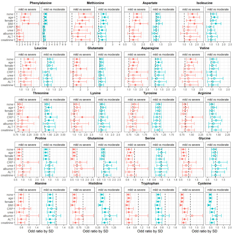
To evaluate the impact of potentially cofounding variables on the association between amino acids and disease severity, we adjusted the logistic models for characteristics found to be significantly different between severity groups. The results of this analysis are presented in Figure 2.
Figure 2.
Odds of having mild vs. severe, or mild vs. moderate COVID-19 symptoms associated with circulating-amino-acid levels unadjusted (none) and adjusted for clinical variables. Results are presented as odds ratios per 1 standard-deviation (SD) increase in amino-acid level. The number of observations for each models varies; see Table 1 for the number of observations available for each variable. The x-axis scale is different for each amino acid. Full circles indicate a significant association and empty circles indicate a non-significant association. BMI: body mass index, CRP: C-reactive protein, ALT: alanine aminotransferase. For CRP, patients receiving tomizumab were excluded because it negates the measurement.
Adjustments for albumin levels had the greatest impact on the association between circulating amino acids and COVID-19 severity; even associations with the most significant amino-acid levels were no longer significant, once adjusted for this covariate. Overall, age, sex and body mass index (BMI) had little impact on the association between amino-acid concentrations and COVID-19-symptom severity.
We also ran models adjusted for comorbidities and medication intake at inclusion (see Supplementary Figures S1 and S2). Overall, adjustment for comorbidities had very little effect on the associations. Medication intake also had little effect, except for oral hypoglycemic agents or insulin, which tended to increase the significance of some associations.
3.2. Are Circulating-Amino-Acid Levels Predictive of Adverse Outcomes?
Among the 476 participants with data on ICU admission and/or vital status at discharge, 26 were admitted to the ICU and 76 died in the hospital, giving a total of 102 patients with adverse outcomes. Blood was drawn on average 19 days before death (range: 1 to 111 days) and 3 days before ICU admission (range: 1 to 21 days).
The characteristics, comorbidities, medication at inclusion and medication during hospitalization, including treatment used for COVID-19, are presented in Supplementary Table S2. There was a greater proportion of men among patients who suffered from an adverse outcome. CRP, creatinine, lactate dehydrogenase and procalcitonin measured at study inclusion were higher in the adverse-outcome group. There was a greater prevalence of atrial fibrillation, arterial hypertension, myocardial infraction, coronary artery disease and chronic kidney disease among the adverse-outcome group.
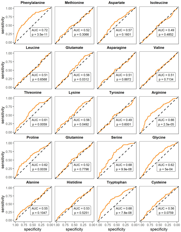
We evaluated the ability of amino acids to distinguish participants who would suffer from adverse outcomes from those who would not, using logistic-regression models and reporting the predictive ability as ROC_AUC. To compare the performance of amino acids to that of variables often measured in a clinical context, we also ran this analysis for clinical variables significantly different across disease-severity categories. Results are presented in Figure 3for amino acids and in Supplementary Table S3 for clinical variables. Circulating-amino-acid levels had a poor-to-average predictive ability, with ROC_AUC ranging from 0.49 (p = 0.6001) for tyrosine to 0.72 (p = 3.5 × 10⁻¹¹) for phenylalanine. Clinical variables had similar or better predictive-abilities, with ROC_AUC ranging from 0.51 for ALT (p = 0.1763) to 0.82 for procalcitonin (p = 0.0019).
Figure 3.
Ability of circulating-amino-acid levels to predict adverse outcomes (intensive-care-unit admission or hospital death). Univariate logistic regressions were used, and areas under the receiving-operator-characteristic curve (ROC_AUC) were computed. A ROC_AUC close to 0.5 means that the predictor is ineffectual, a ROC_AUC over 0.8 means the predictor is good, and a ROC_AUC of 1 means a perfect predictor. Blood was drawn on average 19 days before death and 3 days before ICU admission.
We used the circulating-amino-acid levels and clinical variables significantly associated with adverse outcome in the univariate models as candidate variables for the multivariate model. After removal of variables with ≥50% missing values, scaling and imputation, we used backward and forward stepwise-models to identify 10 variables to include in the final model: CRP, urea, arterial oxygen saturation (SaO2), SBP, phenylalanine, tryptophan, arginine, lysine, glutamate and serine. The final model is presented in Table 3. It had a ROC_AUC of 0.90 (95%CI = 0.86–0.93) for predicting adverse outcomes.
Table 3.
Results from the multivariate logistic-regression model to predict adverse outcomes.
| Term | Estimate | Std. Error | Statistic | df | p Value |
|---|---|---|---|---|---|
| (Intercept) | 8.54 | 3.93 | 2.17 | 355.57 | 0.0306 |
| CRP | 0.01 | 0.00 | 3.58 | 201.67 | 0.0004 |
| Urea | 0.10 | 0.03 | 3.29 | 332.92 | 0.0011 |
| SaO2 | −0.10 | 0.04 | −2.73 | 339.54 | 0.0068 |
| SBP | −0.02 | 0.01 | −2.29 | 417.97 | 0.0228 |
| Phenylalanine | 2.41 | 0.62 | 3.89 | 410.55 | 0.0001 |
| Tryptophan | −1.91 | 0.77 | −2.48 | 401.59 | 0.0136 |
| Arginine | −1.68 | 0.59 | −2.86 | 450.63 | 0.0044 |
| Lysine | 2.83 | 0.94 | 3.00 | 437.35 | 0.0028 |
| Glutamate | −0.65 | 0.37 | −1.73 | 436.87 | 0.0845 |
| Serine | −1.59 | 0.93 | −1.71 | 441.13 | 0.0881 |
Variables were scaled and imputed using multivariate imputation (predictive mean matching for continuous variables and logistic regression for ordinal variables). Variables with ≥50% missing values were dropped. The variables to include in the final model were selected from forward and backward stepwise-regression run on the 50 multiple-imputation datasets. CRP: C-reactive protein, SaO2: arterial oxygen saturation, SBP: systolic blood pressure.
4. Discussion
In this analysis, we aimed to determine whether circulating-amino-acid concentrations were associated with COVID-19-symptom severity, and whether they could predict adverse outcomes in hospitalized patients. We found that 16 out of 20 amino acids were different between severe and mild COVID-19 symptoms. The strongest positive association was observed for phenylalanine, and the strongest inverse association was observed for cysteine. Phenylalanine also had a fair ability to predict adverse outcomes. To our knowledge, this is the largest study investigating circulating amino acids in the context of COVID-19.
A summary of the existing literature on circulating amino acids and COVID-19 severity is provided in Supplementary Table S4. Our results are concordant with previous reports of a significant association between severe COVID-19 and circulating levels of leucine, isoleucine, valine, glutamate and phenylalanine (positively) as well as glutamine, tryptophan, histidine, alanine, proline and cysteine (inversely) (Table S4). We found a larger number of amino acids to be significantly associated with severity than previous studies, which could be explained by our larger sample size. We also confirmed that age, male sex and obesity are significant risk factors for developing severe symptoms when infected with SARS-CoV-2 [25].
Considering that obesity is associated with greater risk of suffering from severe COVID-19 symptoms, we hypothesized that circulating amino acids previously linked to obesity would be the strongest correlates of symptom severity and the best at predicting adverse outcomes. However, this hypothesis did not hold true, as the strongest associations with symptom severity were found for phenylalanine and cysteine, which are not systematically associated with obesity. Moreover, our results showed that the associations between amino-acid levels and disease severity remained significant after adjusting for BMI. Finally, although we would expect amino acids previously reported to be positively associated with adiposity to be also positively associated with COVID-19 severity, this was not always the case. For example, tryptophan is generally positively associated with measurements of adiposity [26], but it was inversely associated with symptom severity in this study. Overall, these results indicate that the state of severe COVID-19 affects amino-acid metabolism beyond what can be observed with elevated adiposity.
We did not find circulating-amino-acid concentrations to be good predictors of ICU admission and hospital death. Indeed, other lab measurements that have been validated in the literature and are much more accessible had similar or better predicting-abilities [27,28,29]. For example, procalcitonin had an accuracy of 82%, whereas the best performing amino acid, phenylalanine, had an accuracy of 72% in identifying patients who would suffer from an adverse outcome. Our multivariate logistic model that included both circulating amino acids and clinical variables had a 90% accuracy for predicting adverse outcomes. This indicates that an index including many relevant variables may perform better than a single biomarker. However, we consider this analysis preliminary, since the model was not validated on an independent cohort and because of the variability of elapsed time between blood draw and adverse-outcome occurrence (from 1 to 111 days). More studies are needed to determine the added value of amino acids to clinical variables for adverse-outcome prediction.
The strengths of this study include the rather large sample-size and the use of the WHO symptom-severity scale. Limitations include the absence of an uninfected control group and the large number of missing data for some variables. Other limitations include the lack of measurement of body-fat distribution and the variability in elapsed time between blood draw and adverse-outcome occurrence. Finally, we acknowledge that amino acids are more easily and frequently measured in metabolomics studies compared to other classes of metabolites, and this has probably contributed to the abundance of studies linking amino acids and COVID-19. Whether amino acids are better than other metabolites at predicting COVID-19 severity or adverse outcomes should be investigated further.
In conclusion, severe COVID-19 symptoms are associated with altered levels of many circulating amino acids, possibly hinting at global changes in nitrogen metabolism. These associations are mostly independent of age, sex and BMI.
Acknowledgments
This work was made possible through open sharing of data and samples from the Biobanque Québécoise COVID-19 (https://www.bqc19.ca/ accessed on 28 September 2022), funded by the Fonds de recherche du Québec—Santé, Génome Québec and the Publich Health Agency of Canada. We thank all participants in BQC19 for their contribution.
Supplementary Materials
The following supporting information can be downloaded at https://www.mdpi.com/article/10.3390/metabo13020201/s1, Figure S1: Odds of having mild vs. severe or moderate COVID-19 symptoms associated with circulating amino acids unadjusted and adjusted for comorbidities associated with disease severity; Figure S2: Odds of having mild vs. severe or mild vs. moderate COVID-19 symptoms associated with circulating amino acids unadjusted and adjusted for medication intake at study inclusion; Table S1: Prevalence of comorbidities by symptom severity in the whole sample (n = 736); Table S2: Characteristics and prevalence of comorbidities by symptom severity in the subsample of 440 patients used to assess the ability of amino-acid levels to predict adverse outcomes; Table S3: Ability of baseline characteristics to predict adverse outcomes; Table S4: Summary of the associations between circulating-amino acid-concentrations and COVID-19-symptom severity reported in the literature.
Author Contributions
Conceptualization, P.P., L.B. and A.T.; methodology, I.M.-P. and F.L.-T.; formal analysis, I.M.-P.; writing—original draft preparation, I.M.-P. and F.L.-T.; writing—review and editing, I.M.-P., F.L.-T., P.P., L.B. and A.T.; supervision, A.T.; funding acquisition, P.P., L.B. and A.T. All authors have read and agreed to the published version of the manuscript.
Institutional Review Board Statement
The study was conducted in accordance with the Declaration of Helsinki, and approved by the Ethics Committee of the Montreal University Hospital Center (Management Framework of the Quebec COVID-19 Biobank—version 8.0, 5 October 2021).
Informed Consent Statement
Informed consent was obtained from all participants involved in the study.
Data Availability Statement
Restrictions apply to the availability of these data. Data was obtained from the Biobanque Québécoise COVID-19. Access to the data can be requested through their website: https://www.bqc19.ca/en accessed on 28 September 2022.
Conflicts of Interest
A.T. and L.B. receive research funding from Johnson & Johnson, Medtronic and GI Windows for studies on bariatric surgery. A.T. and L.B. acted as consultants for Bausch Health and Novo Nordisk. A.T. is a consultant for Biotwin. The other authors had no conflict of interest to disclose.
Funding Statement
The Biobanque Québécoise COVID-19 is funded by the Fonds de recherche du Québec—Santé, Génome Québec and the Publich Health Agency of Canada. This study was funded by a research grant form the Foundation of Institut universitaire de cardiologie et de pneumologie de Québec—Université Laval to A.T., L.B. and P.B. I.M.-P. is the recipient of a scholarship from the Canadian Institutes of Health Research. L.B. and A.T. are co-directors of the Research Chair in Bariatric and Metabolic Surgery at Laval University.
Footnotes
Disclaimer/Publisher’s Note: The statements, opinions and data contained in all publications are solely those of the individual author(s) and contributor(s) and not of MDPI and/or the editor(s). MDPI and/or the editor(s) disclaim responsibility for any injury to people or property resulting from any ideas, methods, instructions or products referred to in the content.
References
- 1.The Novel Coronavirus Pneumonia Emergency Response Epidemiology, T The Epidemiological Characteristics of an Outbreak of 2019 Novel Coronavirus Diseases (COVID-19)—China, 2020. China CDC Wkly. 2020;2:113–122. doi: 10.46234/ccdcw2020.032. [DOI] [PMC free article] [PubMed] [Google Scholar]
- 2.Hasan M.R., Suleiman M., Pérez-López A. Metabolomics in the Diagnosis and Prognosis of COVID-19. Front. Genet. 2021;12:721556. doi: 10.3389/fgene.2021.721556. [DOI] [PMC free article] [PubMed] [Google Scholar]
- 3.Lin B., Liu J., Liu Y., Qin X. Progress in understanding COVID-19: Insights from the omics approach. Crit. Rev. Clin. Lab. Sci. 2021;58:242–252. doi: 10.1080/10408363.2020.1851167. [DOI] [PubMed] [Google Scholar]
- 4.Mussap M., Fanos V. Could metabolomics drive the fate of COVID-19 pandemic? A narrative review on lights and shadows. Clin. Chem. Lab. Med. 2021;59:1891–1905. doi: 10.1515/cclm-2021-0414. [DOI] [PubMed] [Google Scholar]
- 5.Danlos F.X., Grajeda-Iglesias C., Durand S., Sauvat A., Roumier M., Cantin D., Colomba E., Rohmer J., Pommeret F., Baciarello G., et al. Metabolomic analyses of COVID-19 patients unravel stage-dependent and prognostic biomarkers. Cell Death Dis. 2021;12:258. doi: 10.1038/s41419-021-03540-y. [DOI] [PMC free article] [PubMed] [Google Scholar]
- 6.Lee J.W., Su Y., Baloni P., Chen D., Pavlovitch-Bedzyk A.J., Yuan D., Duvvuri V.R., Ng R.H., Choi J., Xie J., et al. Integrated analysis of plasma and single immune cells uncovers metabolic changes in individuals with COVID-19. Nat. Biotechnol. 2022;40:110–120. doi: 10.1038/s41587-021-01020-4. [DOI] [PMC free article] [PubMed] [Google Scholar]
- 7.Atila A., Alay H., Yaman M.E., Akman T.C., Cadirci E., Bayrak B., Celik S., Atila N.E., Yaganoglu A.M., Kadioglu Y., et al. The serum amino acid profile in COVID-19. Amino Acids. 2021;53:1569–1588. doi: 10.1007/s00726-021-03081-w. [DOI] [PMC free article] [PubMed] [Google Scholar]
- 8.Barberis E., Timo S., Amede E., Vanella V.V., Puricelli C., Cappellano G., Raineri D., Cittone M.G., Rizzi E., Pedrinelli A.R., et al. Large-Scale Plasma Analysis Revealed New Mechanisms and Molecules Associated with the Host Response to SARS-CoV-2. Int. J. Mol. Sci. 2020;21:8623. doi: 10.3390/ijms21228623. [DOI] [PMC free article] [PubMed] [Google Scholar]
- 9.Caterino M., Costanzo M., Fedele R., Cevenini A., Gelzo M., Di Minno A., Andolfo I., Capasso M., Russo R., Annunziata A., et al. The Serum Metabolome of Moderate and Severe COVID-19 Patients Reflects Possible Liver Alterations Involving Carbon and Nitrogen Metabolism. Int. J. Mol. Sci. 2021;22:9548. doi: 10.3390/ijms22179548. [DOI] [PMC free article] [PubMed] [Google Scholar]
- 10.Cai Y., Kim D.J., Takahashi T., Broadhurst D.I., Ma S., Rattray N.J.W., Casanovas-Massana A., Israelow B., Klein J., Lucas C., et al. Kynurenic acid underlies sex-specific immune responses to COVID-19. medRxiv. 2020 doi: 10.1101/2020.09.06.20189159. [DOI] [PMC free article] [PubMed] [Google Scholar]
- 11.Páez-Franco J.C., Torres-Ruiz J., Sosa-Hernández V.A., Cervantes-Díaz R., Romero-Ramírez S., Pérez-Fragoso A., Meza-Sánchez D.E., Germán-Acacio J.M., Maravillas-Montero J.L., Mejía-Domínguez N.R., et al. Metabolomics analysis reveals a modified amino acid metabolism that correlates with altered oxygen homeostasis in COVID-19 patients. Sci. Rep. 2021;11:6350. doi: 10.1038/s41598-021-85788-0. [DOI] [PMC free article] [PubMed] [Google Scholar]
- 12.Wu P., Chen D., Ding W., Wu P., Hou H., Bai Y., Zhou Y., Li K., Xiang S., Liu P., et al. The trans-omics landscape of COVID-19. Nat. Commun. 2021;12:4543. doi: 10.1038/s41467-021-24482-1. [DOI] [PMC free article] [PubMed] [Google Scholar]
- 13.Shen B., Yi X., Sun Y., Bi X., Du J., Zhang C., Quan S., Zhang F., Sun R., Qian L., et al. Proteomic and Metabolomic Characterization of COVID-19 Patient Sera. Cell. 2020;182:59–72. doi: 10.1016/j.cell.2020.05.032. [DOI] [PMC free article] [PubMed] [Google Scholar]
- 14.Aghili S.M.M., Ebrahimpur M., Arjmand B., Shadman Z., Pejman Sani M., Qorbani M., Larijani B., Payab M. Obesity in COVID-19 era, implications for mechanisms, comorbidities, and prognosis: A review and meta-analysis. Int. J. Obes. 2021;45:998–1016. doi: 10.1038/s41366-021-00776-8. [DOI] [PMC free article] [PubMed] [Google Scholar]
- 15.Pranata R., Lim M.A., Huang I., Yonas E., Henrina J., Vania R., Lukito A.A., Nasution S.A., Alwi I., Siswanto B.B. Visceral adiposity, subcutaneous adiposity, and severe coronavirus disease-2019 (COVID-19): Systematic review and meta-analysis. Clin. Nutr. ESPEN. 2021;43:163–168. doi: 10.1016/j.clnesp.2021.04.001. [DOI] [PMC free article] [PubMed] [Google Scholar]
- 16.Newgard C.B. Metabolomics and Metabolic Diseases: Where Do We Stand? Cell Metab. 2017;25:43–56. doi: 10.1016/j.cmet.2016.09.018. [DOI] [PMC free article] [PubMed] [Google Scholar]
- 17.Maltais-Payette I., Boulet M.M., Prehn C., Adamski J., Tchernof A. Circulating glutamate concentration as a biomarker of visceral obesity and associated metabolic alterations. Nutr. Metab. 2018;15:78. doi: 10.1186/s12986-018-0316-5. [DOI] [PMC free article] [PubMed] [Google Scholar]
- 18.Maltais-Payette I., Allam-Ndoul B., Perusse L., Vohl M.C., Tchernof A. Circulating glutamate level as a potential biomarker for abdominal obesity and metabolic risk. Nutr. Metab. Cardiovasc. Dis. NMCD. 2019;29:1353–1360. doi: 10.1016/j.numecd.2019.08.015. [DOI] [PubMed] [Google Scholar]
- 19.Maltais-Payette I., Vijay J., Simon M.M., Corbeil J., Brière F., Grundberg E., Tchernof A. Large-scale analysis of circulating glutamate and adipose gene expression in relation to abdominal obesity. Amino Acids. 2022;54:1287–1294. doi: 10.1007/s00726-022-03181-1. [DOI] [PubMed] [Google Scholar]
- 20.Tremblay K., Rousseau S., Zawati M.H., Auld D., Chassé M., Coderre D., Falcone E.L., Gauthier N., Grandvaux N., Gros-Louis F., et al. The Biobanque québécoise de la COVID-19 (BQC19)-A cohort to prospectively study the clinical and biological determinants of COVID-19 clinical trajectories. PLoS ONE. 2021;16:e0245031. doi: 10.1371/journal.pone.0245031. [DOI] [PMC free article] [PubMed] [Google Scholar]
- 21.INSPQ Les Variants du SRAS-CoV-2. [(accessed on 28 September 2022)]. Available online: https://www.inspq.qc.ca/en/node/26927.
- 22.A minimal common outcome measure set for COVID-19 clinical research. Lancet. Infect. Dis. 2020;20:e192–e197. doi: 10.1016/S1473-3099(20)30483-7. [DOI] [PMC free article] [PubMed] [Google Scholar]
- 23.Ford L., Kennedy A.D., Goodman K.D., Pappan K.L., Evans A.M., Miller L.A.D., Wulff J.E., Wiggs B.R., Lennon J.J., Elsea S., et al. Precision of a Clinical Metabolomics Profiling Platform for Use in the Identification of Inborn Errors of Metabolism. J. Appl. Lab. Med. 2020;5:342–356. doi: 10.1093/jalm/jfz026. [DOI] [PubMed] [Google Scholar]
- 24.Carter J.V., Pan J., Rai S.N., Galandiuk S. ROC-ing along: Evaluation and interpretation of receiver operating characteristic curves. Surgery. 2016;159:1638–1645. doi: 10.1016/j.surg.2015.12.029. [DOI] [PubMed] [Google Scholar]
- 25.Mahamat-Saleh Y., Fiolet T., Rebeaud M.E., Mulot M., Guihur A., El Fatouhi D., Laouali N., Peiffer-Smadja N., Aune D., Severi G. Diabetes, hypertension, body mass index, smoking and COVID-19-related mortality: A systematic review and meta-analysis of observational studies. BMJ Open. 2021;11:e052777. doi: 10.1136/bmjopen-2021-052777. [DOI] [PMC free article] [PubMed] [Google Scholar]
- 26.Yamakado M., Tanaka T., Nagao K., Ishizaka Y., Mitushima T., Tani M., Toda A., Toda E., Okada M., Miyano H., et al. Plasma amino acid profile is associated with visceral fat accumulation in obese Japanese subjects. Clin. Obes. 2012;2:29–40. doi: 10.1111/j.1758-8111.2012.00039.x. [DOI] [PubMed] [Google Scholar]
- 27.Lippi G., Plebani M. Procalcitonin in patients with severe coronavirus disease 2019 (COVID-19): A meta-analysis. Clin. Chim. Acta. 2020;505:190–191. doi: 10.1016/j.cca.2020.03.004. [DOI] [PMC free article] [PubMed] [Google Scholar]
- 28.Zhou F., Yu T., Du R., Fan G., Liu Y., Liu Z., Xiang J., Wang Y., Song B., Gu X., et al. Clinical course and risk factors for mortality of adult inpatients with COVID-19 in Wuhan, China: A retrospective cohort study. Lancet. 2020;395:1054–1062. doi: 10.1016/S0140-6736(20)30566-3. [DOI] [PMC free article] [PubMed] [Google Scholar]
- 29.Terpos E., Ntanasis-Stathopoulos I., Elalamy I., Kastritis E., Sergentanis T.N., Politou M., Psaltopoulou T., Gerotziafas G., Dimopoulos M.A. Hematological findings and complications of COVID-19. Am. J. Hematol. 2020;95:834–847. doi: 10.1002/ajh.25829. [DOI] [PMC free article] [PubMed] [Google Scholar]
Associated Data
This section collects any data citations, data availability statements, or supplementary materials included in this article.
Supplementary Materials
Data Availability Statement
Restrictions apply to the availability of these data. Data was obtained from the Biobanque Québécoise COVID-19. Access to the data can be requested through their website: https://www.bqc19.ca/en accessed on 28 September 2022.


