Abstract
Osteoarthritis (OA), a disabling joint inflammatory disease, is characterized by the progressive destruction of cartilage, subchondral bone remodeling, and chronic synovitis. Due to the prolongation of the human lifespan, OA has become a serious public health problem that deserves wide attention. The development of OA is related to numerous factors. Among the factors, nitric oxide (NO) plays a key role in mediating this process. NO is a small gaseous molecule that is widely distributed in the human body, and its synthesis is dependent on NO synthase (NOS). NO plays an important role in various physiological processes such as the regulation of blood volume and nerve conduction. Notably, NO acts as a double-edged sword in inflammatory diseases. Recent studies have shown that NO and its redox derivatives might be closely related to both normal and pathophysiological joint conditions. They can play vital roles as normal bone cell-conditioning agents for osteoclasts, osteoblasts, and chondrocytes. Moreover, they can also induce cartilage catabolism and cell apoptosis. Based on different conditions, the NO/NOS system can act as an anti-inflammatory or pro-inflammatory agent for OA. This review summarizes the studies related to the effects of NO on all normal and OA joints as well as the possible new treatment strategies targeting the NO/NOS system.
Keywords: nitric oxide, nitric oxide synthases, osteoarthritis, osteoclasts, osteoblasts, chondrocytes, inflammation
1. Introduction
Nitric oxide (NO), an endogenous gas, is present in nearly all types of human cells. Initially, researchers did not recognize NO as a component of the human body but rather treated it as a new protein called “endothelium-dependent relaxing factor”. Currently, NO has been found to be a multipurpose intracellular signaling molecule. NO is used as a more sensitive substance for communication between the cells in different tissues and plays important roles in various physiological and pathophysiological responses, including blood pressure, blood circulation, platelet function, inflammation, neurotransmission, and immunity [1]. This review specifically focuses on the physio-pathological mechanism of NO in osteoarthritis (OA) and discusses the treatment potential of NO-related molecular structures for OA.
1.1. Epidemiology of OA
OA is the most common type of arthritis caused by cartilage degradation, which eventually leads to severe pain, limitation of activity, and impaired joint function. According to epidemiological studies, one in every eight people in the United States (27–31 million people among the total US population) is affected by symptomatic OA, while globally, OA affects 250 million people [2,3]. Furthermore, the prolongation of the average human lifespan [4,5] has led to increased OA prevalence and incidence. Recently, OA has become a major cause of impaired mobility and disability, thereby impairing the quality of life and increasing the economic and social disease burden.
1.2. Correlation between OA and NO
OA is caused by abnormal joint loads and mechanical stimulation, leading to a biomechanical process of abrasion [6]. Pathologically, the development of OA depends on chronic inflammatory processes, including immune cell infiltration and the release of cytokines and metalloproteinases into the joints [7,8]. NO is found in the synovial fluid, serum albumin, and urine of OA patients in the form of NO-derived molecules such as sodium nitrite, fluorophenyltryptophan, and N-methyl-L-arginine. The elevated levels of these molecules demonstrate the occurrence of OA, thereby showing the role of NO in bone and joint diseases [9]. The role of NO is essential for the development of OA. Therefore, studying the NO–OA interaction might deepen the comprehensive understanding of OA pathogenesis and provide guidance on OA treatment strategies.
2. NO and NO Synthase (NOS)
NO is an unstable, uncharged free radical, and the N atom in NO with free electrons gets rapidly oxidized. Hence, its biological lifetime is only several seconds; subsequently, NO is active only in the immediate proximity of NO-producing cells. Meanwhile, NO has a small size and favorable lipid solubility, thereby exhibiting high membrane permeability [10]. Therefore, NO can rapidly diffuse out of cells and cross through more than several microns into target cells, acting as a paracrine-signaling molecule.
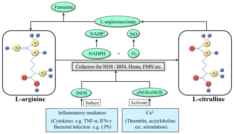
NOS produces NO as a byproduct while oxidizing L-arginine to L-citrulline, using oxygen and nicotinamide adenine dinucleotide phosphate (NADPH) as substrates (Figure 1). In addition, this reaction requires flavin adenine dinucleotide (FAD), flavin mononucleotide (FMN), and (6R-)5,6,7,8-tetrahydro-L-biopterin (BH4) as cofactors. Among these cofactors, FAD and FMN are responsible for the transfer of electrons from NADPH to heme, and BH4 can bind to the oxygenase domain of NOS as an essential cofactor for substrates. Notably, this reaction is stereospecific; NOS can metabolize L-arginine, but not D-arginine [11]. There are three subtypes of NOS present in the body: neuronal NOS (nNOS; NOS1), inducible NOS (iNOS; NOS2), and endothelial NOS (eNOS; NOS3). They have the same NO production process; however, their structures and functions are tailored to the different body parts. nNOS and eNOS are constitutively expressed (thus also called constitutive NOS, cNOS) and can rapidly produce NO based on an increase in cytoplasmatic calcium. In some specific cases, such as oxidation of BH4 or depletion of L-arginine, eNOS can transform from a NO-producing enzyme into an enzyme that generates superoxide anion (O2−), which is called NOS uncoupling. iNOS is only induced when macrophages are stimulated by cytokines including tumor necrosis factor-alpha (TNF-α) and interferon-gamma (IFN-γ) and other factors such as bacterial toxoids. iNOS produces far more NO as compared to that produced by nNOS or eNOS [12]; this increased production might have various pathological effects.
Figure 1.
Formation and synthesis of NO. NAPDH: nicotinamide adenine dinucleotide phosphate; NAPD+: oxidation form of NAPDH; NO: nitric oxide; O2: oxygen; iNOS: inducible nitric oxide synthase; eNOS: endothelial nitric oxide synthase; nNOS: neuronal nitric oxide synthase; TNF: tumor necrosis factor; IFN: interferon; LPS: lipopolysaccharide.
NO is a reactive molecule that can act via numerous pathways, depending on the relative concentrations of NO and the surrounding environment in which NO is produced. The effects of NO can be direct or indirect, depending on the NO concentration—NO can show direct effects (<1 µM NO) as well as indirect effects (>1 µM NO). The indirect effects are induced by the reactive nitrogen species, produced by the interaction of NO with superoxide or oxygen [13].
In inflammatory diseases, NO acts as a double-edged sword. It can produce anti-inflammatory effects under physiological conditions. The agents, such as cytokines, promote iNOS activity, resulting in the synthesis of NO in large amounts by monocytes, macrophages, and granulocytes, among many other cells. NO then scavenges free radicals or kills microbes, thereby preventing cell injury.
NO can also act as a pro-inflammatory mediator. Its overproduction can be cytotoxic [14]. NO can react with superoxide and generate peroxynitrite. The subsequent secondary chain reaction leads to the production of NO2 and hydroxide, which can be even more toxic. This results in damaging the normal tissues, thereby increasing inflammation.
3. Physiological Effects of NO on Cartilage
As a chronic evolutionary disease, OA causes structural changes in the normal articular cavity. In order to comprehensively understand the mechanism of OA, the regulation of osseous tissue under physiological conditions should be explored.
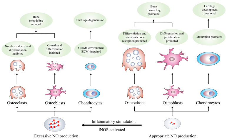
The musculoskeletal system is an essential part of the human body’s functions and is affected by the biomechanical environment. The osseous tissue’s shape and density alter with the changes in the biomechanical environment. Qualitatively, the bone mass and density increase in areas with higher loads and decrease in areas with lower loads; this process is called bone remodeling. Bone remodeling consists of two steps: the stimulation of resorption in response to bone formation and the generation of new bone after bone degradation. Studies have shown the complexity of the process, involving multi-tiered communication networks of osteoclasts, osteoblasts, and other cell types in bone [15,16,17]. In addition, the coordination among endocrine, autocrine, and paracrine signals harmonizes human bone remodeling based on the adjustment of these cells in different stages [18]. Therefore, the recruitment, differentiation, and function of cells in bone remodeling are governed by a series of systemic regulators of bone metabolism (parathyroid hormone, vitamin D, and estrogen), local mediators (receptor of activated nuclear factor kappa-B ligand [RANKL], its antagonist osteoprotegerin, and Wnts/sclerostin), free radicals (superoxide, hydrogen peroxide, and NO), and bone matrix components [19]. The subsequent sections of this review article focus on the effects of the NO/NOS system on bone processes (Table 1 and Figure 2).
Table 1.
Possible effect of NO on different type of cells and its related mechanisms.
| Targeted-Cell | Type of Study | NO Concentration | Related Mechanisms | Effect of NO | Reference |
|---|---|---|---|---|---|
| Osteoclasts | In vivo and in vitro | High | Reduces the number of osteoclasts and inhibits its spread |
Inhibits mature osteoclasts bone resorption |
Kalyanaraman et al., 2017 [20] |
| Osteoclasts | In vitro | High | Induced by inhibited cGMP-degrading activity of PDE | Inhibits precellular osteoclasts differentiation |
Amano et al., 2019 [21] |
| Osteoclasts | In vitro | Low | Induced by RANKL and produces downstream molecular 8-nitro-cGMP |
Promotes osteoclasts differentiation | Kaneko et al., 2018 [22] |
| Osteoclasts | In vitro | Low | Stimulated by cytokines and other mediators such as PG | Promotes osteoclasts bone resorption | Mentaverri et al., 2003 [23] |
| Osteoblasts | In vitro | Low | Induces osteoblast differentiation factor (Cbfa1) expression |
Promotes osteoblasts differentiation | Gloria et al., 2020 [24] |
| Osteoblasts | In vivo and in vitro | Low | Activates Src, Erk-1/2 and Akt signaling pathway through sGC and PKG2 | Promotes osteoblasts proliferation and anti-apoptotic effects |
Cepeda et al., 2020 [25] Ramdani et al., 2018 [26] |
| Osteoblasts | In vivo and in vitro | Low | Stimulated by estrogen | Promotes osteoblasts growth and development |
Gerbarg et al., 2016 [27] Crescitelli et al., 2019 [28] |
| Osteoblasts | In vitro | Low | Responses to mechanical stimulation |
Promotes osteoblasts proliferation and survival |
Wittkowske et al., 2016 [29] Maycas et al., 2017 [30] |
| Chondrocytes | In vitro | High | Induces caspase expression upregulation | Induces chondrocytes apoptosis | Poderoso et al., 2019 [31] Kamm et al., 2019 [32] |
| Chondrocytes | In vivo and in vitro | High | Stimulated by inflammatory mediators | Affects numerous biomolecular processes in chondrocytes |
Wojdasiewicz et al., 2014 [33] |
| Chondrocytes | In vitro | Low | Stimulated chondrocytes hypertrophy and increased the expression of alkaline phosphatase and type X collagen |
Promotes chondrocytes maturation | Teixeira et al., 2005 [34] Drissi et al., 2005 [35] |
Figure 2.
Formation and synthesis of NO. NO: nitric oxide; iNOS: inducible nitric oxide synthase; ECM: extracellular matrix.
3.1. NO and Osteoclasts
Osteoclasts resorb bone and originate from somatic cells related to the single-core macrophage lineage [36]. Although they have unique bone resorption capabilities, they have various characteristics in common with macrophages, some of which are selectively expressed and regulated. This might be due to the adaptation of osteoclasts to osteoclast physiology, thus developing their specific functions. The similarities and nuances that distinguish osteoclasts from non-bone-resorbing macrophages reflect some specific mechanisms in bone remodeling. The NO/NOS system might be one such mechanism.
Thirty years ago, NO was confirmed to regulate the osteoclasts’ roles. Maclntyre reported that in isolated rat osteoclasts, NO could inhibit the spreading of the cells and bone resorption [37]. Moreover, nitrosyl-cobinamide has been recently reported as an immediate NO release agent, which reduces the number of osteoclasts in intact mice and inhibits the increase in osteoclast numbers in ovariectomized mice [20]. Amano found that during osteoclast formation in mouse bone marrow cells, the osteogenic helioxanthin derivative could inhibit the cyclic guanosine monophosphate (cGMP) degradation activity of phosphodiesterase, promote NO production, and inhibit the differentiation of osteoclasts dose-dependently [21]. Other experiments, such as the mouse skull assay and the rat long bone assay, also demonstrated similar results [38,39]. Taken together, these results indicated the inhibitory effects of high NO concentrations on bone resorption via two types of effects: an immediate inhibitory effect on mature osteoclast bone resorption and a reasonable inhibitory effect on precellular osteoclast differentiation. In this pathway, the effects of NO are cGMP-independent and different from the common pathway in most other systems. Notably, NO can induce mature osteoclasts to get rid of bone and reduce their acid secretion, thereby inhibiting bone resorption [40]. This process appears to be mediated by endogenous NO production and requires cGMP and protein kinase G (PKG).
In addition, lower NO concentrations can also promote osteoclast differentiation and survival. Interleukin (IL)-1β and TNF-α are powerful stimulators of bone resorption; they could appropriately enhance NO formation in the organ cultures of bone as well as bone marrow cultures. The addition of NOS inhibitors resulted in inhibiting the induced bone resorption, demonstrating that the NO/NOS system was stimulated by cytokines and other cytokine-induced mediators, such as prostacyclin (PG), to enhance bone resorption [39,41].
Kaneko et al. found that 8-nitro-cGMP, which is a NO derivate that is formed when NO reacts with cGMP in the presence of reactive oxygen species (ROS), could promote RANKL-induced osteoclast differentiation [22]. Moreover, a study assessed the levels of all three NOS modes in bones as well as isolated osteoblasts and osteoclasts using reverse transcription-polymerase chain reaction (RT-PCR) and immunohistochemistry [42]. Knocking out the nNOS gene increased the relative density of trabecular and cortical bone minerals in mice; the accurate measurement of the bone structure indicated a decrease in the total number of osteoclasts and osteoblasts, with worsened bone remodeling, which was reflected by the low mineral accumulation and bone formation rate [43,44,45]. These major manifestations indicated that nNOS might be essential for the differentiation and/or survival of all normal osteoclasts in vivo.
Moreover, nNOS-deficient bone marrow monocytes could produce poorly functional osteoclasts in vitro. Correspondingly, the iNOS-deficient mice showed no significant bone abnormalities, and their femoral length, trabecular bone volume fraction, bone formation rate, and osteoclast surface were normal [46]; this confirmed that the NO production by iNOS was much larger than that produced by nNOS, resulting in the loss of the promotion function. Therefore, in bone, iNOS might limit osteoclast activity by increasing NO levels, thus preventing excessive bone resorption in inflammatory diseases.
In other words, the high NO concentration can reduce the number of osteoclasts and inhibit their differentiation, demonstrating inhibitory effects on osteoclast bone resorption. On the other hand, the low NO concentration, mainly produced by cNOS, can promote the differentiation and survival of osteoclasts. Thus, the osteoclasts produced in nNOS-deficient individuals can lead to poor function. However, these conclusions are based on only in vivo and in vitro studies and lack the support of clinical data.
3.2. NO and Osteoblasts
Similar to their effects on osteoclasts, they might promote the differentiation and survival of osteoblasts and vice versa. The low NO concentration has been confirmed both in vivo and in vitro. Recent studies on human somatic cells and mouse models showed that the osteoblasts lacked argininosuccinate lyase, an enzyme participating in the synthesis of arginine and contributing to NO production, which resulted in low NO production and failure to differentiate [47]. Another study found that the vascular smooth muscle cells from the renal artery of male Wistar rats treated with aminoguanidine (AG; an iNOS inhibitor) showed a reduced expression of osteoblast differentiation factor (Cbfa1) [24]. Moreover, Wei et al. demonstrated that using the biochemical signaling molecules, such as PGs, NO, and insulin-like growth factor-1 (IGF-1) released by osteocytes, could increase osteogenesis, which showed a guiding significance in contemporary clinical treatment [48]. A study investigating the possible underlying mechanism of action showed that the proliferation of osteoblasts was simulated by cell-permeable cGMP analogs and prevented by pharmacological inhibition of soluble guanylyl cyclase (sGC) or protein kinase 2 (PKG2) or siRNA-mediated PKG2 knockdown; this suggests that the positive effects of low NO concentrations on osteoblast proliferation were mediated by sGC and PKG2 [25]. PKG2 exerts its antiapoptotic and proliferation-promoting effects in osteoblasts by activating Src, extracellularly regulated protein kinases-1/2 (Erk-1/2), and Akt [26]. The activated Akt phosphorylates and inactivates glycogen synthase kinase-3β, thereby stabilizing β-catenin and activating the Wnt pathway genes. The activation of the Wnt signaling pathway is a key factor in the differentiation, proliferation, and survival of (pre)osteoblasts and in driving bone formation [49,50]. In addition, a small dose of NO donors could activate the mRNA expression of osteoblast genes, such as alkaline phosphatase, osteocalcin, and collagen-1, and increase bone matrix synthesis and mineralization, thereby enhancing the osteogenic differentiation of (pre)osteoblasts in vitro [51,52,53].
In addition, based on estrogen stimulation, the moderate cNOS expression in osteoblasts can produce NO, which has a substantial role in the growth and development of osteoblasts as well as cytokine production [27,28].
The initiation of mechanical stimulation is highly important for the growth, development, and remodeling of bone [29,30]. When fluid flows through the bone canalicular system, the resulting shear stress stimulates osteoblasts and osteocytes to enhance their anabolism nonspecifically. With the increase in their proliferation and survival, the bone marrow stromal cells, osteoblasts, and osteocyte-like cells respond to fluid shear stress in vitro. This anabolic reaction requires moderate NO production from calcium-mediated eNOS.
The high NO concentrations resulting from NO donors or proinflammatory cytokines can effectively inhibit the growth and differentiation of osteoblasts [54,55]. These conditions often occur in inflammatory disorders and are related to the inhibitory effects of pro-inflammatory cytokines on bone formation. Based on the animal model of inflammation-mediated osteopenia, the active cytokines are the reason for the reduced osteogenesis [56].
In summary, the high production of NO by iNOS in an inflammatory environment can effectively inhibit the growth and differentiation of osteoblasts. In most physiological conditions, the appropriate NO concentrations can promote osteoblast function, which might be mediated by sGC and PKG2. The initiation of mechanical stimulation can also contribute to the proliferation and survival of osteoblasts through moderate NO production.
3.3. NO and Chondrocytes
The effects of NO on chondrocytes in vivo under normal physiological conditions are hard to observe due to the dominance of iNOS in these bone cells. In other words, a necessary stimulation is required for the chondrocytes to produce NO.
A study using the chick cartilage model reported that the NO metabolites played a role in the maturation and differentiation of chondrocytes [34,35]. All three NOS isoforms are expressed and remain active in the growth plate. At least two NO-mediated functions are important in epiphyseal chondrocytes. After the maturation of chondrocytes, NO and related compounds stimulate chondrocyte hypertrophy via the cGMP-dependent pathway, thereby increasing the expression of alkaline phosphatase and type X collagen (maturation markers). In the late stages, inhibiting NO production can inhibit apoptosis, while exposure to NO donors increases apoptosis, suggesting that NO induces this process. This suggested that based on artificial stimulation, moderate NO production might contribute to chondrocyte proliferation in vitro; however, relevant in vivo data are still lacking.
4. Pathological Effects of NO in OA
For a long time, OA pathophysiology was thought to be a process of long-term biomechanical wear caused by abnormal joint load and mechanical stimulation. Therefore, physical factors such as age, sex, obesity, and strain were frequently mentioned in the etiology [2,57]. Trauma was considered to promote this process, causing post-traumatic arthritis or secondary OA.
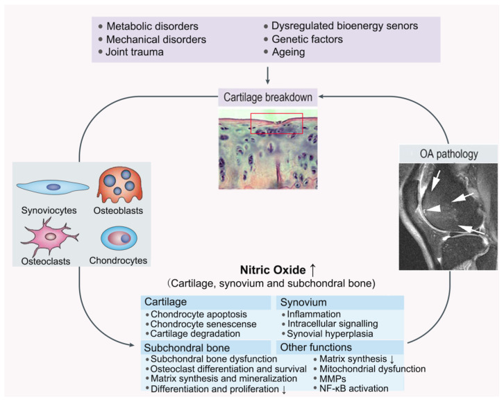
Recently, the importance of metabolic factors has also been reported. Both mechanical and biochemical factors are responsible for OA development [58]. The OA chondrocytes release various inflammatory mediators such as IL-1, TNF-α, and prostaglandin E2 (PGE2), upregulate iNOS and produce excessive NO, which leads to a perpetual release of inflammatory cytokines and other catabolic processes [33]. This affects numerous biomolecular processes in chondrocytes such as proteoglycan and collagen synthesis, as well as metalloproteinase and nuclear factor kappa-B (NF-κB) activation (Figure 3 and Table 2). The specific inhibition of iNOS results in decreasing the production of catabolic factors. In addition, the apoptosis of chondrocytes is also related to these processes [59].
Figure 3.
Joint damage caused through NO pathway in OA. OA: osteoarthritis; MMPs: matrix metalloproteinases; NF-κB: nuclear factor kappa-B.
4.1. Characteristics of OA Chondrocytes
Articular cartilage is a conjunctive tissue, consisting of only one type of cell (chondrocytes), encapsulated in a self-produced extracellular matrix (ECM) [60]. The ECM in cartilage contains water, collagen II, agglomerated proteoglycans, and hydrophilic biological macromolecules. It functions as a mechanical support and a lubricant for bones and joints.
The destruction of cartilage tissue structure in OA is related to changes in the ECM molecular components and can be divided into the following aspects: the formation of chondrocyte clusters, the presence of an irregular surface (fibrillations), the loss of cartilage volume, and matrix calcification [61]. The reduction in proteoglycans typically becomes more obvious with the progression of OA based on the results of optical biomarkers identified using Raman spectroscopy [62]. The distribution of collagen II also changes; it decreases in OA-degenerated areas and increases in chondrocyte cluster areas. The breakdown of ECM components is regulated by a set of aggrecanases such as A disintegrin and metalloproteinase with thrombospondin motifs (ADAMTS)-4 and -5 and collagenases such as matrix metalloproteases (MMPs), which are upregulated by NO [63,64,65]. In addition, the long-term increase in NO levels can significantly inhibit the release of gelatinase and PGs in chondrocytes [66,67]. In other words, the excessive release of NO might destroy the tissue structure of cartilage.
4.2. NO and Increased Matrix Degradation
NO promotes the reduction of the matrix via multiple pathways. NO plays a key role in the synthesis and degradation of proteoglycan and collagen in cartilage. NG-methyl-L-arginine (NMA) and thiocitrulline are potent NOS inhibitors, which can completely inhibit NO production. In a study using rabbit articular cartilage cultures, IL-1β and chondrocyte-activating factors were added to simulate the effects of OA in vitro by inhibiting proteoglycan synthesis and accelerating its breakdown. Both the NOS inhibitors substantially counteracted the suppression of proteoglycan synthesis [68]. The small-molecule inhibitors such as 3-(4-chloro-2-fluorophenyl)-6-(2,4-difluorophenyl)-2H-benzo[e][1,3]oxazine-2,4(3H)-dione (Cm-02) and 6-(2,4-difluorophenyl)-3-(3,4-difluorophenyl)-2H-benzo[e][1,3]oxazine-2,4(3H)-dione (Ck-02) could prevent TNF-α-mediated proteoglycan release into the culture supernatant of cartilage explants [69]. Moreover, luteolin could significantly inhibit IL-1β-induced NO production and reverse the degradation of collagen II [70]. Taken together, these results revealed that proteoglycan and collagen metabolism was related to NO.
NO can upregulate aggrecanases and collagenases. IL-1β could increase the mRNA expression levels of MMP-1, MMP-3, MMP-9, and MMP-13 as well as some aggrecanases in mouse articular chondrocytes. Permine, an inhibitor of the NO/NOS system, could inhibit this upregulation, suggesting that NO was required for the activation of these mRNA expressions. MMP synthesis depends on the active cGMP, which is produced via the canonical signaling pathway of NO; this pathway is also a mechanism by which the increased amount of NO causes cell damage. Notably, the excessive NO concentrations (> 1 mM) can inactivate MMP-9 [71], which is different from the breakdown of the MMP–tissue inhibitor of metalloproteinases (TIMP-1) balance, suggesting that NO might have multiple functions in regulating MMP activity in OA.
In addition, the inhibitory effects of NO on matrix synthesis reflect the NO’s ability to affect the production and expression of autocrine growth factors, such as TGF-β1 and IGF-1 [72]. TGF-β1-neutralizing antibodies can completely block the repair effects of NMA on proteoglycan synthesis, suggesting the role of the TGF-β1 pathway in the ability of NMA to reverse these inhibitory effects. Meanwhile, the ability of IGF-1 to stimulate proteoglycan synthesis can also be antagonized by NO. This suggests that NO can inhibit matrix synthesis by interfering with the key autocrine and paracrine factors.
4.3. NO and Apoptosis
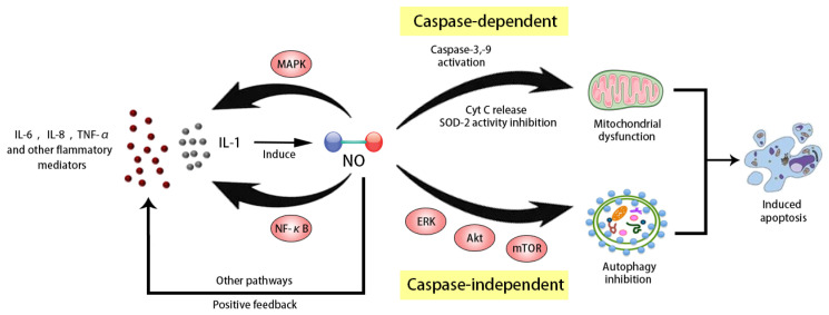
There is little proliferative activity in normal articular chondrocytes, and new cells are supplied from the ECM; therefore, the injury to chondrocytes and difficulty in repairing it are the central damaging features of OA. NO might be a key mediator of chondrocyte apoptosis [73]. Either endogenous or exogenous NO can induce apoptosis in chondrocytes via a mitochondria-dependent mechanism by activating caspase-3 and tyrosine kinases (Figure 4) [31,32]. In an in vitro model of fluid flow shear stress, Ren et al. found that an increase in NO production could reduce mitochondrial membrane potential and promote cytochrome C (Cyt C), another apoptosis mediator, to migrate from the mitochondria to the cytoplasm [74]. In their unilateral anterior crossbite in vivo rat models, the stimulator induced cartilage degeneration along with the upregulation of caspase-3 and caspase-9 expression, suggesting enhanced cell apoptosis. NOS inhibitors can block all these changes. Moreover, the expression of BCL2-associated X (Bax), Cyt C, and caspase-3 was upregulated in Sprague-Dawley rat models of sodium nitroprusside (SNP)-induced OA [75]. The treatment with carboxymethylated chitosan decreased the levels of apoptosis markers such as caspase-3 activation and DNA fragmentation as well as NO levels, suggesting caspase-dependent apoptosis. Another study reported that NO could inhibit autophagy and induce chondrocyte apoptosis [59]. Using SNP in chondrocytes could significantly reduce autophagic activity, autophagic flux, and the expression levels of several autophagy-related genes. This finding was supported by increases in ERK, Akt, and mammalian target of rapamycin (mTOR) phosphorylation. Moreover, the rapamycin-induced autophagy significantly suppressed NO-induced cell apoptosis via the mTOR/p70S6K pathway. In these processes, caspase-3 activation was only weakly detected, suggesting another caspase-independent apoptosis pathway.
Figure 4.
Mechanism of NO inducing apoptosis. IL: interleukin; TNF: tumor necrosis factor; MAPK: mitogen-activated protein kinase; NF-κB: nuclear factor kappa-B; NO: nitric oxide; caspase: cysteinyl aspartate specific proteinase; Cyt: cytochrome; SOD: superoxide dismutase; ERK: extracellular regulated protein kinases; Akt: protein kinase B; mTOR: mammalian target of rapamycin.
Interestingly, Carlo and Loeser found that incubation with NO alone did not induce apoptosis in chondrocytes [76]. The diazeniumdiolates (NOC compounds), which are potent NO donors, did not cause cell death either. Further research found that the conventional treatment with compounds such as SNP and 3-morpholiosydnonimine (SIN-1), produced NO as well as ROS, demonstrating that both the NO and ROS were required to induce cell death [77]. In addition, the NOC compounds can shield against oxidative stress, likely by suppressing the chondrocyte energy metabolism, indicating that NO alone might have beneficial effects on chondrocytes.
4.4. NO and Inflammatory Mediators
As discussed in Section 3, OA chondrocytes spontaneously release various inflammatory mediators such as IL-1β, IL-6, IL-8, TNF-α, and PGE2, while normal chondrocytes do not release these mediators [78]. The induction process involves mechanical and biochemical factors. In general, the occurrence of abnormal mechanical stress causes biochemical and functional changes in cartilage tissues, which induce the production of these inflammatory mediators. Fermor et al. accurately measured the effects of static and intermittent compression physiological levels on NOS activity, NO production, and NOS antigen expression in porcine articular cartilage explants [79]. In the experiment, the static compression on the explant was 0.1 MPa for 24 h, and intermittent compression was 0.1 or 0.5 MPa at 0.5 Hz for 24 h. The results demonstrated both these compressions significantly improved the NO production and NOS activity. Notably, once the initial expression is activated, chondrocytes can release inflammatory mediators spontaneously, even without mechanical stimulation. There is a positive amplification loop in the cartilage tissues, maintaining its catabolic state. Even in the absence of abnormal biomechanical factors, IL-1 and other cytokines will keep on promoting the production of inflammatory molecules, such as NO. In addition, after its production, NO might promote IL-1 synthesis, probably through the mitogen-activated protein kinase (MAPK) and NF-κB signaling pathways [64,80], thereby creating a positive amplification loop that releases NO-mediated inflammatory cytokines. A study showed that NO could increase the synthesis of IL-18 and IL-1-converting enzyme (ICE) to some extent [81]. ICE is a caspase enzyme required for the maturation of IL-1β and IL-18. Feeding the animals with the specific iNOS inhibitor N-iminoethyl-L-dicalcium hydrogen phosphate (L-NIL) could significantly reduce the IL-18 and ICE levels in their femoral condyle and tibial plateau [82]. Besides, NO also participates in the reduction in the levels of trypsin retarder 9 (PI-9), a natural ICE inhibitor, thereby improving IL-1β and IL-18 activity [82].
Another significant cytokine-mediated effect is the activation of the NF-κB signaling pathway, which remains inactive in the cytoplasm due to the inhibitor of NF-κB (IκB) under normal physiological conditions. The stimulation with proinflammatory factors phosphorylates IκB, which then separates from NF-κB. The activated NF-κB migrates to the nucleus and facilitates the transcription of proinflammatory genes, such as IL-1β, TNF-α, iNOS, and other catabolic factors, including MMPs. In this whole process, NO is required for the continuous activation of the NF-κB signaling pathway. Taken together, these results revealed that the interaction between the NO/iNOS system and other inflammatory mediators might jointly lead to a series of pathological changes during OA development.
4.5. NO and ROS/Reactive Nitrogen Oxide Species (RNOS)
NO can combine with superoxide anions (O2−) and generate peroxynitrite, which can promote inflammation and cellular death in cartilage tissues. As compared to non-OA chondrocytes, the OA chondrocytes demonstrated an increase in IL-1-induced ROS. Blanco et al. found that ROS could promote chondrocytes toward necrosis, while NO just induced morphological apoptosis [83]. Both NO and ROS might cause DNA breaks. Chen used a comet assay to determine the level of DNA damage in non-OA and OA cartilage tissues treated with 0–500 mM NO donors (NOC-18 or SIN-1). The result demonstrated that, as compared to non-OA chondrocytes, the OA chondrocytes showed a significant increase in oxidative DNA damage (p < 0.01) [84]. An increase in the NO and ROS concentration is beneficial to increase DNA damage; the specific iNOS inhibitor N-(3-(aminohydroxy)benzyl) acetamidine (1400W) and superoxide scavengers such as superoxide dismutase (SOD) could inhibit this increase. Moreover, the combination of DNA damage and mitogenic stimulation can induce chondrocyte senescence [85]. Different from normal chondrocytes, OA cartilage chondrocytes have a proliferative characteristic, which might be due to the increased access of chondrocytes to proliferative factors in the synovial fluid due to fissuring or loosening of the collagen network or collagen matrix damage [86].
NO and peroxynitrite might have opposite effects on the activation of the NF-κB signaling pathway [87]. Clancy et al. found that incubating the chondrocytes of bovine cartilage tissue with IL-1β resulted in stimulating NF-κB in 40% of the cells, which was shown by the positive immunostaining of NF-κB subunits in the nucleus [87]. The treatment of cells with IL-1β and the NO donor S-nitrosocystine ethyl ester reduced the total number of chondrocytes with active NF-κB to 5%. Moreover, the incubation with IL-1β and peroxynitrite increased the proportion of chondrocytes with active NF-κB from 40% to 73%, indicating the reverse effects of NO. This suggested that NO might not be required for the immediate activation of NF-κB. Notably, the NO catabolic activity is partly mediated by peroxynitrite, and the increase in the NO levels in OA chondrocytes might be related to the elevated RNOS levels.
Peroxynitrite can also induce mitochondrial dysfunction in chondrocytes based on a calcium-dependent pathway, leading to caspase-independent cell apoptosis [31]. These studies once again show that NO and its derivatives fully exert multiple functions in the whole process of OA pathology.
4.6. NO and Pain in the OA Process
Constant and intense pain is a characteristic of severe OA, which can be considered an immediate reason for most patients to seek medical attention. Pain involves complex neural reflex processes [88]. Acute pain can be considered the body’s reaction to a potential injury, stimulating a response to limit such an injury. Pathologically, the release of inflammatory molecules such as IL-1β and IL-6 aggravates inflammatory reactions and cartilage degeneration and causes structural changes, such as osteophyte growth, meniscal injury, and synovitis; thus, they become a direct reason for pain [89]. In this process, NO causes a sustained release of inflammatory factors and is involved in the disease itself. Venkanna et al. conducted a study on S-methylisothiourea (SMT, an iNOS inhibitor) to investigate its role in relieving pain and inflammation. Wistar rats were orally administered with SMT at different concentrations daily after surgery. The mechanical hyperalgesia, thermal hyperalgesia, and tail flick latency after repeated flexion and extension of the OA knee were determined at weekly intervals. The results indicated that SMT could reduce mechanical hyperalgesia and serum levels of IL-1β, TNF-α, and nitrite in rats [90]. On the other hand, NO is also an important neurotransmitter. In persistent pain, NO can cause the central sensitization of the pain pathway, suggesting that nerves might remain sensitized even after the removal of stimulation. This is one of the processes involved in the change from acute pain to chronic pain. A study investigating the role of NO in inflammatory hyperalgesia indicated that the co-injection of PGE2 with an iNOS inhibitor, NMA, resulted in the inhibition of PGE2-induced hyperalgesia in rats, and the high levels of NO produced cGMP-dependent hyperalgesia [91].
Interestingly, recent studies have shown that NO can play a role in pain reduction. The low level of NO produced by cNOS might provide pain relief in OA, which might be related to blood flow, nerve-transmission pathway, opioid-receptor pathway, and anti-inflammatory pathway [92].
Table 2.
Pathological effects of NO in OA and related mechanisms.
| Pathological Effects of NO in OA | Related Mediators or Signaling Pathways | Related Mechanisms | Reference |
|---|---|---|---|
| Increases cartilage matrix degradation |
Aggrecanases and collagenases | NO mediates upregulation of aggrecanases and collagenases |
Wu et al., 2019 [63] Brown et al., 2020 [65] |
| Enhances chondrocyte apoptosis | Caspase-3 and -9, tyrosine kinases, Bax, Cyt C, and ROS |
NO induces ROS generation and co-activates capase-3 to enhance chondrocyte apoptosis through a mitochondria-dependent mechanism |
Poderoso et al., 2019 [31] He et al., 2020 [75] |
| Inhibits chondrocyte autophagy | ERK, Akt and mTOR signaling pathways | NO reduces autophagic activity, autophagic flux, and expression of several autophagy-related genes |
Akaraphutiporn et al., 2020 [59] |
| Induces synthesis and release of inflammatory mediators | Inflammatory mediators and NF-κB signaling | NO interacts with inflammatory mediators which lead to constant inflammatory molecules release and NF-κB activation |
Chen et al., 2008 [84] Moon et al., 2018 [64] |
| Promotes inflammation and cell death | ROS and RNOS | NO combines with O2− to generate peroxynitrite which can cause DNA breaks and chondrocyte senescence, leading to cell necrosis | Copp et al., 2021 [85] |
| Cause central sensitization of the pain pathway | Inflammatory mediators and cGMP-dependent hyperalgesia | NO Promotes inflammation and structural changes of joint and play a role as neurotransmitter | Venkanna et al., 2014 [90] Aley et al., 1998 [91] |
Overall, NO has various roles in OA development, which are far more complex than originally thought. In cartilage matrix, NO can promote matrix degradation as well as inhibition of matrix synthesis, which together cause damage to the matrix environment and thus involve the entire joint cavity. The NO-induced apoptosis further exacerbates the damage to the joint through caspase-dependent and -independent pathways; this condition can be hardly reversed due to the little proliferative activity of chondrocytes. Inflammatory mediators and the ROS/RNOS system are the main triggers of the inflammatory response. They interact with NO to form a positive amplification loop, leading to the continued release of inflammation-related molecules and causing further damage to the cartilage tissues. These factors complement each other, contribute to OA progression, and are eventually characterized by increased pain and limited mobility. This explains why early interventions for OA should be taken; with time, irreversible damage expands gradually. The specific measures are described in the next section.
5. NO in OA Treatment
The traditional OA treatment involves alleviating symptoms and relieving pain mainly using nonsteroidal anti-inflammatory drugs (NSAIDs) in combination with exercise and physiotherapy; sometimes opioids are also used in cases of severe pain [7]. However, the current medication has often limited effects [93] and the adverse effects make long-term treatment compliance difficult. For example, NSAIDS may lead to renal insufficiency, gastritis, and peptic ulcer formation [94,95]. Prolonged NSAIDs usage might also cause rare adverse effects, involving the cardiovascular and cerebrovascular systems [96,97,98]. Moreover, the end-stage OA relies heavily on artificial joint replacement, when the conservative treatments have had little effect [99]. Thus, we have been longing for a novel drug intervention as an early OA treatment. The NO/NOS system is an indispensable part of OA inflammation and might serve as a new target for its clinical treatment. The selective inhibition of iNOS, the major isoform expressed in the OA joint, can significantly decrease NO levels. Some important pharmacotherapeutic agents are listed in Table 3.
Table 3.
Part of the important pharmacotherapy agents of iNOS inhibition.
| Author | Type of Study | Model | Agent | Dose | Method | Outcome | Conclusion |
|---|---|---|---|---|---|---|---|
| Yu et al., 2020 [100] | In vitro | New Zealand White rabbits | N-Monomethyl-L-arginine (L-NMMA) | Not mentioned |
L-NMMA on inhibiting NO to accelerate the influence of simvastatin |
L-NMMA inhibited NO and COX-2 production and NF-κB activation | L-NMMA enhanced the blocking effect of simvastatin on NF-κB activation by inhibiting NO production |
| Lee et al., 2012 [101] | In vitro | New Zealand White rabbits | L-NMMA | 0.5 mM/ 24 h |
L-NMMA on chondrocyte apoptosis | L-NMMA inhibited NO production and NF-kB binding activity | L-NMMA blocks PCB-initiated apoptosis effect |
| Eitner et al., 2021 [102] | In vitro | Human end-stage knee OA chondrocytes |
N-Iminoethyl-L-lysine (L-NIL) | 1, 10, or 20 µM/48 h | L-NIL on preventing release of NO, IL-6, PGE2, and iNOS | L-NIL prevented NO release and mitochondrial dysfunction |
L-NIL improves the impairment of mitochondrial respiration |
| Bentz et al., 2012 [103] | In vitro | Human OA chondrocytes |
L-NIL | 0-20 µM/ 24 h |
L-NIL on chondrocyte oxidative stress, apoptosis, inflammation, and catabolism | L-NIL stifled NO release, iNOS activity, nitrated proteins, and HNE generation and restored both HNE and GSTA4-4 levels |
L-NIL prevents LPO process and ROS production and attenuates cell death, inflammation, and catabolism |
| Castro et al., 2006 [104] | In vivo | OA rats induced by ligament transection surgery |
NG-nitro-L-arginine methyl ester (L-NAME) | 30 mg/kg/bid | L-NAME on joint pain, cell influx, nitrite levels and iNOS expression | L-NAME reduced the time of rats’ right hind paw fails to touch the surface while walking | Prophylactic L-NAME can reduce joint pain |
| Järvinen et al., 2008 [105] | In vitro | Cartilage tissue from OA patients | 1400W | 1 mM/120 h | 1400W on production of inflammatory mediators |
Treatment with 1400W enhanced the production of anti-catabolic IL-10 and reduced MMP-10 | The inhibiting effects of 1400W may point to its anti-inflammatory mechanisms for OA |
| Graverand et al., 2013 [106] | Clinical human study |
Kellgren and Lawrence Grade (KLG) 2 or 3 knee OA patients |
SD-6010 | 50 or 200 mg/day | A 2-year multicenter RCT of SD-6010 in patients with symptomatic knee OA |
In KLG2 patients, JSN after 48 weeks was lower with SD-6010 50 mg/day versus placebo. No improvement in KLG3 patients | SD-6010 may become effective only in “mild to moderate” OA patients, but it cannot slow the rate of JSN |
| Ma et al., 2020 [107] | In vitro | IL-1β induced Sprague-Dawley (SD) rat chondrocytes |
Aminoguanidine (AG) | 0.3, 1 or 3 mM/24,48 or 72 h | AG on COX-2, iNOS, phosphorylated (p)-p65 and NF-κB translocation |
AG downregulated iNOS and COX-2 expression by blocking the NF-κB signaling pathway |
AG may protect chondrocytes and serve as a potential therapeutic for OA |
| Lee et al., 2013 [108] | In vivo | MIA-induced Wistar rat OA model | Coenzyme Q10 (CoQ10) | 100 mg/kg/qd | CoQ10 on inflammatory mediators production and cartilage degradation |
CoQ10 had anti-nociceptive effect and attenuated cartilage degeneration in rat OA model |
CoQ10 exerts a therapeutic effect of OA by inhibiting inflammation |
| Park et al., 2018 [109] | In vitro | SW1353 cells and SD rats | Ethanol extract of sargassum serratifolium (EESS) |
Extract that hard to measure precise concentrations | EESS on inflammatory mediators production and signaling pathways activation |
EESS blocked ROS generation, attenuated NO production, and inhibited MAPK and PI3K/Akt pathways | EESS may have the potential chondroprotection in the prevention and treatment of OA |
| Khan et al., 2017 [110] | In vitro | IL-1β-stimulated human OA chondrocytes and cartilage explants |
Wogonin | 10–50 µM/24 h | Wogonin on inflammatory mediators production and MMPs, s-GAG and COL2A1 levels |
Wogonin mediated Nrf2/ARE pathways, inhibited matrix degradation and suppressed the expression and production of COX-2 and iNOS |
Wogonin exert chondro- and cartilage protection through the suppression of key molecular events |
| Yan et al., 2020 [111] | In vivo and in vitro | C57BL/6 wild-type (WT) rats | Myricitrin (Myr) | 0-100µM/24h in vitro, dose in vivo not mentioned | Myr on inflammatory mediators production and signaling pathway |
Myr suppressed the NF-κB and MAPK signaling pathways and decreased OARSI scores in OA rat models. |
Myr may have therapeutic potential in the treatment of OA |
5.1. iNOS Inhibitors
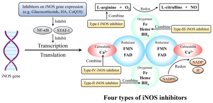
iNOS inhibitors can weaken iNOS activity. Currently, there are four types of iNOS inhibitors (Figure 5 and Table 3), which are the most widely known and are classified based on the inhibitor-binding sites on the NOS [112]. The first type combines with the arginine site, parts of which are reaction-based since they require an active enzyme and NADPH for complete inhibition; most inhibitors belong to the first type. The second type includes a set of compounds that mimic the tetrahydrobiopterin cofactor. The third type interacts directly with heme, and some of them can bind to the heme of the enzyme monomer and prevent the formation of the active dimer; various anti-fungal imidazoles have been shown to inhibit NOS activity via this mechanism. The fourth type interacts with either calmodulin or flavin cofactors; however, this type of inhibitor is not considered a promising means for selective iNOS inhibition.
Figure 5.
Inhibition to NO/NOS system. iNOS gene expression (transcription and translation) can be inhibited through NF-κΒ and STAT-1 signaling pathway. iNOS: inducible nitric oxide synthase; HA: hyaluronic acid; CoQ10: coenzymeQ10; NF-κB: nuclear factor kappa-B; STAT: signal transducer and activator of transcription; O2: oxygen; NO: nitric oxide; BH4: tetrahydrobiopterin; FMN: flavin mononucleotide; FAD: flavin adenine dinucleotide; NAPDH: nicotinamide adenine dinucleotide phosphate; NAPD+: oxidation form of NAPDH.
5.1.1. N-Monomethyl-L-Arginine (L-NMMA)
L-NMMA is one of the first NOS inhibitors that has been widely used to decrease NO bioavailability. It is produced by the degradation of arginine-methylated proteins and exists naturally in living organisms. A study on simvastatin in rabbit articular chondrocytes showed that the L-NMMA treatment could abolish the expression of IL-1β-mediated cyclooxygenase-2 (COX-2) and NF-κB activation and enhance the effects of simvastatin on NF-κB activity, thereby reducing the damage caused by NO and ROS [100]. The percentage of iNOS inhibited by L-NMMA was >95%, leading to the complete inhibition of NO. Meanwhile, it could also increase PGE2 synthesis [113]. L-NMMA could reduce synovial inflammation and tissue damage in vivo. Because L-NMMA has no selectivity for the different subtypes of NOS, the inhibition of eNOS and nNOS might interrupt cellular communication, vascular tone, and neurotransmission. However, such results have not been reported yet.
5.1.2. N-Iminoethyl-L-Lysine (L-NIL)
L-NIL is an arginine-based, moderately selective iNOS inhibitor. As compared to that for nNOS and eNOS, the iNOS selectivity of L-NIL is 23 and 49 times higher, respectively [114]. A lipid peroxidation (LPO) product, 4-hydroxynonenal (HNE), is considered an important mediator in the destruction of cartilage in OA. NO is a key inducer for triggering LPO based on peroxynitrite formation [115]. According to the experiments on inhibiting HNE synthesis in human OA chondrocytes and cartilage tissue explants by NO suppression, L-NIL could dose-dependently inhibit the IL-1β-induced NO release, iNOS activity, nitrated proteins, and HNE synthesis [103]. Moreover, L-NIL can also prevent the inactivation of HNE-metabolizing glutathione-s-transferase formation and inhibit the activation of p47 NADPH oxidase; all these are induced by IL-1β [116]. Furthermore, L-NIL can significantly reduce the makers of cellular death and apoptosis increased by exposure to the cytotoxic amounts of HNE as well as PGE2 and MMP-13 increased by the noncytotoxic amounts of HNE [103]. These findings showed that L-NIL could block the process of LPO and ROS production via NO-dependent and/or -independent pathways and reduce the HNE-induced cellular death, which might alleviate cartilage damage in OA.
5.1.3. NG-nitro-L-Arginine Methyl Ester (L-NAME)
L-NAME is a non-selective NOS inhibitor, having a weaker inhibitory effect on NO production as compared to those of L-NMMA and L-NIL [117]. L-NAME could significantly decrease NO production and inhibit immune suppression during OA initiation in rats, along with decreased levels of MMPs and increased levels of TIMP, thereby protecting cartilage tissues from damage in the early stage of OA [118]. In addition, prophylactic L-NAME could reduce joint pain in rats, which were subjected to the anterior cruciate ligament transection of the right knee for experimental OA [104].
5.1.4. 1400W
1400W is a highly selective iNOS inhibitor with high tissue and cell penetration abilities. It can increase the expression of Dickkopf-1 and frizzled-related proteins, which are attenuated by the IL-1β-induced upregulation of iNOS, which in turn activates the transcription of Wnt-pathway genes in human chondrocytes [119]. 1400W can also inhibit the cytokine-induced expression of MMPs and cellular apoptosis. However, its high dosage has toxic side effects, which might limit its application in patients.
5.1.5. Cindunistat
Cindunistat hydrochloride maleate, also called SD-6010, is a human selective type II iNOS inhibitor that could reduce the production of synovial fluid nitrite as well as osteophyte formation in animal models. It can also relieve inflammation and pain. In a 2-year double-blind randomized controlled trial (RCT), cindunistat improved the radiographic features of Kellgren–Lawrence Grade 2 (KLG2) OA patients in the first 48 weeks; however, the improvement did not last after 96 weeks. Among the patients with KLG3, OA progression persisted, showing no improvement in either 48 or 96 weeks [106]. Meanwhile, a meta-analysis investigating the long-term RCTs of medications for knee OA suggested that cindunistat exhibited little effect [120]. Thus, cindunistat might have effects only on the early stage of OA. This is one of the few cases of NOS inhibitors being used in clinical trials, which revealed that with the progression of OA, inflammation damage cannot be easily reversed.
5.1.6. Aminoguanidine (AG)
AG is an iNOS inhibitor that can inhibit oxidation, apoptosis, and inflammation. It has been reported to reduce the protein expression levels of iNOS and p65 in the liver of a diabetic animal model and reduce osteocyte apoptosis during non-traumatic osteonecrosis [121,122]. According to Ma et al., AG could reduce the gene expression levels of iNOS and COX-2 in IL-1β-induced rat articular chondrocytes by suppressing the NF-κB activity and inhibiting the phosphorylation of IκBα and inhibitor of kappa B kinase-β (IKKβ), thereby showing its anti-inflammatory and cartilage protective effects [107].
5.2. Pharmaceuticals Inhibiting iNOS Expression
NO production can be inhibited by downregulating iNOS expression. Several types of drugs can attenuate iNOS expression through different mechanisms. NF-κΒ and signal transducer and activator of transcription-1 (STAT-1) are the key transcription factors for iNOS [123]. Inhibiting these signaling pathways might be conducive to reducing iNOS expression. Moreover, regulation at the post-transcriptional level also matters. By interfering with the amount of protein modifications, the balance of iNOS synthesis and degradation can be regulated.
5.2.1. Glucocorticoids (GCs)
GCs are steroid hormones that are naturally synthesized in the human body or manufactured synthetically and possess anti-inflammatory effects. Hossein et al., indicated that dexamethasone (DEX; a type of GCs) could reduce the gene expression levels of COX-2, iNOS, IL-1β, IL-18, and TNF-α in synoviocyte as well as the production of NO and PGE2 in monocytes and macrophages, probably by inhibiting the NF-κB and destabilizing the iNOS mRNA [124]. Notably, using excessive GCs can cause serious, life-threatening side effects, thereby restricting their applications.
5.2.2. Hyaluronic Acid (HA)
HA is synthesized by chondrocytes and fibroblasts in the human body as a component of the ECM in articular cartilage tissues. A study on mice subjected to collagen-induced arthritis demonstrated that, as compared to carnosine alone, the combination of HA and carnosine significantly reduced expression levels of iNOS as well as proinflammatory cytokines, chemokines, and COX-2 [125]. The possible mechanism of HA function involves the protection of protein kinase C alpha (PKCa), a factor related to chondrocyte proliferation, against NO inhibition—similar to a PKCa agonist used in articular cartilage.
5.2.3. CoenzymeQ10 (CoQ10)
CoQ10 is a lipid that participates in the electron transport chain and aerobic respiration as a coenzyme for mitochondrial enzymes and exhibits potent antioxidant and anti-inflammatory abilities. CoQ10 could inhibit the NF-κB pathway and its downstream inflammatory mediators, including MMP-9, MMP-13, IL-1β, nitrotyrosine, and receptors for advanced glycation end products [101,126]. It could also significantly reduce the monosodium iodoacetate-induced upregulation of iNOS expression. Treatment with CoQ10 could considerably attenuate pain and cartilage degeneration in animal models [108].
5.2.4. Sargassum serratifolium
Sargassum is a genus of marine brown algae (Phaeophyceae) and is widely distributed in the ocean. It has a highly edible and medicinal value. Current studies have shown that the extracts of Sargassum serratifolium have powerful anti-inflammatory and antioxidant effects [127,128]. Park et al. treated IL-1β-induced human chondrocytes with the ethanol extract of Sargassum serratifolium (EESS); the results showed a reduction in numerous inflammatory markers, including NO, MMPs, and ROS. Their results demonstrated that EESS could inhibit the activation of NF-κΒ, p38 MAPK, and PI3K/Akt signaling pathways, thereby inhibiting iNOS and COX-2 expression [109].
5.2.5. Wogonin
Wogonin (5, 7-dihydroxy-8-methoxyflavone) is a naturally occurring flavonoid derived from the root extracts of Scutellaria baicalensis. According to Khan et al., Wogonin could completely suppress the expression of IL-6, PGE2, iNOS, and COX-2 and reduce the NO production in IL-1β-induced OA chondrocytes, thereby playing an anti-inflammatory role. Wogonin activated the ROS/ERK/nuclear factor erythroid 2-related factor 2 (Nrf2)/heme oxygenase-1 (HO-1)-SOD2-NADPH: Quinone Oxidoreductase-1(NQO1)-glutamate-cysteine ligase catalytic subunit (GCLC) signaling axis to induce low levels of ROS, thereby mediating the Nrf2/ARE pathways and suppressing the molecular events involved in oxidative stress and inflammation [110]. Moreover, wogonin also decreased the release of MMPs and ADAMT-4 and elevated the expression levels of cartilage anabolic factors, including collagen type II alpha 1 (COL2A1) and aggrecan (ACAN), to suppress matrix degradation [129,130].
5.2.6. Myricitrin (Myr)
Myr, a flavonoid compound extracted from Myrica rubra, exhibits anti-inflammatory, antioxidant, and anti-fibrotic effects. Myr can inhibit the expression of iNOS and COX-2, thereby blocking NO and PEG2 production in IL-1β-stimulated OA rats [111,131]. Immunohistochemical analysis showed that the anti-inflammatory effects of Myr were based on the interference of IL-1β-activation of NF-κB and MAPK signaling pathways. The OA Research Society International scores were decreased in surgically induced mouse models in vivo, demonstrating the amelioration of OA development [111].
5.3. Applications of Materials Science
The traditional pharmacotherapeutic strategies have unavoidable flaws; the absorption process of the drug can be affected by various factors, and the patients’ compliance is difficult to guarantee. Therefore, researchers attempt to explore novel approaches in the field of materials science. Wang et al. used umbilical cord mesenchymal stem cells (UCMSCs), loaded with graphene oxide (GO) granular lubricant, for the treatment of knee OA in animal models [132]. In the animal group given the combination of USMSCs and GO, the NO contents decreased significantly in the serum and articular fluid. GO granular lubricant might promote the therapeutic effects of UCMSCs as scaffold-carrying material. Moreover, Shah et al. used growth factors important to chondrogenesis and encapsulated recombinant proteins in poly (lactic-coglycolic acid) 85:15 (PLGA) to fabricate a synthetic artificial stem cell (SASC) system. After injecting SASC, it dramatically reduced the NO contents and gene expression levels of ADAMTS5 and the proteoglycan 4 (PRG4) genes in the OA rodent model, thereby demonstrating significant anti-inflammatory and chondroprotective effects [133].
In another study, nanocomposites showed potent osteoinductive and free radical scavenging abilities. Nano-titanium dioxide (TiO2) with chitosan and chondroitin 4-sulfate (TCG) could reduce the formation of free radicals, such as NO, increase the total antioxidant activity, and upregulate the expression of osteoblast-inducing genes in the MG-63 cell line [134]. In addition, pharmacotherapy can combine with nanocomposites, forming nano-targeted drug delivery systems. By responding to specific internal stimuli (reduction-oxidation, pH, and enzymes) and external stimuli (temperature, ultrasound, magnetic, photo, voltage, and mechanical friction), nano-targeted drug delivery systems can deliver drugs at the proper concentration, time, and space, which overcomes the shortcomings of exceeding scope and premature leakage of traditional drug delivery systems. In comparison, this system greatly improves the efficiency of drug delivery and minimizes the side effects of drugs [135]. These results suggested that the nanocomposites might have a promising application prospect.
5.4. Physical Methods
In addition to the above strategies, some physical methods have also been reported to downregulate NO levels. For instance, hyperbaric oxygen (HBO) can inhibit iNOS expression and chondrocyte apoptosis in rabbit cartilage defects [136]. At higher oxygen tension, the NO, PGE2, and MMP activities decreased in the TNF-α-induced chondrocytes [137]. The combination of HBO with low-intensity pulsed ultrasound can increase matrix and TIMP production and inhibit iNOS expression and NO production [138]. Low-level laser therapy can reduce the expression levels of inflammatory markers, such as TNF-α, iNOS, and IL-1 [139]. Extracorporeal shock-wave therapy could improve pain and motor dysfunction in OA rabbits, in which NO level and chondrocyte apoptosis decreased significantly [140]. In general, these approaches require further clinical research.
6. Discussion and Future Perspectives
Current studies demonstrate that NO plays a critical role in activating various signaling pathways. Among these signaling pathways, NF-κB might be one of the most noteworthy. The activated NF-κB can promote the synthesis of proinflammatory factors, such as IL-1β, TNF-α, and iNOS, thereby aggravating the development of OA. In addition, MAPK, STAT-1, and PI3K/Akt signaling pathways have similar functions. Moreover, NO-activated ERK-Akt-mTOR signaling pathways can inhibit chondrocyte autophagy and promote apoptosis, and MMPs/TGF-β1/IGF-1 signaling pathways can increase matrix degradation, thereby causing cartilage tissue damage. On the other hand, the low NO concentrations can activate the RANKL signaling pathway, which produces downstream molecular 8-nitro-cGMP to promote osteoclast differentiation, as well as the Src-Erk-1/2-Akt signaling pathways, which can promote osteoblast proliferation and has anti-apoptotic effects, thereby enhancing bone remodeling.
Therefore, NO plays not only an inflammatory mediator role in OA but also a normal bone cell-conditioning agent. NO is necessary during the growth, differentiation, and maturation process of several types of bone cells. The pathological increase in NO contents due to stimulatory factors, such as abnormal mechanical stress, results in its constant interaction with various inflammatory mediators and causes severe chondritis and synovitis. In order to break this positive amplification loop, inhibiting the elevated NO levels might be a good approach.
However, in the presence of mature iNOS inhibitors, it might also be a thorny proposition to treat OA through this route due to various reasons. First, the increased dose of inhibitors or duration of treatment will unavoidably inhibit eNOS and nNOS due to the capped selectivity, which can affect the physiological regulation of vascular tone and blood pressure regulation and might lead to complications, such as hypertension [141]. Secondly, most OA patients might not start treatment until the disease has progressed to a certain extent, such as when severe pain occurs; the radiographic performance is often already obvious at this time. The altered mechanics in even radiographic “mild-to-moderate” OA are of such overwhelming significance that the current pharmacological agents might not effectively modify the outcome [142].
Nano-targeted drug delivery systems might be one of the future research directions that might significantly reduce related adverse reactions through precision drug delivery. In addition, it is a desirable prospect if there is the possibility to reverse even mildly altered mechanics by developing new pharmacological agents but not a prosthetic replacement.
7. Conclusions
The biological effects of NO are highly complex in human cartilage and thus in OA. The roles of NO are associated with various factors, such as IL-1β, TNF-α, and PGE2, and multiple signaling pathways, such as NF-κB activation. NO, thus, is a part of the following processes: regulating normal bone cell function and bone remodeling, responding to mechanical stimulation in bone, mediating the effects of sex hormones, inducing catabolic effects, and being an anti-inflammatory or pro-inflammatory factor in bone. Therefore, NO might be a new target for OA treatment. However, the effects of NO on normal and pathological bone and joint functions require further clarification using animal and cell studies. Additional clinical research on the applications of NO inhibitors in OA is also warranted.
Author Contributions
Conceptualization, Y.Z.; writing—original draft preparation, H.J.; writing—review and editing, Y.Z.; visualization, P.J. and X.S.; funding acquisition, Y.Z. All authors have read and agreed to the published version of the manuscript.
Institutional Review Board Statement
Not applicable.
Informed Consent Statement
Not applicable.
Data Availability Statement
Not applicable.
Conflicts of Interest
The authors declare no conflict of interest.
Funding Statement
This project was funded by National Natural Science Foundation of China (grant number: 81802203), Natural Science Foundation of Hubei Province (grant number: 2022CFB117), Fundamental Research Funds for the Central Universities (grant number: 2042018kf0123), Guiding Fund of Renmin Hospital of Wuhan University (grant number: RMYD2018M43).
Footnotes
Disclaimer/Publisher’s Note: The statements, opinions and data contained in all publications are solely those of the individual author(s) and contributor(s) and not of MDPI and/or the editor(s). MDPI and/or the editor(s) disclaim responsibility for any injury to people or property resulting from any ideas, methods, instructions or products referred to in the content.
References
- 1.Lee M., Rey K., Besler K., Wang C., Choy J. Immunobiology of Nitric Oxide and Regulation of Inducible Nitric Oxide Synthase. Results Probl. Cell Differ. 2017;62:181–207. doi: 10.1007/978-3-319-54090-0_8. [DOI] [PubMed] [Google Scholar]
- 2.Vina E.R., Kwoh C.K. Epidemiology of osteoarthritis: Literature update. Curr. Opin. Rheumatol. 2018;30:160–167. doi: 10.1097/BOR.0000000000000479. [DOI] [PMC free article] [PubMed] [Google Scholar]
- 3.O’Neill T.W., McCabe P.S., McBeth J. Update on the epidemiology, risk factors and disease outcomes of osteoarthritis. Best Pract. Res. Clin. Rheumatol. 2018;32:312–326. doi: 10.1016/j.berh.2018.10.007. [DOI] [PubMed] [Google Scholar]
- 4.Beard J.R., Officer A., de Carvalho I.A., Sadana R., Pot A.M., Michel J.P., Lloyd-Sherlock P., Epping-Jordan J.E., Peeters G., Mahanani W.R., et al. The World report on ageing and health: A policy framework for healthy ageing. Lancet. 2016;387:2145–2154. doi: 10.1016/S0140-6736(15)00516-4. [DOI] [PMC free article] [PubMed] [Google Scholar]
- 5.Rahmati M., Nalesso G., Mobasheri A., Mozafari M. Aging and osteoarthritis: Central role of the extracellular matrix. Ageing Res Rev. 2017;40:20–30. doi: 10.1016/j.arr.2017.07.004. [DOI] [PubMed] [Google Scholar]
- 6.Vincent T.L. Mechanoflammation in osteoarthritis pathogenesis. Semin Arthritis Rheum. 2019;49((Suppl. S3)):S36–S38. doi: 10.1016/j.semarthrit.2019.09.018. [DOI] [PubMed] [Google Scholar]
- 7.Abramoff B., Caldera F.E. Osteoarthritis: Pathology, Diagnosis, and Treatment Options. Med. Clin. N. Am. 2020;104:293–311. doi: 10.1016/j.mcna.2019.10.007. [DOI] [PubMed] [Google Scholar]
- 8.Robinson W.H., Lepus C.M., Wang Q., Raghu H., Mao R., Lindstrom T.M., Sokolove J. Low-grade inflammation as a key mediator of the pathogenesis of osteoarthritis. Nat. Rev. Rheumatol. 2016;12:580–592. doi: 10.1038/nrrheum.2016.136. [DOI] [PMC free article] [PubMed] [Google Scholar]
- 9.Ersoy Y., Ozerol E., Baysal O., Temel I., MacWalter R.S., Meral U., Altay Z.E. Serum nitrate and nitrite levels in patients with rheumatoid arthritis, ankylosing spondylitis, and osteoarthritis. Ann. Rheum. Dis. 2002;61:76–78. doi: 10.1136/ard.61.1.76. [DOI] [PMC free article] [PubMed] [Google Scholar]
- 10.Subczynski W.K., Lomnicka M., Hyde J.S. Permeability of nitric oxide through lipid bilayer membranes. Free Radic. Res. 1996;24:343–349. doi: 10.3109/10715769609088032. [DOI] [PubMed] [Google Scholar]
- 11.Moncada S., Higgs A. The L-arginine-nitric oxide pathway. N. Engl. J. Med. 1993;329:2002–2012. doi: 10.1056/NEJM199312303292706. [DOI] [PubMed] [Google Scholar]
- 12.Koppenol W.H., Traynham J.G. Say NO to nitric oxide: Nomenclature for nitrogen- and oxygen-containing compounds. Methods Enzymol. 1996;268:3–7. doi: 10.1016/s0076-6879(96)68004-5. [DOI] [PubMed] [Google Scholar]
- 13.Davis K.L., Martin E., Turko I.V., Murad F. Novel effects of nitric oxide. Annu. Rev. Pharmacol. Toxicol. 2001;41:203–236. doi: 10.1146/annurev.pharmtox.41.1.203. [DOI] [PubMed] [Google Scholar]
- 14.Korhonen R., Lahti A., Kankaanranta H., Moilanen E. Nitric oxide production and signaling in inflammation. Curr. Drug Targets Inflamm. Allergy. 2005;4:471–479. doi: 10.2174/1568010054526359. [DOI] [PubMed] [Google Scholar]
- 15.Kim J.M., Lin C., Stavre Z., Greenblatt M.B., Shim J.H. Osteoblast-Osteoclast Communication and Bone Homeostasis. Cells. 2020;9:2073. doi: 10.3390/cells9092073. [DOI] [PMC free article] [PubMed] [Google Scholar]
- 16.Shirazi S., Ravindran S., Cooper L.F. Topography-mediated immunomodulation in osseointegration; Ally or Enemy. Biomaterials. 2022;291:121903. doi: 10.1016/j.biomaterials.2022.121903. [DOI] [PMC free article] [PubMed] [Google Scholar]
- 17.Morimoto A., Kikuta J., Nishikawa K., Sudo T., Uenaka M., Furuya M., Hasegawa T., Hashimoto K., Tsukazaki H., Seno S., et al. SLPI is a critical mediator that controls PTH-induced bone formation. Nat. Commun. 2021;12:2136. doi: 10.1038/s41467-021-22402-x. [DOI] [PMC free article] [PubMed] [Google Scholar]
- 18.Srivastava R.K., Sapra L., Mishra P.K. Osteometabolism: Metabolic Alterations in Bone Pathologies. Cells. 2022;11:3943. doi: 10.3390/cells11233943. [DOI] [PMC free article] [PubMed] [Google Scholar]
- 19.Siddiqui J.A., Partridge N.C. Physiological Bone Remodeling: Systemic Regulation and Growth Factor Involvement. Physiology. 2016;31:233–245. doi: 10.1152/physiol.00061.2014. [DOI] [PMC free article] [PubMed] [Google Scholar]
- 20.Kalyanaraman H., Ramdani G., Joshua J., Schall N., Boss G.R., Cory E., Sah R.L., Casteel D.E., Pilz R.B. A Novel, Direct NO Donor Regulates Osteoblast and Osteoclast Functions and Increases Bone Mass in Ovariectomized Mice. J. Bone Miner. Res. 2017;32:46–59. doi: 10.1002/jbmr.2909. [DOI] [PMC free article] [PubMed] [Google Scholar]
- 21.Amano H., Iwaki F., Oki M., Aoki K., Ohba S. An osteogenic helioxanthin derivative suppresses the formation of bone-resorbing osteoclasts. Regen. Ther. 2019;11:290–296. doi: 10.1016/j.reth.2019.08.007. [DOI] [PMC free article] [PubMed] [Google Scholar]
- 22.Kaneko K., Miyamoto Y., Tsukuura R., Sasa K., Akaike T., Fujii S., Yoshimura K., Nagayama K., Hoshino M., Inoue S., et al. 8-Nitro-cGMP is a promoter of osteoclast differentiation induced by RANKL. Nitric Oxide. 2018;72:46–51. doi: 10.1016/j.niox.2017.11.006. [DOI] [PubMed] [Google Scholar]
- 23.Mentaverri R., Kamel S., Wattel A., Prouillet C., Sevenet N., Petit J.P., Tordjmann T., Brazier M. Regulation of bone resorption and osteoclast survival by nitric oxide: Possible involvement of NMDA-receptor. J. Cell Biochem. 2003;88:1145–1156. doi: 10.1002/jcb.10463. [DOI] [PubMed] [Google Scholar]
- 24.Gloria M.A.D., Mouro M.G., Geraldini S., Higa E.M.S., Carvalho A.B. Cbfa1 expression in vascular smooth muscle cells may be elevated by increased nitric oxide/iNOS. J. Bras. Nefrol. 2020;42:300–306. doi: 10.1590/2175-8239-JBN-2019-0166. [DOI] [PMC free article] [PubMed] [Google Scholar]
- 25.Cepeda S.B., Sandoval M.J., Crescitelli M.C., Rauschemberger M.B., Massheimer V.L. The isoflavone genistein enhances osteoblastogenesis: Signaling pathways involved. J. Physiol. Biochem. 2020;76:99–110. doi: 10.1007/s13105-019-00722-3. [DOI] [PubMed] [Google Scholar]
- 26.Ramdani G., Schall N., Kalyanaraman H., Wahwah N., Moheize S., Lee J.J., Sah R.L., Pfeifer A., Casteel D.E., Pilz R.B. cGMP-dependent protein kinase-2 regulates bone mass and prevents diabetic bone loss. J. Endocrinol. 2018;238:203–219. doi: 10.1530/JOE-18-0286. [DOI] [PMC free article] [PubMed] [Google Scholar]
- 27.Gerbarg P.L., Brown R.P. Pause menopause with Rhodiola rosea, a natural selective estrogen receptor modulator. Phytomedicine. 2016;23:763–769. doi: 10.1016/j.phymed.2015.11.013. [DOI] [PubMed] [Google Scholar]
- 28.Crescitelli M.C., Rauschemberger M.B., Cepeda S., Sandoval M., Massheimer V.L. Role of estrone on the regulation of osteoblastogenesis. Mol. Cell. Endocrinol. 2019;498:110582. doi: 10.1016/j.mce.2019.110582. [DOI] [PubMed] [Google Scholar]
- 29.Wittkowske C., Reilly G.C., Lacroix D., Perrault C.M. In Vitro Bone Cell Models: Impact of Fluid Shear Stress on Bone Formation. Front. Bioeng. Biotechnol. 2016;4:87. doi: 10.3389/fbioe.2016.00087. [DOI] [PMC free article] [PubMed] [Google Scholar]
- 30.Maycas M., Esbrit P., Gortázar A.R. Molecular mechanisms in bone mechanotransduction. Histol. Histopathol. 2017;32:751–760. doi: 10.14670/HH-11-858. [DOI] [PubMed] [Google Scholar]
- 31.Poderoso J.J., Helfenberger K., Poderoso C. The effect of nitric oxide on mitochondrial respiration. Nitric Oxide. 2019;88:61–72. doi: 10.1016/j.niox.2019.04.005. [DOI] [PubMed] [Google Scholar]
- 32.Kamm A., Przychodzen P., Kuban-Jankowska A., Jacewicz D., Dabrowska A.M., Nussberger S., Wozniak M., Gorska-Ponikowska M. Nitric oxide and its derivatives in the cancer battlefield. Nitric Oxide. 2019;93:102–114. doi: 10.1016/j.niox.2019.09.005. [DOI] [PubMed] [Google Scholar]
- 33.Wojdasiewicz P., Poniatowski Ł.A., Szukiewicz D. The role of inflammatory and anti-inflammatory cytokines in the pathogenesis of osteoarthritis. Mediat. Inflamm. 2014;2014:561459. doi: 10.1155/2014/561459. [DOI] [PMC free article] [PubMed] [Google Scholar]
- 34.Teixeira C.C., Ischiropoulos H., Leboy P.S., Adams S.L., Shapiro I.M. Nitric oxide-nitric oxide synthase regulates key maturational events during chondrocyte terminal differentiation. Bone. 2005;37:37–45. doi: 10.1016/j.bone.2005.03.010. [DOI] [PubMed] [Google Scholar]
- 35.Drissi H., Zuscik M., Rosier R., O'Keefe R. Transcriptional regulation of chondrocyte maturation: Potential involvement of transcription factors in OA pathogenesis. Mol. Aspects Med. 2005;26:169–179. doi: 10.1016/j.mam.2005.01.003. [DOI] [PubMed] [Google Scholar]
- 36.Ono T., Nakashima T. Recent advances in osteoclast biology. Histochem. Cell Biol. 2018;149:325–341. doi: 10.1007/s00418-018-1636-2. [DOI] [PubMed] [Google Scholar]
- 37.MacIntyre I., Zaidi M., Alam A.S., Datta H.K., Moonga B.S., Lidbury P.S., Hecker M., Vane J.R. Osteoclastic inhibition: An action of nitric oxide not mediated by cyclic GMP. Proc. Natl. Acad. Sci. USA. 1991;88:2936–2940. doi: 10.1073/pnas.88.7.2936. [DOI] [PMC free article] [PubMed] [Google Scholar]
- 38.Löwik C.W., Nibbering P.H., van de Ruit M., Papapoulos S.E. Inducible production of nitric oxide in osteoblast-like cells and in fetal mouse bone explants is associated with suppression of osteoclastic bone resorption. J. Clin. Investig. 1994;93:1465–1472. doi: 10.1172/JCI117124. [DOI] [PMC free article] [PubMed] [Google Scholar]
- 39.Ralston S.H., Ho L.P., Helfrich M.H., Grabowski P.S., Johnston P.W., Benjamin N. Nitric oxide: A cytokine-induced regulator of bone resorption. J. Bone Miner. Res. 1995;10:1040–1049. doi: 10.1002/jbmr.5650100708. [DOI] [PubMed] [Google Scholar]
- 40.Yaroslavskiy B.B., Li Y., Ferguson D.J., Kalla S.E., Oakley J.I., Blair H.C. Autocrine and paracrine nitric oxide regulate attachment of human osteoclasts. J. Cell. Biochem. 2004;91:962–972. doi: 10.1002/jcb.20009. [DOI] [PubMed] [Google Scholar]
- 41.Ralston S.H., Grabowski P.S. Mechanisms of cytokine induced bone resorption: Role of nitric oxide, cyclic guanosine monophosphate, and prostaglandins. Bone. 1996;19:29–33. doi: 10.1016/8756-3282(96)00101-9. [DOI] [PubMed] [Google Scholar]
- 42.Helfrich M.H., Evans D.E., Grabowski P.S., Pollock J.S., Ohshima H., Ralston S.H. Expression of nitric oxide synthase isoforms in bone and bone cell cultures. J. Bone Miner. Res. 1997;12:1108–1115. doi: 10.1359/jbmr.1997.12.7.1108. [DOI] [PubMed] [Google Scholar]
- 43.Yan Q., Feng Q., Beier F. Endothelial nitric oxide synthase deficiency in mice results in reduced chondrocyte proliferation and endochondral bone growth. Arthritis Rheum. 2010;62:2013–2022. doi: 10.1002/art.27486. [DOI] [PubMed] [Google Scholar]
- 44.Hefler L.A., Reyes C.A., O'Brien W.E., Gregg A.R. Perinatal development of endothelial nitric oxide synthase-deficient mice. Biol. Reprod. 2001;64:666–673. doi: 10.1095/biolreprod64.2.666. [DOI] [PubMed] [Google Scholar]
- 45.Afzal F., Polak J., Buttery L. Endothelial nitric oxide synthase in the control of osteoblastic mineralizing activity and bone integrity. J. Pathol. 2004;202:503–510. doi: 10.1002/path.1536. [DOI] [PubMed] [Google Scholar]
- 46.Watanuki M., Sakai A., Sakata T., Tsurukami H., Miwa M., Uchida Y., Watanabe K., Ikeda K., Nakamura T. Role of inducible nitric oxide synthase in skeletal adaptation to acute increases in mechanical loading. J. Bone Miner. Res. 2002;17:1015–1025. doi: 10.1359/jbmr.2002.17.6.1015. [DOI] [PubMed] [Google Scholar]
- 47.Liu H., Rosen C.J. Nitric oxide and bone: The phoenix rises again. J. Clin. Investig. 2021;131:e147072. doi: 10.1172/JCI147072. [DOI] [PMC free article] [PubMed] [Google Scholar]
- 48.Cao W., Helder M.N., Bravenboer N., Wu G., Jin J., Ten Bruggenkate C.M., Klein-Nulend J., Schulten E. Is There a Governing Role of Osteocytes in Bone Tissue Regeneration? Curr. Osteoporos. Rep. 2020;18:541–550. doi: 10.1007/s11914-020-00610-6. [DOI] [PMC free article] [PubMed] [Google Scholar]
- 49.Deng S., Dai G., Chen S., Nie Z., Zhou J., Fang H., Peng H. Dexamethasone induces osteoblast apoptosis through ROS-PI3K/AKT/GSK3β signaling pathway. Biomed. Pharmacother. 2019;110:602–608. doi: 10.1016/j.biopha.2018.11.103. [DOI] [PubMed] [Google Scholar]
- 50.Nishimura R., Hata K., Kida J. Regulation of osteoblasts and chondrocytes by Wnt signaling. Clin. Calcium. 2019;29:299–307. doi: 10.20837/4201903299. [DOI] [PubMed] [Google Scholar]
- 51.Otsuka E., Hirano K., Matsushita S., Inoue A., Hirose S., Yamaguchi A., Hagiwara H. Effects of nitric oxide from exogenous nitric oxide donors on osteoblastic metabolism. Eur. J. Pharmacol. 1998;349:345–350. doi: 10.1016/s0014-2999(98)00190-3. [DOI] [PubMed] [Google Scholar]
- 52.Mancini L., Moradi-Bidhendi N., Becherini L., Martineti V., MacIntyre I. The biphasic effects of nitric oxide in primary rat osteoblasts are cGMP dependent. Biochem. Biophys. Res. Commun. 2000;274:477–481. doi: 10.1006/bbrc.2000.3164. [DOI] [PubMed] [Google Scholar]
- 53.Hikiji H., Shin W.S., Oida S., Takato T., Koizumi T., Toyo-Oka T. Direct action of nitric oxide on osteoblastic differentiation. FEBS Lett. 1997;410:238–242. doi: 10.1016/s0014-5793(97)00597-8. [DOI] [PubMed] [Google Scholar]
- 54.Damoulis P.D., Hauschka P.V. Cytokines induce nitric oxide production in mouse osteoblasts. Biochem. Biophys. Res. Commun. 1994;201:924–931. doi: 10.1006/bbrc.1994.1790. [DOI] [PubMed] [Google Scholar]
- 55.Ralston S.H., Todd D., Helfrich M., Benjamin N., Grabowski P.S. Human osteoblast-like cells produce nitric oxide and express inducible nitric oxide synthase. Endocrinology. 1994;135:330–336. doi: 10.1210/endo.135.1.7516867. [DOI] [PubMed] [Google Scholar]
- 56.Minne H.W., Pfeilschifter J., Scharla S., Mutschelknauss S., Schwarz A., Krempien B., Ziegler R. Inflammation-mediated osteopenia in the rat: A new animal model for pathological loss of bone mass. Endocrinology. 1984;115:50–54. doi: 10.1210/endo-115-1-50. [DOI] [PubMed] [Google Scholar]
- 57.Sacitharan P.K. Ageing and Osteoarthritis. Subcell Biochem. 2019;91:123–159. doi: 10.1007/978-981-13-3681-2_6. [DOI] [PubMed] [Google Scholar]
- 58.Grässel S., Zaucke F., Madry H. Osteoarthritis: Novel Molecular Mechanisms Increase Our Understanding of the Disease Pathology. J. Clin. Med. 2021;10:1938. doi: 10.3390/jcm10091938. [DOI] [PMC free article] [PubMed] [Google Scholar]
- 59.Akaraphutiporn E., Sunaga T., Bwalya E.C., Yanlin W., Carol M., Okumura M. An Insight into the Role of Apoptosis and Autophagy in Nitric Oxide-Induced Articular Chondrocyte Cell Death. Cartilage. 2020;13:826S–838S. doi: 10.1177/1947603520976768. [DOI] [PMC free article] [PubMed] [Google Scholar]
- 60.Krishnan Y., Grodzinsky A.J. Cartilage diseases. Matrix Biol. 2018;71–72:51–69. doi: 10.1016/j.matbio.2018.05.005. [DOI] [PMC free article] [PubMed] [Google Scholar]
- 61.Charlier E., Deroyer C., Ciregia F., Malaise O., Neuville S., Plener Z., Malaise M., de Seny D. Chondrocyte dedifferentiation and osteoarthritis (OA) Biochem. Pharmacol. 2019;165:49–65. doi: 10.1016/j.bcp.2019.02.036. [DOI] [PubMed] [Google Scholar]
- 62.Casal-Beiroa P., Balboa-Barreiro V., Oreiro N., Pértega-Díaz S., Blanco F.J., Magalhães J. Optical Biomarkers for the Diagnosis of Osteoarthritis through Raman Spectroscopy: Radiological and Biochemical Validation Using Ex Vivo Human Cartilage Samples. Diagnostics. 2021;11:546. doi: 10.3390/diagnostics11030546. [DOI] [PMC free article] [PubMed] [Google Scholar]
- 63.Wu Y., Lin Z., Yan Z., Wang Z., Fu X., Yu K. Sinomenine contributes to the inhibition of the inflammatory response and the improvement of osteoarthritis in mouse-cartilage cells by acting on the Nrf2/HO-1 and NF-κB signaling pathways. Int. Immunopharmacol. 2019;75:105715. doi: 10.1016/j.intimp.2019.105715. [DOI] [PubMed] [Google Scholar]
- 64.Moon S.M., Lee S.A., Han S.H., Park B.R., Choi M.S., Kim J.S., Kim S.G., Kim H.J., Chun H.S., Kim D.K., et al. Aqueous extract of Codium fragile alleviates osteoarthritis through the MAPK/NF-κB pathways in IL-1β-induced rat primary chondrocytes and a rat osteoarthritis model. Biomed. Pharmacother. 2018;97:264–270. doi: 10.1016/j.biopha.2017.10.130. [DOI] [PubMed] [Google Scholar]
- 65.Brown S.B., Hornyak J.A., Jungels R.R., Shah Y.Y., Yarmola E.G., Allen K.D., Sharma B. Characterization of Post-Traumatic Osteoarthritis in Rats Following Anterior Cruciate Ligament Rupture by Non-Invasive Knee Injury (NIKI) J. Orthop. Res. 2020;38:356–367. doi: 10.1002/jor.24470. [DOI] [PMC free article] [PubMed] [Google Scholar]
- 66.Sun E.Y., Fleck A.K.M., Abu-Hakmeh A.E., Kotsakis A., Leonard G.R., Wan L.Q. Cartilage Metabolism is Modulated by Synovial Fluid Through Metalloproteinase Activity. Ann. Biomed. Eng. 2018;46:810–818. doi: 10.1007/s10439-018-2010-1. [DOI] [PubMed] [Google Scholar]
- 67.Goldring M.B., Berenbaum F. The regulation of chondrocyte function by proinflammatory mediators: Prostaglandins and nitric oxide. Clin. Orthop. Relat. Res. 2004;427:S37–S46. doi: 10.1097/01.blo.0000144484.69656.e4. [DOI] [PubMed] [Google Scholar]
- 68.Stefanovic-Racic M., Möllers M.O., Miller L.A., Evans C.H. Nitric oxide and proteoglycan turnover in rabbit articular cartilage. J. Orthop. Res. 1997;15:442–449. doi: 10.1002/jor.1100150318. [DOI] [PubMed] [Google Scholar]
- 69.Ho Y.-J., Lu J.-W., Ho L.-J., Lai J.-H., Huang H.-S., Lee C.-C., Lin T.-Y., Lien S.-B., Lin L.-C., Chen L.W., et al. Anti-inflammatory and anti-osteoarthritis effects of Cm-02 and Ck-02. Biochem. Biophys. Res. Commun. 2019;517:155–163. doi: 10.1016/j.bbrc.2019.07.036. [DOI] [PubMed] [Google Scholar]
- 70.Fei J., Liang B., Jiang C., Ni H., Wang L. Luteolin inhibits IL-1β-induced inflammation in rat chondrocytes and attenuates osteoarthritis progression in a rat model. Biomed. Pharmacother. 2019;109:1586–1592. doi: 10.1016/j.biopha.2018.09.161. [DOI] [PubMed] [Google Scholar]
- 71.O’Sullivan S., Medina C., Ledwidge M., Radomski M.W., Gilmer J.F. Nitric oxide-matrix metaloproteinase-9 interactions: Biological and pharmacological significance--NO and MMP-9 interactions. Biochim. Biophys. Acta. 2014;1843:603–617. doi: 10.1016/j.bbamcr.2013.12.006. [DOI] [PubMed] [Google Scholar]
- 72.Studer R., Jaffurs D., Stefanovic-Racic M., Robbins P.D., Evans C.H. Nitric oxide in osteoarthritis. Osteoarthr. Cartil. 1999;7:377–379. doi: 10.1053/joca.1998.0216. [DOI] [PubMed] [Google Scholar]
- 73.Charlier E., Relic B., Deroyer C., Malaise O., Neuville S., Collée J., Malaise M.G., De Seny D. Insights on Molecular Mechanisms of Chondrocytes Death in Osteoarthritis. Int. J. Mol. Sci. 2016;17:2146. doi: 10.3390/ijms17122146. [DOI] [PMC free article] [PubMed] [Google Scholar]
- 74.Ren H., Yang H., Xie M., Wen Y., Liu Q., Li X., Liu J., Xu H., Tang W., Wang M. Chondrocyte apoptosis in rat mandibular condyles induced by dental occlusion due to mitochondrial damage caused by nitric oxide. Arch. Oral Biol. 2019;101:108–121. doi: 10.1016/j.archoralbio.2019.03.006. [DOI] [PubMed] [Google Scholar]
- 75.He B., Wu F., Li X., Liu Y., Fan L., Li H. Mitochondrial dependent pathway is involved in the protective effects of carboxymethylated chitosan on nitric oxide-induced apoptosis in chondrocytes. BMC Complement. Med. Ther. 2020;20:23. doi: 10.1186/s12906-019-2808-x. [DOI] [PMC free article] [PubMed] [Google Scholar]
- 76.Del Carlo M., Jr., Loeser R.F. Nitric oxide-mediated chondrocyte cell death requires the generation of additional reactive oxygen species. Arthritis Rheum. 2002;46:394–403. doi: 10.1002/art.10056. [DOI] [PubMed] [Google Scholar]
- 77.Whiteman M., Armstrong J.S., Cheung N.S., Siau J.L., Rose P., Schantz J.T., Jones D.P., Halliwell B. Peroxynitrite mediates calcium-dependent mitochondrial dysfunction and cell death via activation of calpains. FASEB J. 2004;18:1395–1397. doi: 10.1096/fj.03-1096fje. [DOI] [PubMed] [Google Scholar]
- 78.Abramson S.B., Attur M., Amin A.R., Clancy R. Nitric oxide and inflammatory mediators in the perpetuation of osteoarthritis. Curr. Rheumatol. Rep. 2001;3:535–541. doi: 10.1007/s11926-001-0069-3. [DOI] [PubMed] [Google Scholar]
- 79.Fermor B., Weinberg J.B., Pisetsky D.S., Misukonis M.A., Banes A.J., Guilak F. The effects of static and intermittent compression on nitric oxide production in articular cartilage explants. J. Orthop. Res. 2001;19:729–737. doi: 10.1016/S0736-0266(00)00049-8. [DOI] [PubMed] [Google Scholar]
- 80.Lou Y., Wang C., Zheng W., Tang Q., Chen Y., Zhang X., Guo X., Wang J. Salvianolic acid B inhibits IL-1β-induced inflammatory cytokine production in human osteoarthritis chondrocytes and has a protective effect in a mouse osteoarthritis model. Int. Immunopharmacol. 2017;46:31–37. doi: 10.1016/j.intimp.2017.02.021. [DOI] [PubMed] [Google Scholar]
- 81.Kitaura H., Fujimura Y., Yoshimatsu M., Kohara H., Morita Y., Aonuma T., Fukumoto E., Masuyama R., Yoshida N., Takano-Yamamoto T. IL-12- and IL-18-mediated, nitric oxide-induced apoptosis in TNF-α-mediated osteoclastogenesis of bone marrow cells. Calcif. Tissue Int. 2011;89:65–73. doi: 10.1007/s00223-011-9494-0. [DOI] [PubMed] [Google Scholar]
- 82.Boileau C., Martel-Pelletier J., Moldovan F., Jouzeau J.Y., Netter P., Manning P.T., Pelletier J.P. The in situ up-regulation of chondrocyte interleukin-1-converting enzyme and interleukin-18 levels in experimental osteoarthritis is mediated by nitric oxide. Arthritis Rheum. 2002;46:2637–2647. doi: 10.1002/art.10518. [DOI] [PubMed] [Google Scholar]
- 83.Blanco F.J., Ochs R.L., Schwarz H., Lotz M. Chondrocyte apoptosis induced by nitric oxide. Am. J. Pathol. 1995;146:75–85. [PMC free article] [PubMed] [Google Scholar]
- 84.Chen A.F., Davies C.M., De Lin M., Fermor B. Oxidative DNA damage in osteoarthritic porcine articular cartilage. J. Cell Physiol. 2008;217:828–833. doi: 10.1002/jcp.21562. [DOI] [PMC free article] [PubMed] [Google Scholar]
- 85.Copp M.E., Flanders M.C., Gagliardi R., Gilbertie J.M., Sessions G.A., Chubinskaya S., Loeser R.F., Schnabel L.V., Diekman B.O. The combination of mitogenic stimulation and DNA damage induces chondrocyte senescence. Osteoarthr. Cartil. 2021;29:402–412. doi: 10.1016/j.joca.2020.11.004. [DOI] [PMC free article] [PubMed] [Google Scholar]
- 86.Sandell L.J., Aigner T. Articular cartilage and changes in arthritis. An introduction: Cell biology of osteoarthritis. Arthritis Res. 2001, 3, 107–113. [DOI] [PMC free article] [PubMed]
- 87.Clancy R.M., Gomez P.F., Abramson S.B. Nitric oxide sustains nuclear factor kappaB activation in cytokine-stimulated chondrocytes. Osteoarthr. Cartil. 2004;12:552–558. doi: 10.1016/j.joca.2004.04.003. [DOI] [PubMed] [Google Scholar]
- 88.Morgan M., Nazemian V., Harrington K., Ivanusic J.J. Mini review: The role of sensory innervation to subchondral bone in osteoarthritis pain. Front. Endocrinol. 2022;13:1047943. doi: 10.3389/fendo.2022.1047943. [DOI] [PMC free article] [PubMed] [Google Scholar]
- 89.Coaccioli S., Sarzi-Puttini P., Zis P., Rinonapoli G., Varrassi G. Osteoarthritis: New Insight on Its Pathophysiology. J. Clin. Med. 2022;11:6013. doi: 10.3390/jcm11206013. [DOI] [PMC free article] [PubMed] [Google Scholar]
- 90.Balaganur V., Pathak N.N., Lingaraju M.C., More A.S., Latief N., Kumari R.R., Kumar D., Tandan S.K. Effect of S-methylisothiourea, an inducible nitric oxide synthase inhibitor, in joint pain and pathology in surgically induced model of osteoarthritis. Connect. Tissue Res. 2014;55:367–377. doi: 10.3109/03008207.2014.953629. [DOI] [PubMed] [Google Scholar]
- 91.Aley K.O., McCarter G., Levine J.D. Nitric oxide signaling in pain and nociceptor sensitization in the rat. J. Neurosci. 1998;18:7008–7014. doi: 10.1523/JNEUROSCI.18-17-07008.1998. [DOI] [PMC free article] [PubMed] [Google Scholar]
- 92.Hancock C.M., Riegger-Krugh C. Modulation of pain in osteoarthritis: The role of nitric oxide. Clin. J. Pain. 2008;24:353–365. doi: 10.1097/AJP.0b013e31815e5418. [DOI] [PubMed] [Google Scholar]
- 93.Weng Q., Goh S.L., Wu J., Persson M.S.M., Wei J., Sarmanova A., Li X., Hall M., Doherty M., Jiang T., et al. Comparative efficacy of exercise therapy and oral non-steroidal anti-inflammatory drugs and paracetamol for knee or hip osteoarthritis: A network meta-analysis of randomised controlled trials. Br. J. Sports Med 2023, bjsports-2022-105898. [DOI] [PMC free article] [PubMed]
- 94.Bindu S., Mazumder S., Bandyopadhyay U. Non-steroidal anti-inflammatory drugs (NSAIDs) and organ damage: A current perspective. Biochem. Pharmacol. 2020;180:114147. doi: 10.1016/j.bcp.2020.114147. [DOI] [PMC free article] [PubMed] [Google Scholar]
- 95.Panchal N.K., Prince Sabina E. Non-steroidal anti-inflammatory drugs (NSAIDs): A current insight into its molecular mechanism eliciting organ toxicities. Food Chem. Toxicol. 2023;172:113598. doi: 10.1016/j.fct.2022.113598. [DOI] [PubMed] [Google Scholar]
- 96.Richards M.M., Maxwell J.S., Weng L., Angelos M.G., Golzarian J. Intra-articular treatment of knee osteoarthritis: From anti-inflammatories to products of regenerative medicine. Phys. Sportsmed. 2016;44:101–108. doi: 10.1080/00913847.2016.1168272. [DOI] [PMC free article] [PubMed] [Google Scholar]
- 97.Richard M.J., Driban J.B., McAlindon T.E. Pharmaceutical treatment of osteoarthritis. Osteoarthr. Cartil. 2022, S1063-4584(1022)00928-00921. [DOI] [PubMed]
- 98.Schjerning A.M., McGettigan P., Gislason G. Cardiovascular effects and safety of (non-aspirin) NSAIDs. Nat. Rev. Cardiol. 2020;17:574–584. doi: 10.1038/s41569-020-0366-z. [DOI] [PubMed] [Google Scholar]
- 99.Szponder T., Latalski M., Danielewicz A., Krać K., Kozera A., Drzewiecka B., Nguyen Ngoc D., Dobko D., Wessely-Szponder J. Osteoarthritis: Pathogenesis, Animal Models, and New Regenerative Therapies. J. Clin. Med. 2022;12:5. doi: 10.3390/jcm12010005. [DOI] [PMC free article] [PubMed] [Google Scholar]
- 100.Yu S.M., Han Y., Kim S.J. Simvastatin abolishes nitric oxide- and reactive oxygen species-induced cyclooxygenase-2 expression by blocking the nuclear factor κB pathway in rabbit articular chondrocytes. Cell Biol. Int. 2020;44:2153–2162. doi: 10.1002/cbin.11424. [DOI] [PubMed] [Google Scholar]
- 101.Tsai K.L., Huang Y.H., Kao C.L., Yang D.M., Lee H.C., Chou H.Y., Chen Y.C., Chiou G.Y., Chen L.H., Yang Y.P., et al. A novel mechanism of coenzyme Q10 protects against human endothelial cells from oxidative stress-induced injury by modulating NO-related pathways. J. Nutr. Biochem. 2012;23:458–468. doi: 10.1016/j.jnutbio.2011.01.011. [DOI] [PubMed] [Google Scholar]
- 102.Eitner A., Müller S., König C., Wilharm A., Raab R., Hofmann G.O., Kamradt T., Schaible H.G. Inhibition of Inducible Nitric Oxide Synthase Prevents IL-1β-Induced Mitochondrial Dysfunction in Human Chondrocytes. Int. J. Mol. Sci. 2021;22:2477. doi: 10.3390/ijms22052477. [DOI] [PMC free article] [PubMed] [Google Scholar]
- 103.Bentz M., Zaouter C., Shi Q., Fahmi H., Moldovan F., Fernandes J.C., Benderdour M. Inhibition of inducible nitric oxide synthase prevents lipid peroxidation in osteoarthritic chondrocytes. J. Cell Biochem. 2012;113:2256–2267. doi: 10.1002/jcb.24096. [DOI] [PubMed] [Google Scholar]
- 104.Castro R.R., Cunha F.Q., Silva F.S., Jr., Rocha F.A. A quantitative approach to measure joint pain in experimental osteoarthritis—Evidence of a role for nitric oxide. Osteoarthr. Cartil. 2006;14:769–776. doi: 10.1016/j.joca.2006.01.013. [DOI] [PubMed] [Google Scholar]
- 105.Järvinen K., Vuolteenaho K., Nieminen R., Moilanen T., Knowles R.G., Moilanen E. Selective iNOS inhibitor 1400W enhances anti-catabolic IL-10 and reduces destructive MMP-10 in OA cartilage. Survey of the effects of 1400W on inflammatory mediators produced by OA cartilage as detected by protein antibody array. Clin. Exp. Rheumatol. 2008;26:275–282. [PubMed] [Google Scholar]
- 106.Hellio le Graverand M.P., Clemmer R.S., Redifer P., Brunell R.M., Hayes C.W., Brandt K.D., Abramson S.B., Manning P.T., Miller C.G., Vignon E. A 2-year randomised, double-blind, placebo-controlled, multicentre study of oral selective iNOS inhibitor, cindunistat (SD-6010), in patients with symptomatic osteoarthritis of the knee. Ann. Rheum. Dis. 2013;72:187–195. doi: 10.1136/annrheumdis-2012-202239. [DOI] [PubMed] [Google Scholar]
- 107.Ma Y., Song X., Ma T., Li Y., Bai H., Zhang Z., Hu H., Yuan R., Wen Y., Gao L. Aminoguanidine inhibits IL-1β-induced protein expression of iNOS and COX-2 by blocking the NF-κB signaling pathway in rat articular chondrocytes. Exp. Ther. Med. 2020;20:2623–2630. doi: 10.3892/etm.2020.9021. [DOI] [PMC free article] [PubMed] [Google Scholar]
- 108.Lee J., Hong Y.S., Jeong J.H., Yang E.J., Jhun J.Y., Park M.K., Jung Y.O., Min J.K., Kim H.Y., Park S.H., et al. Coenzyme Q10 ameliorates pain and cartilage degradation in a rat model of osteoarthritis by regulating nitric oxide and inflammatory cytokines. PLoS ONE. 2013;8:e69362. doi: 10.1371/journal.pone.0069362. [DOI] [PMC free article] [PubMed] [Google Scholar]
- 109.Park C., Jeong J.W., Lee D.S., Yim M.J., Lee J.M., Han M.H., Kim S., Kim H.S., Kim G.Y., Park E.K., et al. Sargassum serratifolium Extract Attenuates Interleukin-1β-Induced Oxidative Stress and Inflammatory Response in Chondrocytes by Suppressing the Activation of NF-κB, p38 MAPK, and PI3K/Akt. Int. J. Mol. Sci. 2018;19:2308. doi: 10.3390/ijms19082308. [DOI] [PMC free article] [PubMed] [Google Scholar]
- 110.Khan N.M., Haseeb A., Ansari M.Y., Devarapalli P., Haynie S., Haqqi T.M. Wogonin, a plant derived small molecule, exerts potent anti-inflammatory and chondroprotective effects through the activation of ROS/ERK/Nrf2 signaling pathways in human Osteoarthritis chondrocytes. Free Radic. Biol. Med. 2017;106:288–301. doi: 10.1016/j.freeradbiomed.2017.02.041. [DOI] [PMC free article] [PubMed] [Google Scholar]
- 111.Yan Z., Lin Z., Wu Y., Zhan J., Qi W., Lin J., Shen J., Xue X., Pan X. The protective effect of myricitrin in osteoarthritis: An in vitro and in vivo study. Int. Immunopharmacol. 2020;84:106511. doi: 10.1016/j.intimp.2020.106511. [DOI] [PubMed] [Google Scholar]
- 112.Leonidou A., Lepetsos P., Mintzas M., Kenanidis E., Macheras G., Tzetis M., Potoupnis M., Tsiridis E. Inducible nitric oxide synthase as a target for osteoarthritis treatment. Expert. Opin. Ther. Targets. 2018;22:299–318. doi: 10.1080/14728222.2018.1448062. [DOI] [PubMed] [Google Scholar]
- 113.Kakita H., Aoyama M., Nagaya Y., Asai H., Hussein M.H., Suzuki M., Kato S., Saitoh S., Asai K. Diclofenac enhances proinflammatory cytokine-induced phagocytosis of cultured microglia via nitric oxide production. Toxicol. Appl. Pharmacol. 2013;268:99–105. doi: 10.1016/j.taap.2013.01.024. [DOI] [PubMed] [Google Scholar]
- 114.Alderton W.K., Cooper C.E., Knowles R.G. Nitric oxide synthases: Structure, function and inhibition. Biochem. J. 2001;357 Pt 3:593–615. doi: 10.1042/0264-6021:3570593. [DOI] [PMC free article] [PubMed] [Google Scholar]
- 115.Abusarah J., Bentz M., Benabdoune H., Rondon P.E., Shi Q., Fernandes J.C., Fahmi H., Benderdour M. An overview of the role of lipid peroxidation-derived 4-hydroxynonenal in osteoarthritis. Inflamm. Res. 2017;66:637–651. doi: 10.1007/s00011-017-1044-4. [DOI] [PubMed] [Google Scholar]
- 116.Kaur J., Dhaunsi G.S., Turner R.B. Interleukin-1 and nitric oxide increase NADPH oxidase activity in human coronary artery smooth muscle cells. Med. Princ. Pract. 2004;13:26–29. doi: 10.1159/000074047. [DOI] [PubMed] [Google Scholar]
- 117.Vuolteenaho K., Moilanen T., Al-Saffar N., Knowles R.G., Moilanen E. Regulation of the nitric oxide production resulting from the glucocorticoid-insensitive expression of iNOS in human osteoarthritic cartilage. Osteoarthr. Cartil. 2001;9:597–605. doi: 10.1053/joca.2001.0431. [DOI] [PubMed] [Google Scholar]
- 118.Hsu C.C., Lin C.L., Jou I.M., Wang P.H., Lee J.S. The protective role of nitric oxide-dependent innate immunosuppression in the early stage of cartilage damage in rats: Role of nitric oxide in cartilage damage. Bone Jt. Res. 2017;6:253–258. doi: 10.1302/2046-3758.64.BJJ-2016-0161.R1. [DOI] [PMC free article] [PubMed] [Google Scholar]
- 119.Zhong L., Schivo S., Huang X., Leijten J., Karperien M., Post J.N. Nitric Oxide Mediates Crosstalk between Interleukin 1β and WNT Signaling in Primary Human Chondrocytes by Reducing DKK1 and FRZB Expression. Int. J. Mol. Sci. 2017;18:2491. doi: 10.3390/ijms18112491. [DOI] [PMC free article] [PubMed] [Google Scholar]
- 120.Gregori D., Giacovelli G., Minto C., Barbetta B., Gualtieri F., Azzolina D., Vaghi P., Rovati L.C. Association of Pharmacological Treatments with Long-term Pain Control in Patients With Knee Osteoarthritis: A Systematic Review and Meta-analysis. JAMA. 2018;320:2564–2579. doi: 10.1001/jama.2018.19319. [DOI] [PMC free article] [PubMed] [Google Scholar]
- 121.Di Naso F.C., Rodrigues G., Simões Dias A., Porawski M., Fillmann H., Marroni N.P. Hepatic nitrosative stress in experimental diabetes. J. Diabetes Complicat. 2012;26:378–381. doi: 10.1016/j.jdiacomp.2012.04.015. [DOI] [PubMed] [Google Scholar]
- 122.Wang J., Kalhor A., Lu S., Crawford R., Ni J.D., Xiao Y. iNOS expression and osteocyte apoptosis in idiopathic, non-traumatic osteonecrosis. Acta Orthop. 2015;86:134–141. doi: 10.3109/17453674.2014.960997. [DOI] [PMC free article] [PubMed] [Google Scholar]
- 123.Lee W., Yang S., Lee C., Park E.K., Kim K.M., Ku S.K., Bae J.S. Aloin reduces inflammatory gene iNOS via inhibition activity and p-STAT-1 and NF-κB. Food Chem. Toxicol. 2019;126:67–71. doi: 10.1016/j.fct.2019.02.025. [DOI] [PubMed] [Google Scholar]
- 124.Maghsoudi H., Hallajzadeh J., Rezaeipour M. Evaluation of the effect of polyphenol of escin compared with ibuprofen and dexamethasone in synoviocyte model for osteoarthritis: An in vitro study. Clin. Rheumatol. 2018;37:2471–2478. doi: 10.1007/s10067-018-4097-z. [DOI] [PubMed] [Google Scholar]
- 125.Impellizzeri D., Siracusa R., Cordaro M., Peritore A.F., Gugliandolo E., D’Amico R., Fusco R., Crupi R., Rizzarelli E., Cuzzocrea S., et al. Protective effect of a new hyaluronic acid-carnosine conjugate on the modulation of the inflammatory response in mice subjected to collagen-induced arthritis. Biomed. Pharmacother. 2020;125:110023. doi: 10.1016/j.biopha.2020.110023. [DOI] [PubMed] [Google Scholar]
- 126.Li X., Guo Y., Huang S., He M., Liu Q., Chen W., Liu M., Xu D., He P. Coenzyme Q10 Prevents the Interleukin-1 Beta Induced Inflammatory Response via Inhibition of MAPK Signaling Pathways in Rat Articular Chondrocytes. Drug Dev. Res. 2017;78:403–410. doi: 10.1002/ddr.21412. [DOI] [PubMed] [Google Scholar]
- 127.Lim S., Choi A.H., Kwon M., Joung E.J., Shin T., Lee S.G., Kim N.G., Kim H.R. Evaluation of antioxidant activities of various solvent extract from Sargassum serratifolium and its major antioxidant components. Food Chem. 2019;278:178–184. doi: 10.1016/j.foodchem.2018.11.058. [DOI] [PubMed] [Google Scholar]
- 128.Joung E.J., Kwon M., Gwon W.G., Cao L., Lee S.G., Utsuki T., Wakamatsu N., Kim J.I., Kim H.R. Meroterpenoid-Rich Fraction of the Ethanol Extract of Sargassum Serratifolium Suppresses Collagen-Induced Rheumatoid Arthritis in DBA/1J Mice Via Inhibition of Nuclear Factor κB Activation. Mol. Nutr. Food Res. 2020;64:e1900373. doi: 10.1002/mnfr.201900373. [DOI] [PubMed] [Google Scholar]
- 129.Park J.S., Lee H.J., Lee D.Y., Jo H.S., Jeong J.H., Kim D.H., Nam D.C., Lee C.J., Hwang S.C. Chondroprotective Effects of Wogonin in Experimental Models of Osteoarthritis in vitro and in vivo. Biomol. Ther. 2015;23:442–448. doi: 10.4062/biomolther.2015.045. [DOI] [PMC free article] [PubMed] [Google Scholar]
- 130.Abusarah J., Benabdoune H., Shi Q., Lussier B., Martel-Pelletier J., Malo M., Fernandes J.C., de Souza F.P., Fahmi H., Benderdour M. Elucidating the Role of Protandim and 6-Gingerol in Protection Against Osteoarthritis. J. Cell Biochem. 2017;118:1003–1013. doi: 10.1002/jcb.25659. [DOI] [PubMed] [Google Scholar]
- 131.Lei Y. Myricitrin decreases traumatic injury of the spinal cord and exhibits antioxidant and anti‑inflammatory activities in a rat model via inhibition of COX‑2, TGF‑β1, p53 and elevation of Bcl‑2/Bax signaling pathway. Mol Med Rep. 2017;16:7699–7705. doi: 10.3892/mmr.2017.7567. [DOI] [PubMed] [Google Scholar]
- 132.Wang X.D., Wan X.C., Liu A.F., Li R., Wei Q. Effects of umbilical cord mesenchymal stem cells loaded with graphene oxide granular lubrication on cytokine levels in animal models of knee osteoarthritis. Int. Orthop. 2021;45:381–390. doi: 10.1007/s00264-020-04584-z. [DOI] [PubMed] [Google Scholar]
- 133.Shah S., Esdaille C.J., Bhattacharjee M., Kan H.M., Laurencin C.T. The synthetic artificial stem cell (SASC): Shifting the paradigm of cell therapy in regenerative engineering. Proc. Natl. Acad. Sci. USA. 2022;119:e2116865118. doi: 10.1073/pnas.2116865118. [DOI] [PMC free article] [PubMed] [Google Scholar]
- 134.Kandiah K., Duraisamy N., Amirthalingam V., Ramasamy B. Scavenging free radicals and soaring osteoinduction by extra cellular matrix protein-based nanocomposites on degenerative bone treatments. Mater. Sci. Eng. C Mater. Biol. Appl. 2017;77:1189–1195. doi: 10.1016/j.msec.2017.03.223. [DOI] [PubMed] [Google Scholar]
- 135.Zhang M., Hu W., Cai C., Wu Y., Li J., Dong S. Advanced application of stimuli-responsive drug delivery system for inflammatory arthritis treatment. Mater. Today Bio. 2022;14:100223. doi: 10.1016/j.mtbio.2022.100223. [DOI] [PMC free article] [PubMed] [Google Scholar]
- 136.Yuan L.J., Ueng S.W., Lin S.S., Yeh W.L., Yang C.Y., Lin P.Y. Attenuation of apoptosis and enhancement of proteoglycan synthesis in rabbit cartilage defects by hyperbaric oxygen treatment are related to the suppression of nitric oxide production. J. Orthop. Res. 2004;22:1126–1134. doi: 10.1016/j.orthres.2004.01.006. [DOI] [PubMed] [Google Scholar]
- 137.Tilwani R.K., Vessillier S., Pingguan-Murphy B., Lee D.A., Bader D.L., Chowdhury T.T. Oxygen tension modulates the effects of TNFα in compressed chondrocytes. Inflamm. Res. 2017;66:49–58. doi: 10.1007/s00011-016-0991-5. [DOI] [PMC free article] [PubMed] [Google Scholar]
- 138.Yuan L.J., Niu C.C., Lin S.S., Yang C.Y., Chan Y.S., Chen W.J., Ueng S.W. Effects of low-intensity pulsed ultrasound and hyperbaric oxygen on human osteoarthritic chondrocytes. J. Orthop. Surg. Res. 2014;9:5. doi: 10.1186/1749-799X-9-5. [DOI] [PMC free article] [PubMed] [Google Scholar]
- 139.Huang T.H., Lu Y.C., Kao C.T. Low-level diode laser therapy reduces lipopolysaccharide (LPS)-induced bone cell inflammation. Lasers Med. Sci. 2012;27:621–627. doi: 10.1007/s10103-011-1006-y. [DOI] [PubMed] [Google Scholar]
- 140.Zhao Z., Ji H., Jing R., Liu C., Wang M., Zhai L., Bai X., Xing G. Extracorporeal shock-wave therapy reduces progression of knee osteoarthritis in rabbits by reducing nitric oxide level and chondrocyte apoptosis. Arch. Orthop. Trauma Surg. 2012;132:1547–1553. doi: 10.1007/s00402-012-1586-4. [DOI] [PubMed] [Google Scholar]
- 141.Melikian N., Seddon M.D., Casadei B., Chowienczyk P.J., Shah A.M. Neuronal nitric oxide synthase and human vascular regulation. Trends. Cardiovasc. Med. 2009;19:256–262. doi: 10.1016/j.tcm.2010.02.007. [DOI] [PMC free article] [PubMed] [Google Scholar]
- 142.Felson D.T., Kim Y.J. The futility of current approaches to chondroprotection. Arthritis Rheum. 2007;56:1378–1383. doi: 10.1002/art.22526. [DOI] [PubMed] [Google Scholar]
Associated Data
This section collects any data citations, data availability statements, or supplementary materials included in this article.
Data Availability Statement
Not applicable.


