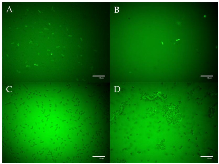
Figure 5.
Images of bacterial cells observed using fluorescence microscopy. E. coli incubated with (A) F-PEP (B) F-P-PEP, and (C) F-P. Fluorescently-labeled PEP and polymer construct F-P-PEP but not the F-P polymer showed significant accumulation on the bacterial cell walls. (D) F-P-PEP interacted with S. aureus extracellular structures.

