Abstract
In November 2017, Biomphalaria pfeifferi, the key intermediate host for Schistosoma mansoni in Africa, was first reported in Lake Malawi, Mangochi District. Two subsequent malacological surveys in 2018 and 2019 confirmed its lacustrine presence, as well as its presence along the Upper Shire River. These surveys provided sufficient specimens for analyses of the genetic structure and a transmission assessment for intestinal schistosomiasis. A total of 76 collected snails were characterized by a DNA sequence analysis of a 650 bp fragment of the mitochondrial cytochrome oxidase subunit 1 (cox1); by size fractionation of six fluorescently labelled microsatellite loci (Bgμl16, Bgμl, Bpf8, rg6, U-7, and rg9);by denaturing PAGE; and by detection of pre-patent Schistosoma infection by real-time PCR with a TaqMan® probe. Five closely related cox1 haplotypes were identified, all present within a single location, with only one haplotype common across all the other locations sampled. No allelic size variation was detected with the microsatellites and all loci were monomorphic. Overall, the pre-patent prevalence of Schistosoma spp. was 31%, with infected snails found at several sampling locations. In this part of Lake Malawi, Bi. pfeifferi exhibits low genetic diversity and is clearly being exposed to the miracidia of S. mansoni, which is likely facilitating the autochthonous transmission of this parasite.
Keywords: intermediate snail hosts, DNA barcoding, microsatellites, Schistosoma mansoni, intestinal schistosomiasis
1. Introduction
The freshwater pulmonate snail Biomphalaria pfeifferi (Gastropoda: Planorbidae) is a key intermediate host for Schistosoma mansoni (Trematoda: Schistosomatidae) [1]. The latter helminth species is a parasitic blood fluke responsible for intestinal schistosomiasis. This fluke can be found across much of Africa and South America [1], and broadly speaking, the endemic zone of intestinal schistosomiasis is delineated by the underlying distribution of permissive snail populations. As the intermediate snail host populations change or vary, the actual or at-risk areas for disease transmission may also change; these dynamics, or fluctuations, are driven in part by ongoing and/or future climate change [2]. Whilst schistosomiasis transmission typically takes place within small water contact foci, even very large water bodies, such as Lake Malawi, are not invulnerable to environmental or biological transformations. For example, within this lake, there have been several significant ecological changes during the past four decades, most probably arising from environmental determinants, actions of humans, and the host–parasite evolution of snails and schistosomes [3].
Lake Malawi occupies about one-fifth of Malawi, representing a unique natural resource for water provision, fishing, agriculture, and international tourism [1]. In the 1980s, for the first-time, urogenital schistosomiasis started to be demonstrated among international tourists, with the later incrimination of sandy beaches with open waters as transmission foci, where deep water Bulinus nyssanus could be found [4]. Contrary to commonly held assumptions that Bulinus globosus was the only local host for Schistosoma haematobium, it was demonstrated experimentally and observed from natural infections that Bu. nyassanus was a permissive host. Indeed, several lacustrine factors influence the transmission of urogenital and, latterly, intestinal schistosomiasis within the lake. About 1000 fish species are estimated to be in the lake with many being molluscivorous [5]; however, with continuous over-fishing, fish predation was relaxed and thought to allow the expansion of snail populations, particularly the softer-shelled intermediate snail hosts of schistosomes [6]. At the same time, several environmental factors, either individually or in combination, are changing the lacustrine environment. There is increasing local pollution from an expanding local human population, reducing water quality, alongside the extraction of substantial amounts of lake water for irrigation purposes which run back into the lake creating more permanent water bodies around its periphery. These wet points would have otherwise dried out during the year and now, if colonized by snails, provide local refugia [5,6,7].
While urogenital schistosomiasis is considered the most common form of schistosomiasis along the Lake Malawi shoreline, there have been dramatic changes in the epidemiology of both forms of schistosomiasis locally [8]. This includes the detection of hybrid schistosomes within the S. haematobium group and the first report of Bu. africanus and Bu. angolensis within the lake, with currently unknown transmission potentials with S. haematobium [8,9]. More importantly, there was the first detection of Bi. pfeifferi within the lake in November 2017 [9,10]. Historically, Bi. pfeifferi was not considered endemic within the lake and is acting as a biological facilitator of a fulminating outbreak of intestinal schistosomiasis within Mangochi District [11]. Two later malacological surveys undertaken in 2018 and 2019 collected additional snail material sufficient for population genetic analyses. In this paper, we seek to determine the genetic structure of Bi. pfeifferi, alongside an assessment of pre-patent infection levels with Schistosoma spp. by molecular xenomonitoring.
2. Materials and Methods
2.1. Malacological Surveillance
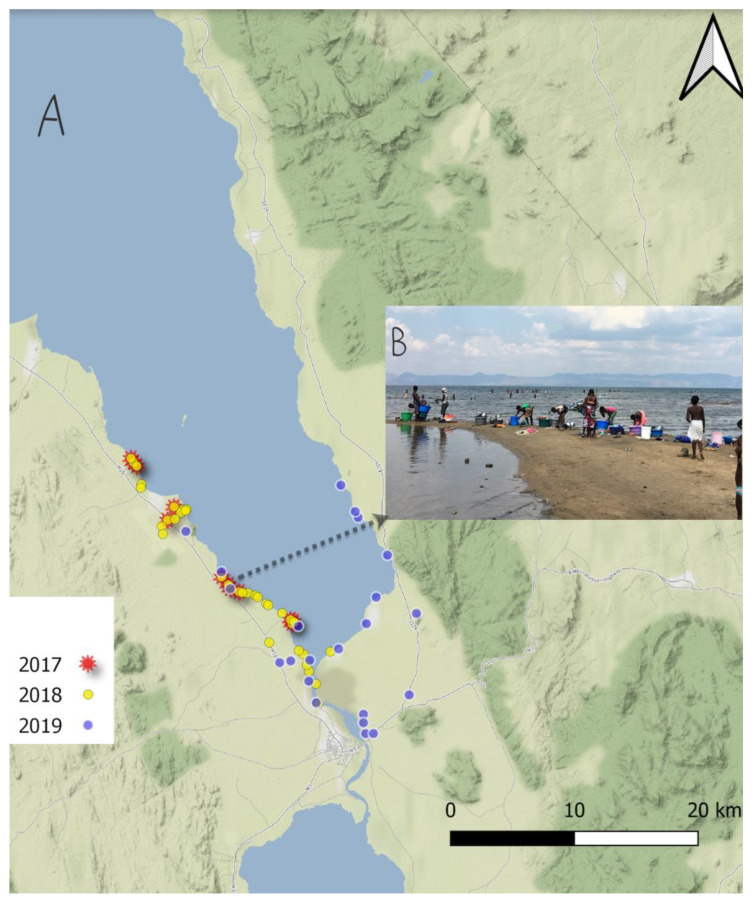
The malacological surveys comprised three directed surveys, pilot, exhaustive, and confirmation studies, carried out in 2017, 2018, and 2019, respectively, along the southern shoreline of Lake Malawi, and nearby water bodies, in Mangochi District (Figure 1). Based on the unexpected finding of Biomphalaria in the pilot study in 2017, we conducted a more exhaustive sampling study in May 2018 (after the rainy season) and visited 43 habitats in the southern region of the lake. One year after, in May 2019, we conducted a confirmation study, including 22 sites distributed in the southern and the eastern region of Lake Malawi. In all the surveys, we considered the collection of snails at known water contact sites, where human activities such as swimming, fishing and, washing take place (Figure 1B).
Figure 1.
(A) The sample sites of visited locations in Mangochi District during each of the November 2017, May 2018, and May 2019 surveys. (B) Picture from a typical lakeshore site, displaying the extensive water contact by local people.
The average time for visiting each snail sampling site was 20 min, during which active searching using slotted scoops took place. According to the habitat type, the scoops were either long- or short-handled, depending on the water depth of the collection site. Then, all the collected snails were sorted by site and transported to the field laboratory. All the snails were kept in glass or plastic bottles in the dark for several hours before being exposed to light to stimulate cercarial shedding. Two days after the collection, this inspection was performed by visually checking the cercariae under the dissecting microscope. Finally, all the snails were preserved in 70% ethanol in universal glass bottles with aluminum caps and labelled with location code and date for transportation back to the Liverpool School of Tropical Medicine (LSTM), UK. Research authorizations were approved in the UK by the LSTM Research Ethics Committee (application 17-018) and in Malawi by the National Health Sciences Research Committee (1805).
2.2. Morphological Analyses, Sample Preparation and DNA Extraction
On arrival at the LSTM, all the samples collected in 2017, 2018, and 2019 were identified to genus level by morphological characteristics [1] and recounted per location prior to DNA extraction. Total genomic DNA was extracted from head–foot snail tissue, according to the methodology of Al-Harbi et al. (2022), utilizing the DNeasy Blood and Tissue Kit (Qiagen™, Hilden, Germany).
2.3. PCR and DNA Sequencing Analysis
A partial region of the mitochondrial cytochrome oxidase subunit 1 (cox1) was amplified using LC1490 and HCO2198 [12] forward and reverse primers, respectively, in a 25 μL PCR reaction for later sequence analysis. PCR was performed using Illustra puReTaq Ready-To-Go PCR beads (GE Healthcare), containing of 0.2 μM of forward and reverse primers, 2 μL of genomic DNA, and 21 μL of nuclease-free water. Successful amplification was verified by running 5 μL of PCR products across a 1% agarose gel for 40 min and visualizing a 650 bp length fragment. The PCR products that had successfully amplified the target region were then purified utilizing the MinElute PCR Purification Kit (Qiagen™, Hilden, Germany) according to the manufacturer’s instructions. The purified PCR products were then shipped to Source Bioscience (www.sourcebioscience.com, accessed on 1 October 2022), Cambridge, UK, for Sanger sequencing. All the sequence and chromatogram data retrieved were then analyzed and trimmed using Geneious 11.1 (Geneious). Using the NCBI database (https://blast.ncbi.nlm.nih.gov/Blast.cgi, accessed on 12 February 2022), all the trimmed sequences were individually subjected to a nucleotide BLAST search to speciate each isolate and investigate any genetic variation from previously deposited sequences.
2.4. Microsatellite Markers Used to Characterize Biomphalaria spp. Specimens
Six microsatellite markers were used to characterize Bi. Pfeifferi: Bgμl6, Bpf8, rg6, μBG1, U7, and RG9, according to the methodology of Jones et al. (1999) [13,14,15] (Table 1). These loci were purposefully chosen based on their wide-ranging amplicon sizes. The microsatellite markers were pooled into two groups for each DNA specimen in the analysis. Moreover, different dye labels were attached to the forward primers. Then, the samples were sent to Macrogen Europe (https://www.macrogen-europe.com, accessed on 2 December 2022) for fragment analysis. Moreover, two PCR products from each microsatellite marker were produced and, after being purified, were Sanger sequenced, as above, to confirm the amplification of the intended loci.
Table 1.
Primers used for six chosen microsatellite marker loci (Jones et al. 1999).
| Locus | Primer Sequence (5′-3′) | Source |
|---|---|---|
| Bpf8 | F:GGTTCCCATAGCATACAGTGC R:GGCTTACAAAGAAACAGGCATAC |
[13] |
| U-7 | F: TCATGACATCTAATGGGAGAAG R:GGGCTCAGAGAAATGAATGG |
Unpublished data |
| rg6 | F: GATTTTGTCTCACGGAAACG R:GCGTGCTTATGTAGCAAAGG |
[14] |
| rg9 | F:GGAGCTGTCGTAATTATAGTTCA R:TTGAGGTGTCATGGTTCTAGG |
[14] |
| Bgµ16 | F:CTGTTATTCATTATTTCATAGAGC R: GGGGATCTAACACATCAG |
[15] |
| µBg1 | F:TTAATTCTACTGGACTCACATGG R:CTGCCAATGTTTACATGCTG |
[15] |
2.5. Real-Time PCR to Detect Pre-Patent Schistosoma spp. Infections within Snails
Real-time PCR was carried out to detect any pre-patent Schistosoma spp. DNA within the Bi. pfeifferi extractions. This was conducted by detecting and amplifying a Schistosoma spp. genus-specific 77 bp fragment of the internal transcribed spacer-2 (ITS-2) sub-unit, as described previously [8]. The PCR cycling conditions were as follows: 3 min initialization at 95 °C, followed by 50 cycles of 15 s denaturation at 95 °C, 20 s annealing at 60 °C, and 45 s extension at 72 °C. The real-time PCR was conducted on a Chromo4 version 3.1 (Bio Rad, 2015) thermal cycler.
2.6. Data Handling and Statistical Analysis
Cox1 chromatograms were obtained via Sanger sequencing, and the microsatellite data were analyzed using Geneious® (11.1.5). Haplotype network analysis was carried out using PopART software (version 1.7).
3. Results
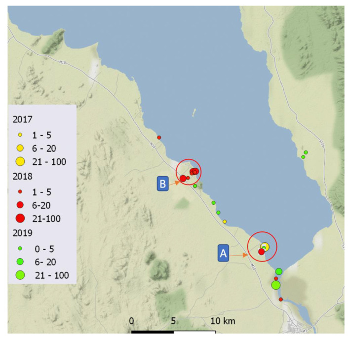
In total, 201 Bi. pfeifferi specimens were collected from 20 locations across the malacological surveys conducted in 2017, 2018, and 2019 (Figure 2). Only one snail was found to be actively shedding S. mansoni cercariae in 2018, and just under half of all the collected snails were subjected to all the outlined molecular analyses.
Figure 2.
Map (www.qgis.org, accessed on 22 November 2022) shows a summary of colonization sites of Bi. pfeifferi in the lake across 2017, 2018, and 2019. (A) Palm Beach, where all cox1 haplotypes were identified as present. (B) A small canal irrigation channel leading to the lake from an inland artificial lake fish hatchery.
3.1. Molecular Characterisation
The Sanger sequence analysis revealed five different haplotypes characterized by five polymorphic sites (Table 2). Of the 76 Bi. pfeifferi specimens successfully sequenced, 69 snails were haplotype 1, 5 were haplotype 2, 1 was haplotype 3, and 1 was haplotype 4. Of note, the distribution of haplotype H1 was detected in all the locations surveyed across the three years, whereas haplotypes H2, H4, and H5 were confined to one collection site in 2017 in Palm Beach (Figure 2).
Table 2.
Nucleotide variation within each of the cox1 haplotypes against NC_038059 from GenBank, which was used as a reference DNA sequence for comparisons. The alignments between Bi. pfeifferi show five single nucleotide polymorphism (SNP) point mutations on the mitochondrial cox1 gene of Bi. pfeifferi at positions 169, 187, 250, 537, and 569.
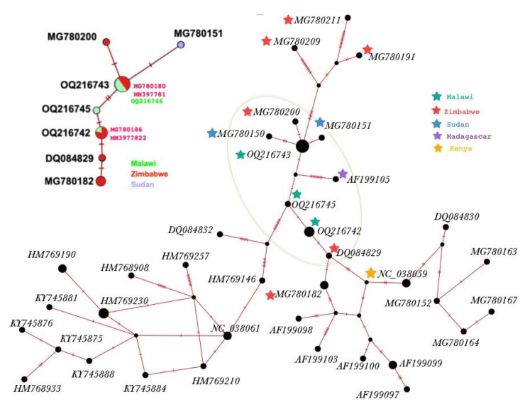
All of the cox1 haplotypes identified in this study (Table 2, Figure 3) are closely genetically related to two haplotypes of Bi. pfeifferi identified from Zimbabwe. Moreover, the alignments between the Bi. pfeifferi samples show the pairwise variation of the haplotypes (Table 2); four SNPs were identified between H1 and H5.
Figure 3.
Network of COX1 gene haplotypes of fifty Bi. pfeifferi from GenBank, including the five cox1 haplotypes from Malawi that were used in this study. In addition, COX1 haplotype network of samples selected based on genetic pairwise distance to samples from Malawi (similarity > 99%). Hatch mark indicates the different nucleotides in the pairwise alignment.
3.2. Microsatellite Data Analysis
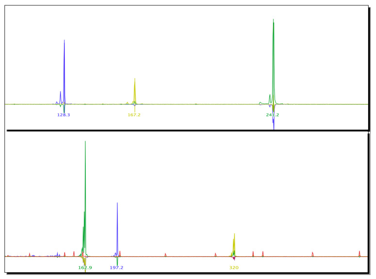
The microsatellite data analysis showed no variation at these loci. Interestingly, the length of the allele amplicons of Bfp8 and U7 was slightly shorter than expected. Moreover, the examination of the six markers shows that significant heterozygote deficiencies are observed within the sample, as shown by one single peak for each (Figure A1 in Appendix A). The peaks of all the samples showed that all the alleles were homozygous (Table 3). The results of the microsatellite identity analyses were confirmed by sequence analysis via BLAST.
Table 3.
Details six microsatellite markers, size of amplicons, and allele frequency.
| N | Marker | Expected Amplicon Size Range (bp) | Size of Amplicon | Allele Frequency |
|---|---|---|---|---|
| 1 | Bgμl6 | 113–135 | 128 bp | 1.00 |
| 2 | Bpf8 | 193–225 | 167 bp | 1.00 |
| 3 | rg6 | 233–244 | 241 bp | 1.00 |
| 4 | μBG1 | 165–167 | 162 bp | 1.00 |
| 5 | U7 | 222–246 | 197 bp | 1.00 |
| 6 | RG9 | 313–321 | 320 bp | 1.00 |
3.3. Detection of Schistosoma spp. DNA within Collected Bi. pfeifferi Specimens
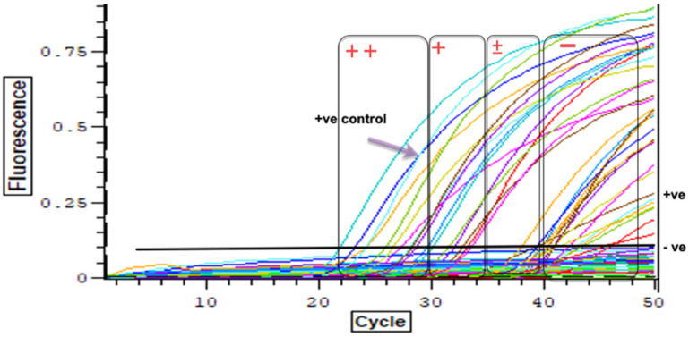
In total, Schistosoma DNA was detected within 31% of the examined Bi. pfeifferi specimens. Of the 57 snails collected within the lake, only ten samples were infected (13%), whereas 74% of the 19 snails collected from waterbodies nearby the lake were positive by real-time PCR analysis (Table 4) (Figure 4).
Table 4.
Summary of collection data and molecular investigation of Bi. pfeifferi.
| Location | Type | Year | Latitude | Longitude | Number of Snails Examined | Infected | Haplotype and Acc.No |
|---|---|---|---|---|---|---|---|
| 1 | Lake | 2017 | −14.36919 | 35.17629 | 2 | 0 | H1 OQ216742 |
| 2 | Lake | −14.39363 | 35.22104 | 17 | 2 | H1 OQ216742 H2 OQ216743 H3 OQ216744 H4 OQ216745 H5 OQ216746 |
|
| 3 | Lake | 2018 | −14.27752 | 35.10419 | 1 | 0 | H1 OQ216742 |
| 4 | Stream | −14.32120 | 35.13072 | 5 | 4 | ||
| 5 | Lake | −14.39363 | 35.22104 | 22 | 5 | ||
| 6 | Lake | −14.44975 | 35.24018 | 3 | 3 | ||
| 7 | Lake | −14.42708 | 35.23349 | 1 | 0 | ||
| 8 | Stream | −14.31371 | 35.14174 | 4 | 4 | ||
| 9 | Stream | −14.31568 | 35.14030 | 2 | 2 | ||
| 10 | Stream | −14.32033 | 35.13613 | 2 | 1 | ||
| 11 | Stream | −14.31338 | 35.14449 | 1 | 0 | ||
| 12 | Lake | −14.31437 | 35.14376 | 2 | 0 | ||
| 13 | Lake | 2019 | −14.44930 | 35.23871 | 1 | 0 | |
| 14 | River | −14.34131 | 35.28897 | 2 | 1 | ||
| 15 | River | −14.41937 | 35.23394 | 2 | 1 | ||
| 16 | River | −14.32895 | 35.14481 | 1 | 1 | ||
| 17 | Lake | −14.39545 | 35.22569 | 3 | 0 | ||
| 18 | Lake | −14.29649 | 35.25654 | 2 | 0 | ||
| 19 | Lake | −14.31494 | 35.26697 | 2 | 0 | ||
| 20 | Lake | −14.36913 | 35.17623 | 1 | 0 |
Figure 4.
Real-time PCR results show the gradient, strong positive Ct ≤ 30 (++), positive 30 < Ct ≤ 35 (+), trace positive 35 < Ct ≤ 40 (±), negative Ct > 40 (−).
4. Discussion
The recent surveys conducted in Malawi since 2017 have revealed significant changes in the epidemiology of schistosomiasis in Malawi, particularly in Lake Malawi, including the first report of Bi. pfeifferi [9,10]. The Biomphalaria species have exhibited strong local and global spreading capabilities that may advance because of global warming and expansion in the development of the water and agricultural projects [16]. Following a recent invasion and colonization by this species in this area, we report here the invasion patterns and steady expansion of Bi. pfeifferi snails from the southern to the eastern region of the lake between 2017 and 2019. In addition, we report, for the first time, a population genetic analysis and dynamic of the transmission of Bi. pfeifferi along the southern shoreline of Lake Malawi, Mangochi District. It is worth mentioning that the colonization of Biomphalaria was followed by the finding of the S. mansoni infection among local people in 2018 [10], before the situation advanced into an outbreak of intestinal schistosomiasis, as reported in 2020 [11]. It is noteworthy that the geographic distribution of S. mansoni is reliant on the distribution of the Biomphalaria species that play key roles in the parasite transmission [16].
The Sanger sequencing and fragment analyses of the cox1 gene inferred a genetic lineage of Bi. pfeifferi likely originating from Zimbabwe. Furthermore, through microsatellite analyses, five Bi. pfeifferi haplotypes were identified in this area. Interestingly, of the five haplotypes, only one haplotype has colonized the lake and water bodies in Mangochi District through 2018 and 2019. Likewise, only Palm Beach contained all five haplotypes in 2017, while all the other sites contained haplotype H1, which is suggestive of a single expansion of snails from Palm Beach with multiple introductions into this particular site. This population of Bi. pfeifferi from Lake Malawi has never before been assessed using microsatellite markers, and so, no direct comparisons between these data and other studies have been made until now.
Our data are in agreement with similar previous studies demonstrating low genetic variation across the Biomphalaria populations with these loci [17]. For example, Angers et al. (2003) carried out a genetic study of Bi. pfeifferi using Bfp8 loci for samples from various areas: Ivory Coast, Senegal, Ethiopia, Niger and Cameroon, Zimbabwe, Oman, and Madagascar. They found low variation within an individual population, including a population from Zimbabwe, which is further suggestive of an original source being found there. A lack of genetic variation supports our hypothesis that Bi. pfeifferi has likely colonized this area of Lake Malawi very recently and has likely driven the recent outbreak of intestinal schistosomiasis locally [11]. In addition, the genetic population structure could have contributed to the dramatic increase in S. mansoni, as low genetic variation with putative high schistosome compatibility is known to raise the transmission of S. mansoni [18,19,20].
This study has also, for the first time, by way of detecting Schistosoma spp. DNA within collected Bi. pfeifferi specimens, incriminated the recently established Bi. pfeifferi populations in this area as being those actively transmitting intestinal schistosomiasis. In addition, the analysis of these data appears to suggest that transmission of S. mansoni is occurring at a higher intensity within the temporal waterbodies surrounding the lake shore than within the lake itself. This may be a result of more frequent human water contact with such waterbodies and may also be because these waterbodies provide a more suitable habitat for Bi. pfeifferi populations when compared to the lake. For example, the environmental factors known to hinder Biomphalaria population dynamics and population expansion, such as waves and predatory fish, may be lacking within these peripheral waterbodies. It is worthwhile noting that understanding the local biological factors in active transmission areas is essential for snail control [21]. Furthermore, an understanding of the mode of invasion and distribution of Bi. pfeifferi in addition to the experimental clarification of the susceptibility to S. mansoni would forecast the potential spread of schistosomiasis [16].
The detection of schistosome infections in snails is a promising monitoring tool for successful control and elimination because it helps in the understanding of the local transmission dynamics [22]. However, extra developments are still needed and essential for long-term success [23]. The challenges include the identification of the ideal marker to detect the exact species of schistosome within snails and more field testing alongside a more rigorous standardization of protocols [23]. With the development of molecular methods, several molecular techniques have been used for Schistosoma transmission mapping by detecting parasite DNA in snails that were not actively shedding cercariae [22,24,25]. For example, in our study cercarial shedding analyses would have missed new epidemiological developments regarding snail–Schistosoma dynamics in the lake without detecting Schistosoma infections. In 2022, malacological surveys conducted by a HUGS team in Mangochi District upon examination for shedding cercariae found patently infected Bi. pfeifferi from sites, and upon microscopy these shedding cercariae were confirmed to be of human infecting species, with later ongoing DNA barcoding studies of S. mansoni to confirm. Although detecting snails shedding cercariae is the only way to fully incriminate an active intermediate host definitively, a quantitative step has been attempted to facilitate analysis beyond a simple positive or negative result. Nonetheless, there is a lack of standardization in the interpretation of Ct values, which makes the results of real-time PCR assays comparable, although real-time PCR assays can measure relative infection intensities [23]. Although it is a limitation in this study, mapping pre-patent infections in snails provides a promising tool for transmission assessment(s) prior to revision of the current control and future prevention. We recommend the expansion of future malacological surveillance with expanded molecular xenomonitoring to better monitor the environmental epidemiology.
In conclusion, this study aimed to understand the dynamics, molecular identification, and genetic structure of Biomphalaria, alongside detection of the schistosome parasites within the snails. We observed a low genetic diversity of Bi. pfeifferi, which might enhance the transmission of intestinal schistosomiasis. Looking to the future, we expect larger zones of the lake to be at risk of intestinal schistosomiasis, which calls for closer epidemiological and malacological surveillance, with future molecular xenomonitoring inspections.
Appendix A
Figure A1.
Diagram showing the results of microsatellite data analysis of six loci, indicating the peak of each locus. In order: Bgμl6 (128 bp); Bpf8 (167 bp); rg6 (241 bp); μBG1(162 bp); U7 (197 bp); and RG9 (320 bp).
Author Contributions
Methodology, M.H.A., C.C., T.M.A., and S.J.; validation, J.A. and J.M.; formal analysis, M.H.A.; investigation, M.H.A., C.C., and J.H.; resources, S.A.K., E.J.L., and P.M.; writing—original draft, M.H.A.; writing—review and editing, J.A., S.J., and J.R.S.; supervision, J.R.S.; funding acquisition, J.R.S. All authors have read and agreed to the published version of the manuscript.
Institutional Review Board Statement
Research authorizations were approved in the UK by LSTM Research Ethics Committee (application 17-018) and in Malawi by the National Health Sciences Research Committee (1805).
Informed Consent Statement
Not applicable.
Data Availability Statement
Not applicable.
Conflicts of Interest
The authors declare no conflict of interest.
Funding Statement
Mohammad H. Alharbi, Sam Jones, Seke Kayuni receive salary support from the Wellcome Trust; these surveys were therefore supported, in part, by the National Institute for Health Research (NIHR) (using the UK’s Official Development Assistance (ODA) Funding) and the Wellcome Trust [220818/Z/20/Z] under the NIHR-Wellcome Partnership for Global Health Research. The views expressed are those of the authors and not necessarily those of Wellcome, the NIHR, or the Department of Health and Social Care. John Archer funding from Medical Research Council Doctoral Training Partnership (MRC-DTP).
Footnotes
Disclaimer/Publisher’s Note: The statements, opinions and data contained in all publications are solely those of the individual author(s) and contributor(s) and not of MDPI and/or the editor(s). MDPI and/or the editor(s) disclaim responsibility for any injury to people or property resulting from any ideas, methods, instructions or products referred to in the content.
References
- 1.Brown D.S. Freshwater Snails of Africa and Their Medical Importance. CRC Press; London, UK: 1994. [Google Scholar]
- 2.Stensgaard A.-S., Utzinger J., Vounatsou P., Hürlimann E., Schur N., Saarnak C.F., Simoonga C., Mubita P., Kabatereine N.B., Tchuenté L.-A.T. Large-scale determinants of intestinal schistosomiasis and intermediate host snail distribution across Africa: Does climate matter? Acta Trop. 2013;128:378–390. doi: 10.1016/j.actatropica.2011.11.010. [DOI] [PubMed] [Google Scholar]
- 3.Madsen H., Stauffer J.R. Schistosomiasis Control Under Changing Ecological Settings in Lake Malawi. EcoHealth. 2022;19:320–323. doi: 10.1007/s10393-022-01606-7. [DOI] [PubMed] [Google Scholar]
- 4.Cetron M.S., Chitsulo L., Sullivan J.J., Pilcher J., Wilson M., Noh J., Tsang V.C., Hightower A.W., Addiss D.G. Schistosomiasis in lake Malawi. Lancet. 1996;348:1274–1278. doi: 10.1016/S0140-6736(96)01511-5. [DOI] [PubMed] [Google Scholar]
- 5.Chidammodzi C., Muhandiki V. Development of indicators for assessment of Lake Malawi Basin in an Integrated Lake Basin Management (ILBM) framework. Int. J. Commons. 2015;9:209–236. doi: 10.18352/ijc.479. [DOI] [Google Scholar]
- 6.Stauffer J.R., Arnegard M.E., Cetron M., Sullivan J.J., Chitsulo L.A., Turner G.F., Chiotha S., McKaye K. Controlling vectors and hosts of parasitic diseases using fishes. BioScience. 1997;47:41–49. doi: 10.2307/1313005. [DOI] [Google Scholar]
- 7.Bootsma H.A., Jorgensen S.E. Lake Malawi/Nyasa: Experience and Lessons Learned Brief. 2013. [(accessed on 12 December 2022)]. Available online: https://www.semanticscholar.org/paper/Lake-Malawi%2FNyasa%3A-Experience-and-lessons-learned-Bootsma-J%C3%B8rgensen/4eeb195d7d53a1fe2f8afc8ee77b310503d79804.
- 8.Alharbi M.H., Iravoga C., Kayuni S.A., Cunningham L., LaCourse E.J., Makaula P., Stothard J.R. First Molecular Identification of Bulinus africanus in Lake Malawi Implicated in Transmitting Schistosoma Parasites. Trop. Med. Infect. Dis. 2022;7:195. doi: 10.3390/tropicalmed7080195. [DOI] [PMC free article] [PubMed] [Google Scholar]
- 9.Webster B.L., Alharbi M., Kayuni S., Makaula P., Halstead F., Christiansen R., Juziwelo L., Stanton M., LaCourse J., Rollinson D., et al. Schistosome Interactions within the Schistosoma Haematobium Group, Malawi. National Center for Infectious Diseases; Atlanta, GA, USA: 2019. [DOI] [PMC free article] [PubMed] [Google Scholar]
- 10.Alharbi M.H., Condemine C., Christiansen R., LaCourse E.J., Makaula P., Stanton M.C., Juziwelo L., Kayuni S., Stothard J.R. Biomphalaria pfeifferi snails and intestinal schistosomiasis, Lake Malawi, Africa, 2017–2018. Emerg. Infect. Dis. 2019;25:613. doi: 10.3201/eid2503.181601. [DOI] [PMC free article] [PubMed] [Google Scholar]
- 11.Kayuni S.A., O’Ferrall A.M., Baxter H., Hesketh J., Mainga B., Lally D., Al-Harbi M.H., LaCourse E.J., Juziwelo L., Musaya J. An outbreak of intestinal schistosomiasis, alongside increasing urogenital schistosomiasis prevalence, in primary school children on the shoreline of Lake Malawi, Mangochi District, Malawi. Infect. Dis. Poverty. 2020;9:121. doi: 10.1186/s40249-020-00736-w. [DOI] [PMC free article] [PubMed] [Google Scholar]
- 12.Vrijenhoek R. DNA primers for amplification of mitochondrial cytochrome c oxidase subunit I from diverse metazoan invertebrates. Mol. Mar. Biol. Biotechnol. 1994;3:294–299. [PubMed] [Google Scholar]
- 13.Charbonnel N., Angers B., Razatavonjizay R., Bremond P., Jarne P. Microsatellite variation in the freshwater snail Biomphalaria pfeifferi. Mol. Ecol. 2000;9:1006–1007. doi: 10.1046/j.1365-294x.2000.00939-9.x. [DOI] [PubMed] [Google Scholar]
- 14.Nguema R.M., Langand J., Galinier R., Idris M.A., Shaban M.A., Al Yafae S., Moné H., Mouahid G. Genetic diversity, fixation and differentiation of the freshwater snail Biomphalaria pfeifferi (Gastropoda, Planorbidae) in arid lands. Genetica. 2013;141:171–184. doi: 10.1007/s10709-013-9715-8. [DOI] [PubMed] [Google Scholar]
- 15.Jones C.S., Lockyer A.E., Rollinson D., Piertney S.B., Noble L.R. Isolation and characterization of microsatellite loci in the freshwater gastropod, Biomphalaria glabrata, an intermediate host for Schistosoma mansoni. Mol. Ecol. 1999;8:2149–2151. doi: 10.1046/j.1365-294x.1999.00802-5.x. [DOI] [PubMed] [Google Scholar]
- 16.Habib M.R., Lv S., Rollinson D., Zhou X.-N. Invasion and Dispersal of Biomphalaria Species: Increased Vigilance Needed to Prevent the Introduction and Spread of Schistosomiasis. Front. Med. 2021;8:614797. doi: 10.3389/fmed.2021.614797. [DOI] [PMC free article] [PubMed] [Google Scholar]
- 17.Angers B., Charbonnel N., Galtier N., Jarne P. The influence of demography, population structure and selection on molecular diversity in the selfing freshwater snail Biomphalaria pfeifferi. Genet. Res. 2003;81:193–204. doi: 10.1017/S0016672303006219. [DOI] [PubMed] [Google Scholar]
- 18.Webster J., Davies C., Hoffman J., Ndamba J., Noble L., Woolhouse M. Population genetics of the schistosome intermediate host Biomphalaria pfeifferi in the Zimbabwean highveld: Implications for co-evolutionary theory. Ann. Trop. Med. Parasitol. 2001;95:203–214. doi: 10.1080/00034983.2001.11813630. [DOI] [PubMed] [Google Scholar]
- 19.Sandland G.J., Wethington A.R., Foster A.V., Minchella D.J. Effects of host outcrossing on the interaction between an aquatic snail and its locally adapted parasite. Parasitol. Res. 2009;105:555–561. doi: 10.1007/s00436-009-1428-7. [DOI] [PubMed] [Google Scholar]
- 20.Sandland G.J., Foster A.V., Zavodna M., Minchella D.J. Interplay between host genetic variation and parasite transmission in the Biomphalaria glabrata–Schistosoma mansoni system. Parasitol. Res. 2007;101:1083–1089. doi: 10.1007/s00436-007-0593-9. [DOI] [PubMed] [Google Scholar]
- 21.Jamieson B.G. Schistosoma: Biology, Pathology and Control. CRC Press; London, UK: 2017. [Google Scholar]
- 22.Pennance T., Archer J., Lugli E.B., Rostron P., Llanwarne F., Ali S.M., Amour A.K., Suleiman K.R., Li S., Rollinson D. Development of a molecular snail xenomonitoring assay to detect Schistosoma haematobium and Schistosoma bovis infections in their Bulinus snail hosts. Molecules. 2020;25:4011. doi: 10.3390/molecules25174011. [DOI] [PMC free article] [PubMed] [Google Scholar]
- 23.Kamel B., Laidemitt M.R., Lu L., Babbitt C., Weinbaum O.L., Mkoji G.M., Loker E.S. Detecting and identifying Schistosoma infections in snails and aquatic habitats: A systematic review. PLoS Negl. Trop. Dis. 2021;15:e0009175. doi: 10.1371/journal.pntd.0009175. [DOI] [PMC free article] [PubMed] [Google Scholar]
- 24.Rollinson D., Knopp S., Levitz S., Stothard J.R., Tchuenté L.-A.T., Garba A., Mohammed K.A., Schur N., Person B., Colley D.G. Time to set the agenda for schistosomiasis elimination. Acta Trop. 2013;128:423–440. doi: 10.1016/j.actatropica.2012.04.013. [DOI] [PubMed] [Google Scholar]
- 25.Born-Torrijos A., Poulin R., Raga J.A., Holzer A.S. Estimating trematode prevalence in snail hosts using a single-step duplex PCR: How badly does cercarial shedding underestimate infection rates? Parasites Vectors. 2014;7:243. doi: 10.1186/1756-3305-7-243. [DOI] [PMC free article] [PubMed] [Google Scholar]
Associated Data
This section collects any data citations, data availability statements, or supplementary materials included in this article.
Data Availability Statement
Not applicable.


