Abstract
The human gut microbiota is the community of microorganisms living in the human gut. This microbial ecosystem contains bacteria beneficial to their host and plays important roles in human physiology, participating in energy harvest from indigestible fiber, vitamin synthesis, and regulation of the immune system, among others. Accumulating evidence suggests a possible link between compositional and metabolic aberrations of the gut microbiota and coronary artery disease in humans. Manipulating the gut microbiota through targeted interventions is an emerging field of science, aiming at reducing the risk of disease. Among the interventions with the most promising results are probiotics, prebiotics, synbiotics, and trimethylamine N-oxide (TMAO) inhibitors. Contemporary studies of probiotics have shown an improvement of inflammation and endothelial cell function, paired with attenuated extracellular matrix remodeling and TMAO production. Lactobacilli, Bifidobacteria, and Bacteroides are some of the most well studied probiotics in experimental and clinical settings. Prebiotics may also decrease inflammation and lead to reductions in blood pressure, body weight, and hyperlipidemia. Synbiotics have been associated with an improvement in glucose homeostasis and lipid abnormalities. On the contrary, no evidence yet exists on the possible benefits of postbiotic use, while the use of antibiotics is not warranted, due to potentially deleterious effects. TMAO inhibitors such as 3,3-dimethyl-1-butanol, iodomethylcholine, and fluoromethylcholine, despite still being investigated experimentally, appear to possess anti-inflammatory, antioxidant, and anti-fibrotic properties. Finally, fecal transplantation carries conflicting evidence, mandating the need for further research. In the present review we summarize the links between the gut microbiota and coronary artery disease and elaborate on the varied therapeutic measures that are being explored in this context.
Keywords: gut microbiota, TMAO, probiotics, prebiotics, synbiotics, coronary artery disease, TMAO inhibitors
1. Introduction
The human gut microbiota is the microbial community residing in the human gut. It comprises around 50 trillion microorganisms [1], a number roughly equal to the number of human cells. These organisms are overwhelmingly anaerobic bacteria, averaging a few hundred species per host [2]. Humans are generally colonized at birth, with a microbiota that increasingly diversifies until it reaches an adult configuration around age 3, remaining relatively stable until old age [2,3,4,5,6]. The richness and diversity of the gut microbiota is determined by numerous factors, such as geography, host genetics, and age, but primarily by host dietary habits [7,8,9]. Although not all gut bacteria are beneficial to their host, several species of mutualistic microorganisms are not only tolerated by the human immune system, but constitute metabolic participants to various host-related physiological functions. Among others, such functions include vitamin synthesis, regulation of the immune system, and the production of energy from sources which cannot be digested in the human intestinal tract [4,5]. Of particular importance is the production by the microbiota of the short-chain fatty acids (SCFA) propionate, acetate, and butyrate. Butyrate has a strong anti-inflammatory potential and constitutes the main energy source for gut epithelial cells, while SCFA as a whole may be partly responsible for the benefits derived from consumption of the fiber-rich Mediterranean diet [10,11].
Despite the fact that modern microbiota research is relatively young, mounting evidence already suggests links of microbial community compositional and/or functional aberrations to human disease, including coronary artery disease (CAD) [12,13,14]. Such aberrations (relative to control groups of subjects) are generally termed “dysbiosis”, although the lack of consensus on what is considered a normal gut microbiota makes the application of this term somewhat arbitrary. In the following sections, we will briefly summarize the links between gut bacterial dysbiosis and CAD, and focus on possible therapeutic interventions which might reduce the associated risk.
2. The Association of Gut Bacterial Dysbiosis with CAD
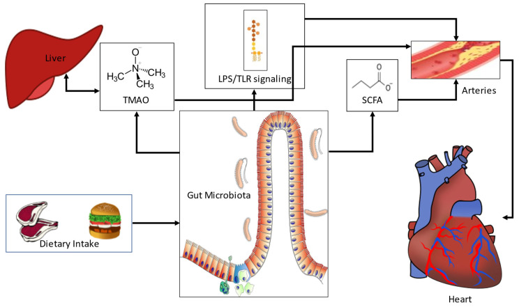
The pathophysiological basis of the association between bacterial dysbiosis and CAD may involve multiple cellular and molecular pathways that mainly relate to infection, changes in host bile acid and lipid metabolic profiles, leakage of harmful endotoxins through the gut epithelial barrier, and generation of potentially atherogenic bacterial metabolites (Figure 1).
Figure 1.
The pathophysiology of gut microbial associations with coronary artery disease. Diet critically shapes the gut microbiota, providing substrates for fermentation. Undigested fiber consumed by bacteria leads to the production of SCFA. Low levels of SCFA have been linked to host inflammation, which may influence atherosclerosis. Many bacterial species also produce TMA using choline as a source. TMA is metabolized to TMAO in the liver by flavin monooxygenases, and is associated with atherosclerosis. Bacterial LPS leaking into the circulation from the gut initiates TLR-mediated systemic inflammation, also linked to atherosclerosis. LPS: Lipopolysaccharide, SCFA: short-chain fatty acids, TLR: toll-like receptor, TMAO: trimethylamine N-oxide.
Infection causes systemic inflammation, which may destabilize atherosclerotic plaques. This is particularly true for respiratory pathogens [15], but it is less clear whether gut infections also lead to similar results [16]. Atherosclerotic plaques contain bacteria which may play some role in promoting atherosclerosis and could have translocated from the gut, although evidence is scarce [12].
Bile acid metabolism involves bacteria-mediated deconjugation and formation of secondary products, as well as bacterial influence on the bile acid receptor (farnesoid X receptor, FXR). FXR-deficient mice develop hypercholesterolemia [17], although evidence in humans is limited.
Gut bacterial functions also affect lipid metabolism [18], but their role on the key low-density lipoprotein (LDL) molecule is obscure at best. Host-microbiota crosstalk concerning lipid metabolism is at least in part mediated by nuclear peroxisome proliferator-activated receptors (PPAR), with species-specific interactions [19]. As an example, PPARγ responds to butyrate production by gut bacteria and facilitates β-oxidation, while suppressing Nitric Oxide (NO) synthesis, maintaining the anaerobic milieu in the colonic environment and preventing dysbiosis [19,20,21]. Further, a high fat diet has been shown to procure spatial and compositional alterations of the gut microbiota, together with dysregulation of PPARγ signaling, which seems to control the spatial distribution of bacteria in the ileum of mice [22]. These phenomena were reversed by administration of a PPARγ agonist.
Gram negative bacterial wall component lipopolysaccharide (LPS), an endotoxin, has strong connections to low grade systemic inflammation in humans and its leakage into the bloodstream through a compromised or even intact epithelial barrier may contribute to atherosclerosis [23,24,25]. LPS initiates Toll-like receptor (TLR) signaling and influences circulating cytokine levels, including Tumor Necrosis Factor (TNF)—α.
Finally, certain bacterial metabolites have received attention for their potentially proatherogenic properties. Trimethylamine (TMA), a bacterial derivative of choline, is metabolized to trimethylamine N-oxide (TMAO) by flavin-containing monooxygenases (FMO) in the liver and is arguably the single most researched bacterial metabolite in relation to CAD. It was originally associated with promotion of atherosclerosis in rodents [26], and has since been extensively studied, with conflicting results. Increased TMAO levels have also been detected in non-alcoholic fatty liver disease [27], which is a known risk factor for CAD [28]. Bacterial species known to produce TMA include Escherichia coli, Clostridium XIVa strains, and Eubacterium species strain AB3007 [29], as well as Anaerococcus hydrogenalis, Escherichia fergusonii, Proteus penneri, Providencia rettgeri, and Edwardsiella tarda [30]. Many more are expected to exist, to the point that TMA production can be considered functionally redundant in the mammalian gut [31]. An additional metabolite is tryptophan; a reduced capacity of bacterial tryptophan metabolism has been linked to host metabolic dysregulation [32].
Based on the pathophysiological rationale, numerous clinical studies have been conducted in humans, mostly of an observational design (summarized in a recent position paper of the European Society of Cardiology Working Group on coronary pathophysiology and microcirculation) [33]. Multiple studies have found altered microbial diversity in subjects with atherosclerosis or atherosclerotic-related cardiovascular events. Although results are often not corroborated between studies, perhaps due to technical issues or patient background differences, an overall shift in ecological metrics such as alpha and beta diversity has been noted and individual microbial taxa differing in relative abundance have been pinpointed. As examples, stroke patients had a greater abundance of opportunistic pathogens Enterobacter, Megasphaera, Oscillibacter, and Desulfovibrio, and a smaller abundance of generally beneficial genera, such as Bacteroides, Prevotella, and Faecalibacterium [34]. Similarly, shotgun sequencing of the gut microbiome in a large cohort revealed a reduction in the Bacteroides and Prevotella genera, and an enrichment in the Streptococcus and Escherichia genera in atherosclerotic patients [35]. Patients in this cohort also had differences in other bacterial species and strains, such as more Klebsiella spp. and Enterobacter aerogenes, and fewer butyrate-producing bacteria, including Roseburia intestinalis and Faecalibacterium cf. prausnitzii.
More importantly, metabolic characteristics of the gut microbiota of atherosclerotic patients were shown to differ in comparison to healthy control subjects, pointing towards a larger inflammatory effect [35]. In yet another large cohort study, Enterococcus was significantly enriched, while Faecalibacterium, Subdoligranulum, Roseburia, and Eubacterium rectale were significantly depleted in CAD patients [36]. This study also verified metabolic shifts in CAD related bacterial communities, including enhanced LPS biosynthesis and increased protein and tryptophan metabolism. Lastly, TMAO has been linked to an increased cardiovascular risk in many studies and meta-analyses [37,38,39], although conflicting results exist and causality has not been proven in humans [40,41].
It is not apparent why differences exist between healthy subjects and CAD patients. The observational nature of the vast majority of the relative studies precludes any inference of causality and since a single standardized research protocol is generally not implemented internationally, comparison between studies is challenging at best.
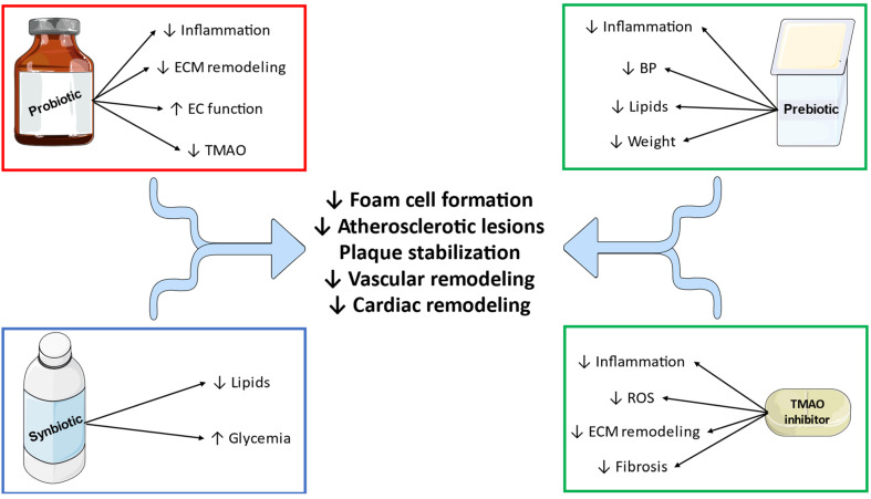
Conceivably, the links between gut bacterial dysbiosis and CAD have generated interest in therapeutic interventions that may modulate the possible risk (Table 1 and Figure 2). The following section will focus on the relative research efforts.
Table 1.
Selected preclinical and clinical studies of gut microbiome modulation demonstrating the effect of interventions in experimental, surrogate, or clinical markers of atherosclerosis.
| Study | Year | Population | Intervention | Outcome |
|---|---|---|---|---|
| Probiotics | ||||
| Malik et al. [42] | 2018 | Male CAD patients | Lactobacillus plantarum | ↓Inflammatory markers ↑Endothelial function No change in TMAO |
| Moludi et al. [43] | 2021 | CAD patients | Lactobacillus rhamnosus | ↓Body weight ↓Inflammatory markers |
| Koppinger et al. [44] | 2020 | LDLr−/− mice with ischemia/reperfusion injury | Lactobacillus reuteri | ↓Infarct size |
| Sun et al. [45] | 2022 | CAD patients | Bifidobacterium lactis Probio-M8 | Improvement in anginal, anxiety, and depressive symptoms ↓Inflammatory markers ↓TMAO ↓Proatherogenic aminoacids |
| Yoshida et al. [46] | 2018 | Female Wistar rats on a HFD |
Bacteroides vulgatus
Bacteroides dorei |
Prevention of atherosclerotic plaque formation |
| O’Morain et al. [47] | 2021 | Male LDLr−/− mice on a HFD |
Lactobacillus acidophilus Bifidobacterium bifidum Bifidobacterium animalis subsp. Lactis Lactobacillus plantarum |
↓Aortic root occlusion Atherosclerotic plaque stabilization ↓Inflammatory-, extracellular matrix remodeling-, and apoptosis-related gene expression |
| Prebiotics | ||||
| Aarsaether et al. [48] | 2006 | Patients scheduled for CABG | Β-1,3/1,6 glucan | ↓CK-MB and cTnT |
| Merino-Aguilar et al. [49] | 2014 | Obese male Wistar rats | Fructooligosaccharides | ↓Body weight ↓Inflammatory markers Improved lipid profile |
| Dehghan et al. [50] | 2016 | Female patients with DM | Oligofructose-enriched inulin | ↓Inflammatory markers ↓Blood pressure Improved lipid profile |
| Parnell et al. [51] | 2009 | Overweight patients | Oligofructose | ↓Body weight |
| Synbiotics | ||||
| Tajabadi-Ebrahimi et al. [52] | 2017 | Diabetic patients with CAD |
Lactobacillus acidophilus 2 × 10⁹ CFU/g Lactobacillus casei 2 × 10⁹ CFU/g Bifidobacterium bifidum 2 × 10⁹ CFU/g 800 mg inulin |
Improved insulin-glucose homeostasis Improved lipid profile |
| TMAO Inhibitors | ||||
| Wang et al. [53] | 2015 | ApoE−/− mice | DMB | ↓Foam cell formation ↓Atherosclerotic lesion development |
| Chen et al. [54] | 2022 | Wild type mice with partial carotid artery ligation | DMB | ↓Vascular remodeling ↓NLRP3 inflammasome expression ↓Endoplasmic reticulum stress ↓Reactive oxygen species formation |
| Organ et al. [55] | 2020 | Wild type mice with transient aortic constriction | Iodomethylcholine | ↓Adverse cardiac remodeling ↓Inflammation, fibrosis, and extracellular matrix remodeling |
| Witkowski et al. [56] | 2021 | Mouse model of arterial injury | Fluoromethylcholine | ↓Tissue factor expression |
| Fecal Transplantation | ||||
| Smits et al. [57] | 2018 | Male patients with MetSy | Fecal transplantation from vegan donors | No alterations in TMAO or vascular inflammation |
CAD: coronary artery disease, TMAO: trimethylamine N-oxide, LDL: low-density lipoprotein, HFD: high-fat diet, CABG: coronary artery bypass grafting, CK-MB: creatine kinase-myocardial bound, cTnT: cardiac troponin T, DM: diabetes mellitus, CFU: colony-forming unit, DMB: 3,3-dimethyl-1-butanol, MetSy: metabolic syndrome, ↓: decreased, ↑ increased.
Figure 2.
Beneficial effects of gut microbiota modulators in coronary artery disease. ECM: extracellular matrix, EC: endothelial cell, TMAO: trimethylamine N-oxide, BP: blood pressure, ROS: reactive oxygen species. ↓: decreased, ↑ increased.
3. Current Therapeutic Interventions
3.1. Probiotics
Probiotics are ‘live microorganisms that, when administered in adequate amounts, confer a health benefit on the host’ [58]. The most common probiotics are Lactobacilli and Bifidobacteria. Many preparations are commercially available and are mostly used as dietary supplements. Administration of probiotics may confer beneficial cardiovascular actions, according to findings from several mechanistic studies. More specifically, reports of anti-inflammatory, antioxidant, antithrombotic, and endothelium-protective effects have been published [59]. Moreover, probiotics could restore and strengthen the intestinal barrier integrity, limit hazardous LPS leakage, and suppress TMAO formation [59]. They may also promote bile acid deconjugation, thus increasing bile acid excretion and cholesterol utilization [33]. Hence, they may possess anti-atherosclerotic potential. A previous review of their potentially anti-atherosclerotic effects can be found elsewhere [60].
Lactobacilli are among the most well-studied probiotics with regards to their cardiovascular benefit. In a mechanistic study in a swine model of CAD, an anti-inflammatory and antioxidant effect mediated by the induction of NF-E2-related factor 2 in the ischemic myocardium could have been the outcome of Lactobacillus plantarum supplementation [61]. This has been further tested in men with CAD, with Lactobacillus plantarum administration resulting in anti-inflammatory and endothelium-protective effects, without affecting metabolic parameters or TMAO concentration, however [42,62]. Further, in a recently reported open-label, randomized trial of 77 dyslipidemic patients, administration of Lactobacillus plantarum together with a lipid-lowering agent (simvastatin 20 mg) resulted in significant improvement of the lipid profile and reduction of the calculated cardiovascular risk, compared to simvastatin 20 mg alone [63]. Concerning Lactobacillus rhamnosus, a randomized, double-blind trial of 44 CAD patients on a 12-week supplementation with this probiotic, together with caloric restriction, showed anti-inflammatory effects and significant weight loss, compared to caloric restriction alone [43]. Other Lactobacilli, such as Lactobacillus paracasei DTA81, have also been tested in experimental atherosclerotic conditions, with initial reports indicating an anti-inflammatory effect together with improved lipid and glucose homeostasis [64]. Lactobacillus reuteri administration in LDLr−/− mice has also produced anti-inflammatory actions that attenuated cardiac ischemia/reperfusion injury, evidenced by reduced infarct size, without affecting cholesterol levels [44]. Oral administration of Lactobacillus rhamnosus GR-1 ameliorated cardiac remodelling and pump failure in rats with induced acute myocardial infarction [65]. A recent meta-analysis of studies found that Lactobacillus acidophilus may have a greater effect in lowering cholesterol than other probiotics [66].
Bifidobacteria also yield probiotic strains with potential benefit in atherosclerosis. Patients with CAD were enrolled in a randomized, double-blind, placebo-controlled trial examining the efficacy of the probiotic strain Bifidobacterium lactis Probio-M8 in combination with conventional treatment involving lipid-lowering (atorvastatin) and β-blockade (metoprolol) [45]. Based on the study results, the study group reported significant improvement in anginal, anxiety, and depressive symptoms assessed through the Seattle Angina Questionnaire, Self-Rating Anxiety Scale, and the Self-Rating Depression Scale, respectively, compared to the control group. Moreover, the investigators documented a greater reduction in interleukin-6 and LDL with the probiotic strain at 6 months, compared to placebo. Regarding gut microbial composition, an abundance of Bifidobacterium adolescentis, Bifidobacterium animalis, Bifidobacterium bifidum, and Butyricicoccus porcorum, together with decreased Flavonifractor plautii and Parabacteroides johnsonii was detected in probiotic-treated subjects. Interestingly, the study group participants exhibited an increase in bioactive microbial metabolites and a decrease in TMAO and proatherogenic amino acids (l-leucine, l-valine). This modulation of gut microbiota and gut metabolome profile through the probiotic strain might have been responsible for the improvement in quality of life, as well as for the reported anti-inflammatory and hypolipidemic effects.
Less evidence is available for other probiotic strains, such as Limosilactobacillus fermentum. Administration of a probiotic formulation containing Liminosilactobacillus fermentum in Wistar rats fed with a high fat diet restored gut microbial composition, which was further accompanied by attenuated metabolic and blood pressure abnormalities [67]. Moreover, the same formulation promoted anti-inflammatory and antioxidant actions at the level of the heart in female Wistar rats consuming a high fat diet [68]. Probiotics Bacteroides vulgatus and Bacteroides dorei inhibited atherosclerotic plaque formation in ApoE-deficient mice [46].
Some of the above-mentioned probiotics have also been administered in combinations. Preclinical studies have provided important insight in this regard. Male LDLr−/− mice consuming a high fat diet were treated with or without a combination of probiotics (Lactobacillus acidophilus, Bifidobacterium bifidum, Bifidobacterium animalis subsp. lactis, Lactobacillus plantarum) for 12 weeks [47]. The investigators noted a significant reduction in the degree of aortic root occlusion paired with features of plaque stabilization (decreased plaque lipid content and macrophages, increase in α-smooth muscle cell actin) in mice receiving the probiotic combination. Moreover, a downregulated expression of several genes was seen in the study group, involving critical pathways (inflammation, lipid transport and metabolism, cell adhesion, extracellular matrix remodeling, apoptosis). Additionally, the probiotic formulation resulted in attenuated monocyte chemoattractant protein-1-induced monocyte migration, monocyte and macrophage proliferation, foam cell formation, and smooth muscle cell proliferation and migration. Furthermore, in an experimental study involving male Wistar rats fed with a high fat diet, treatment with Lactobacillus rhamnosus FM9 and Limosilactobacillus fermentum Y57 had similar efficacy in increasing high-density lipoprotein cholesterol levels compared to a lipid-lowering agent [69]. At the same time, the researchers documented a greater reduction in total cholesterol and LDL with the above-mentioned probiotics. In a human clinical study of young and middle-aged women with arterial hypertension, 8-week treatment with the combination of Lactobacillus paracasei LPC-37, Lactobacillus rhamnosus HN001, Lactobacillus acidophilus NCFM, and Bifidobacterium lactis HN019 significantly reduced fasting glucose and improved the lipid profile and autonomic modulation [70]. At the same time, a trend towards a significant blood pressure-lowering effect (systolic blood pressure reduction ~5 mmHg, diastolic blood pressure reduction ~2 mmHg) was observed.
Although many of the above studies are of promising potential, it should be stressed that most probiotic health claims generally rely on poor evidence. The ideal probiotic strain(s) and/or dose in the context of CAD are still unspecified.
3.2. Prebiotics
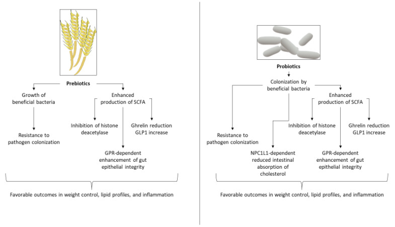
Prebiotics are indigestible food substances that are selectively utilized by and can promote the growth of beneficial gut bacteria, conferring a health benefit on the host [71]. Some of the most common prebiotics that have been tested in the context of cardiovascular disease (CVD) are fructooligosaccharides, inulin, galactooligosaccharides (galactans), beta glucan, Minolest, pectin polysaccharides, and chitosan oligosaccharides. Evidence from human trials suggest that they might be of value in CAD. Possible underlying mechanisms include increased production of SCFA, enhancement of gut epithelial tight junctions, and enhanced growth of beneficial bacteria [72]. More specifically, prebiotics may favor growth and metabolic functions of known beneficial bacterial genera, such as those of Lactobacillus and Bifidobacterium, some of which are themselves used as probiotics, as described above, and may antagonize growth of or colonization by pathogenic species [73]. Production of SCFA by such bacteria may promote gut epithelial integrity through G-protein coupled receptor 43 (GPR43) signaling [74]. Further, SCFA-dependent inhibition of histone deacetylase results in a robust anti-inflammatory gut environment [75,76]. Prebiotic administration may also lead to a reduction of ghrelin release and an increase of glucagon-like peptide-1 (GLP-1) production [51], favorably affecting weight control and fat accumulation (Figure 3).
Figure 3.
Examples of cellular and molecular mechanisms underlying the potential effects of pre- and probiotic administration in metabolic and coronary artery disease. GLP1: glucagon-like peptide-1, GPR: G-protein coupled receptor, NPC1L1: Niemann-Pick C1-like 1 protein, SCFA: short-chain fatty acids.
However, it must be stressed that the European Food and Safety Authority generally refutes any health claim related to prebiotic preparations [77].
There are very few clinical studies directly addressing the impact of prebiotics on human CAD, although evidence exists in animal models [72,78]. One study in humans showed that treatment with beta glycans prevented ischemia/reperfusion injury in patients who underwent coronary artery bypass grafting [48]. The administration of chitosan oligosaccharides in another human trial of CAD patients improved lipid profiles, and increased the abundance of the genera Faecalibacterium, Alistipes, Escherichia, Lactobacillus, Lactococcus, and Phascolarctobacterium [79].
Generally prebiotic-specific and influenced by host dietary background, results from both animal and human studies are promising in high CAD risk disorders, such as obesity and the metabolic syndrome (for a comprehensive review see Santos-Marcos et al. [80]), as well as in healthy subjects [80]. As an example, fructooligosaccharides significantly reduce body weight and inflammatory cytokines in obese rats [49]. A reduction of inflammatory cytokines and an improvement in lipid profiles has also been reported in a randomized controlled trial of human diabetic patients taking inulin vs. placebo [50]. Also in humans, oligofructose consumption was associated with a decrease in ghrelin and weight loss, evidence that again come from a randomized controlled trial [51]. Inulin use in a human randomized control trial led to an increase of Bifidobacterium [81], a result that was also seen with the use of galactooligosaccharides in another randomized controlled trial that recruited obese, prediabetic humans [82]. Moreover, the use of agave fructans has been shown to increase both Bifidobacteria and Lactobacilli in yet another randomized controlled trial [83]. Further, administration of guar gum led to increases in Bifidobacterium, the Clostridium coccoides group, the Roseburia/Eubacterium rectale group, Eubacterium hallii, and butyrate-producing bacterium strain SS2/1, generally enhancing butyrate production in a study of healthy human subjects [84].
3.3. Synbiotics
Synbiotics are ‘a mixture comprising live microorganisms and substrate(s) selectively utilized by host microorganisms that confers a health benefit on the host’ [85]. Synbiotics have shown promise as interventions in human CAD and CAD related disorders, such as obesity and diabetes mellitus (reviewed in Saez-Lara et al.) [86]. As expected, the molecular mechanisms underlying the beneficial effect are those concerned with the pre- and probiotic components of the mixture, as described above. In a randomized controlled trial of diabetic patients with CAD, a synbiotic mixture of Lactobacillus acidophilus, Lactobacillus casei, Bifidobacterium bifidum and inulin led to improvements in glycemia and HDL [52], while another randomized controlled trial showed improvements in lipid levels in patients with type 2 diabetes consuming a mixture of Lactobacillus sporogenes and inulin [87]. A meta-analysis of randomized controlled trials found that synbiotic supplementation in patients with metabolic syndrome significantly reduced serum insulin levels, triglycerides, total cholesterol, LDL, waist circumference, body weight, systolic blood pressure, and serum interleukin-6, and increased HDL [88]. The most effective synbiotic in CAD is currently unknown.
3.4. Antibiotics
The use of antibiotics targeting atherosclerotic plaque microorganisms in CAD has failed to show any benefit and has recently been proven harmful to humans [89,90]. This approach is also harmful to scores of beneficial gut bacteria and should be abandoned.
Microbial investigation of atherosclerotic plaques has shown the existence of a diverse community of more than 50 bacterial species in coronary lesions, including Staphylococcus species, Proteus vulgaris, Klebsiella pneumoniae, Chlamydia pneumoniae, Chlamydia trachomatis, and Streptococcus species [91]. Most of these bacteria probably originate from the oral and gut flora, and translocate to the coronary arteries through bloodstream circulation [91].
An intriguing hypothesis had been formulated, that bacteria may at least influence the progression of atherosclerosis by induction of inflammation in the atherosclerotic plaque. One of the earliest attempts to address the possible association of a microorganism with CAD showed that increased antibodies against Chlamydia pneumoniae predicted cardiovascular events in patients with myocardial infarction, and that administration of azithromycin reduced this risk [92]. However, subsequent and considerably larger studies failed to show any benefit with azithromycin or gatifloxacin in the secondary prevention of events [93,94]. Finally, a comprehensive Cochrane meta-analysis involving more than 26,000 patients receiving macrolides or quinolones conclusively showed that antibiotic use has no benefit in secondary prevention and that it also has a strong trend for being harmful to humans, tending to increase all-cause mortality [89].
3.5. Postbiotics
Postbiotics have been recently defined as ‘preparations of inanimate microorganisms and/or their components that confer a health benefit on the host’ [95]. Using this definition, there is currently no evidence of any postbiotic associated effect on human CAD patients [95]. Purified bacterial metabolites that have shown effects in various health conditions do not fall under the term “postbiotic” [95]. However, components of bacterial cell walls do fall under this term and some evidence suggests a beneficial metabolic effect of purified Akkermansia muciniphila membrane protein Amuc_1100 in diabetic mice, implying a possible antiatherogenic role [96].
3.6. Additional Interventions
3.6.1. TMAO Inhibition
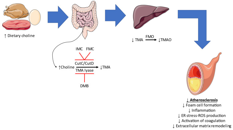
As mentioned, TMAO has garnered significant scientific attention and studies aiming to inhibit its production and investigate the result have been conducted. In the earliest study, by Wang et al., a structural analog of choline, 3,3-dimethyl-1-butanol (DMB), inhibited TMA-lyase activity, decrease TMA levels, and ultimately lowered TMAO levels in mice under a high-choline or L-carnitine diet [53]. Interestingly, this was accompanied by reduced foam cell formation and diminished atherosclerotic lesion development in ApoE−/− mice. In another study, DMB ameliorated cardiac dysfunction in male CD1 mice fed with a high fat diet, without inducing alterations in body weight or cholesterol levels [97]. The cardioprotective effect could be attributed to the anti-inflammatory and anti-fibrotic effects observed histologically. Such observations were reported in a rat model of type 2 cardiorenal syndrome [98]. Anti-inflammatory, antioxidant, and endothelial protective actions were further noted in aged Fischer-344 rats and aged C57BL/6N treated with DMB [99,100]. In a recently reported study, DMB was administered in a wild-type mouse model of partial carotid artery ligation together with a high choline diet or a high TMAO diet [54]. Compared to the control groups, DMB treatment regulated the diet-induced adverse vascular remodeling by attenuating the flow-induced atherosclerotic lesion formation and the expression of NLRP3 inflammasome, the endoplasmic reticulum stress burden, and reactive oxygen species formation [54]. Roberts et al. assessed the importance of a crucial microbial TMA-generating enzyme pair, CutC/CutD [101]. In a well-designed study, they proposed multiple halomethylcholines with the ability to prevent platelet activation and thrombus formation in vitro, without noting an increased bleeding risk [101]. Among the mentioned molecules, iodomethylcholine and fluoromethylcholine, which inhibit TMA-lyase activity similarly to DBM, have been assessed in other studies [55]. According to the histologic examinations, there was a reduction in the expression of inflammatory, fibrotic, and extracellular matrix remodeling markers in animals treated with iodomethylcholine. Apart from these effects, iodomethylcholine led to beneficial alterations in cholesterol and bile acid metabolism in wild-type C57BL/6J mice fed with a high cholesterol diet [102]. Fluoromethylcholine has been shown to reverse TMAO-induced tissue factor expression in a mouse model of arterial injury [56], indicating a possible anti-thrombotic role for this agent. Summarizing the findings mentioned above, inhibition of TMAO formation may have a role in the attenuation of atherosclerosis progression via reducing foam cell formation, inflammation, endoplasmic reticulum and oxidative stress, coagulation, and extracellular matrix remodeling (Figure 4).
Figure 4.
The effect of TMAO inhibition in atherosclerosis. Consumption of a high-choline diet leads to its conversion to trimethylamine (TMA) in the gut, with the aid of CutC/CutD and TMA-lyase. Inhibition of these enzymes by iodomethylcholine (IMC)/fluoromethylcholine (FMC) and 3,3-dimethyl-1-butanol (DMB) leads to lower trimethylamine N-Oxide (TMAO) production in the liver. As a result, atherosclerosis may be attenuated through multiple mechanisms. FMO: flavin-containing monooxygenase, ER: endoplasmic reticulum, ROS: reactive oxygen species.
Flavonoids, a large class of polyphenolic compounds, are present in tea, citrus fruit, citrus fruit juices, berries, red wine, apples, and legumes. In flavonoid aglycones, baicalein, fisetin, acacetin, and myricetin exhibited a decent binding effect with TMA-lyase, as did baicalin, naringin, and hesperidin in flavonoid glycosides [103]. Therefore, inhibition of TMA-lyase activity is another method of reducing TMAO concentration. This observation could partly explain the beneficial effects of flavonoids in CAD prevention [104]. Regarding other forms of diet, such as the Mediterranean diet, the findings are controversial, and sex-specific associations may be present [105,106]. Further research in this field is required to cement the role of specific dietary patterns in TMAO modulation.
To sum up, evidence in humans is all but lacking, and the use of TMAO inhibitors, or any other means of gut microbial modulation, is currently investigational. It is still too early to recommend microbiota measurements or modulation, by TMAO inhibitors or otherwise, for the specific purpose of preventing CAD.
3.6.2. Fecal Transplantation
Fecal transplantation, a technique that has been of clinical use in the management of Clostridioides difficile infection [107], has also been evaluated concerning its potential cardiometabolic benefits in atherosclerosis and possibly CAD. An improvement in insulin sensitivity following the administration of gut microbiota from lean donors to men with metabolic syndrome has been reported, accompanied by an increase in the concentration of butyrate-producing intestinal bacteria (Roseburia intestinalis, Eubacterium hallii) [108]. A subsequent small scale, randomized controlled trial assigned 20 male patients with metabolic syndrome to vegan donor or autologous fecal transplantation [57]. Even though an alteration of host gut microbiota towards a vegan gut microbiota type was documented, this was not associated with reductions in TMAO or vascular inflammation through imaging or ex vivo peripheral blood monocyte production of pro-inflammatory cytokines [57]. These controversial human studies are accompanied by preclinical experiments. In a mouse model of autoimmune myocarditis, fecal microbiota transplantation promoted anti-inflammatory actions through restoration of the Firmicutes/Bacteroidetes ratio [109]. This is especially important, since a pro-inflammatory gut microbiome may be detrimental towards the development and progression of atherosclerosis [110]. In a recently reported experimental study involving the atherosclerosis-prone C1q/TNF-related protein 9-knockout mice, fecal microbiota transplantation from wild-type mice altered the composition of the host gut microbiota and abrogated the atherosclerotic lesions in the carotid artery following partial ligation [111].
4. Conclusions
There is currently an abundance of evidence that gut microbial dysbiosis is associated with CAD and cardiovascular risk. Low grade systemic inflammation induced by gut bacterial components or functions is probably the main underlying pathophysiological mechanism. Moreover, metabolites produced by the gut microbiota serve as markers of increased risk in cardiovascular patients, but causality in humans has not been proven yet. Numerous therapeutic interventions targeting the microbiota have been suggested and tried, with modest effect. Although some of the evidence is relatively strong, based on small randomized controlled trials and even meta-analyses, until larger randomized controlled trials are designed and conducted, manipulation of the gut microbiota in the context of CAD will remain far from clinical practice.
Author Contributions
Conceptualization: T.K. and D.T. Writing—original draft preparation: T.K. and P.T. Writing—review and editing: T.K., P.T., K.T. and D.T. All authors have read and agreed to the published version of the manuscript.
Conflicts of Interest
The authors declare no conflict of interest.
Funding Statement
This research received no external funding.
Footnotes
Disclaimer/Publisher’s Note: The statements, opinions and data contained in all publications are solely those of the individual author(s) and contributor(s) and not of MDPI and/or the editor(s). MDPI and/or the editor(s) disclaim responsibility for any injury to people or property resulting from any ideas, methods, instructions or products referred to in the content.
References
- 1.Sender R., Fuchs S., Milo R. Are We Really Vastly Outnumbered? Revisiting the Ratio of Bacterial to Host Cells in Humans. Cell. 2016;164:337–340. doi: 10.1016/j.cell.2016.01.013. [DOI] [PubMed] [Google Scholar]
- 2.Eckburg P.B., Bik E.M., Bernstein C.N., Purdom E., Dethlefsen L., Sargent M., Gill S.R., Nelson K.E., Relman D.A. Diversity of the human intestinal microbial flora. Science. 2005;308:1635–1638. doi: 10.1126/science.1110591. [DOI] [PMC free article] [PubMed] [Google Scholar]
- 3.Koenig J.E., Spor A., Scalfone N., Fricker A.D., Stombaugh J., Knight R., Angenent L.T., Ley R.E. Succession of microbial consortia in the developing infant gut microbiome. Proc. Natl. Acad. Sci. USA. 2011;108((Suppl. S1)):4578–4585. doi: 10.1073/pnas.1000081107. [DOI] [PMC free article] [PubMed] [Google Scholar]
- 4.Faith J.J., Guruge J.L., Charbonneau M., Subramanian S., Seedorf H., Goodman A.L., Clemente J.C., Knight R., Heath A.C., Leibel R.L., et al. The long-term stability of the human gut microbiota. Science. 2013;341:1237439. doi: 10.1126/science.1237439. [DOI] [PMC free article] [PubMed] [Google Scholar]
- 5.Guarner F., Malagelada J.R. Gut flora in health and disease. Lancet. 2003;361:512–519. doi: 10.1016/S0140-6736(03)12489-0. [DOI] [PubMed] [Google Scholar]
- 6.Clemente J.C., Ursell L.K., Parfrey L.W., Knight R. The impact of the gut microbiota on human health: An integrative view. Cell. 2012;148:1258–1270. doi: 10.1016/j.cell.2012.01.035. [DOI] [PMC free article] [PubMed] [Google Scholar]
- 7.David L.A., Maurice C.F., Carmody R.N., Gootenberg D.B., Button J.E., Wolfe B.E., Ling A.V., Devlin A.S., Varma Y., Fischbach M.A., et al. Diet rapidly and reproducibly alters the human gut microbiome. Nature. 2014;505:559–563. doi: 10.1038/nature12820. [DOI] [PMC free article] [PubMed] [Google Scholar]
- 8.Yatsunenko T., Rey F.E., Manary M.J., Trehan I., Dominguez-Bello M.G., Contreras M., Magris M., Hidalgo G., Baldassano R.N., Anokhin A.P., et al. Human gut microbiome viewed across age and geography. Nature. 2012;486:222–227. doi: 10.1038/nature11053. [DOI] [PMC free article] [PubMed] [Google Scholar]
- 9.Turnbaugh P.J., Ridaura V.K., Faith J.J., Rey F.E., Knight R., Gordon J.I. The effect of diet on the human gut microbiome: A metagenomic analysis in humanized gnotobiotic mice. Sci. Transl. Med. 2009;1:6ra14. doi: 10.1126/scitranslmed.3000322. [DOI] [PMC free article] [PubMed] [Google Scholar]
- 10.Garcia-Mantrana I., Selma-Royo M., Alcantara C., Collado M.C. Shifts on Gut Microbiota Associated to Mediterranean Diet Adherence and Specific Dietary Intakes on General Adult Population. Front. Microbiol. 2018;9:890. doi: 10.3389/fmicb.2018.00890. [DOI] [PMC free article] [PubMed] [Google Scholar]
- 11.Salvi P.S., Cowles R.A. Butyrate and the Intestinal Epithelium: Modulation of Proliferation and Inflammation in Homeostasis and Disease. Cells. 2021;10:1775. doi: 10.3390/cells10071775. [DOI] [PMC free article] [PubMed] [Google Scholar]
- 12.Jonsson A.L., Backhed F. Role of gut microbiota in atherosclerosis. Nat. Rev. Cardiol. 2017;14:79–87. doi: 10.1038/nrcardio.2016.183. [DOI] [PubMed] [Google Scholar]
- 13.Lynch S.V., Pedersen O. The Human Intestinal Microbiome in Health and Disease. N. Engl. J. Med. 2016;375:2369–2379. doi: 10.1056/NEJMra1600266. [DOI] [PubMed] [Google Scholar]
- 14.Witkowski M., Weeks T.L., Hazen S.L. Gut Microbiota and Cardiovascular Disease. Circ. Res. 2020;127:553–570. doi: 10.1161/CIRCRESAHA.120.316242. [DOI] [PMC free article] [PubMed] [Google Scholar]
- 15.Smeeth L., Thomas S.L., Hall A.J., Hubbard R., Farrington P., Vallance P. Risk of myocardial infarction and stroke after acute infection or vaccination. N. Engl. J. Med. 2004;351:2611–2618. doi: 10.1056/NEJMoa041747. [DOI] [PubMed] [Google Scholar]
- 16.Hizo-Abes P., Clark W.F., Sontrop J.M., Young A., Huang A., Thiessen-Philbrook H., Austin P.C., Garg A.X., Walkerton Health Study I. Cardiovascular disease after Escherichia coli O157:H7 gastroenteritis. CMAJ. 2013;185:E70–E77. doi: 10.1503/cmaj.112161. [DOI] [PMC free article] [PubMed] [Google Scholar]
- 17.Lambert G., Amar M.J., Guo G., Brewer H.B., Jr., Gonzalez F.J., Sinal C.J. The farnesoid X-receptor is an essential regulator of cholesterol homeostasis. J. Biol. Chem. 2003;278:2563–2570. doi: 10.1074/jbc.M209525200. [DOI] [PubMed] [Google Scholar]
- 18.Fu J., Bonder M.J., Cenit M.C., Tigchelaar E.F., Maatman A., Dekens J.A., Brandsma E., Marczynska J., Imhann F., Weersma R.K., et al. The Gut Microbiome Contributes to a Substantial Proportion of the Variation in Blood Lipids. Circ. Res. 2015;117:817–824. doi: 10.1161/CIRCRESAHA.115.306807. [DOI] [PMC free article] [PubMed] [Google Scholar]
- 19.Hasan A.U., Rahman A., Kobori H. Interactions between Host PPARs and Gut Microbiota in Health and Disease. Int. J. Mol. Sci. 2019;20:387. doi: 10.3390/ijms20020387. [DOI] [PMC free article] [PubMed] [Google Scholar]
- 20.Byndloss M.X., Olsan E.E., Rivera-Chavez F., Tiffany C.R., Cevallos S.A., Lokken K.L., Torres T.P., Byndloss A.J., Faber F., Gao Y., et al. Microbiota-activated PPAR-gamma signaling inhibits dysbiotic Enterobacteriaceae expansion. Science. 2017;357:570–575. doi: 10.1126/science.aam9949. [DOI] [PMC free article] [PubMed] [Google Scholar]
- 21.Montaigne D., Butruille L., Staels B. PPAR control of metabolism and cardiovascular functions. Nat. Rev. Cardiol. 2021;18:809–823. doi: 10.1038/s41569-021-00569-6. [DOI] [PubMed] [Google Scholar]
- 22.Tomas J., Mulet C., Saffarian A., Cavin J.B., Ducroc R., Regnault B., Kun Tan C., Duszka K., Burcelin R., Wahli W., et al. High-fat diet modifies the PPAR-gamma pathway leading to disruption of microbial and physiological ecosystem in murine small intestine. Proc. Natl. Acad. Sci. USA. 2016;113:E5934–E5943. doi: 10.1073/pnas.1612559113. [DOI] [PMC free article] [PubMed] [Google Scholar]
- 23.Wiedermann C.J., Kiechl S., Dunzendorfer S., Schratzberger P., Egger G., Oberhollenzer F., Willeit J. Association of endotoxemia with carotid atherosclerosis and cardiovascular disease: Prospective results from the Bruneck Study. J. Am. Coll. Cardiol. 1999;34:1975–1981. doi: 10.1016/S0735-1097(99)00448-9. [DOI] [PubMed] [Google Scholar]
- 24.Stoll L.L., Denning G.M., Weintraub N.L. Potential role of endotoxin as a proinflammatory mediator of atherosclerosis. Arterioscler. Thromb. Vasc. Biol. 2004;24:2227–2236. doi: 10.1161/01.ATV.0000147534.69062.dc. [DOI] [PubMed] [Google Scholar]
- 25.Van den Munckhof I.C.L., Kurilshikov A., Ter Horst R., Riksen N.P., Joosten L.A.B., Zhernakova A., Fu J., Keating S.T., Netea M.G., de Graaf J., et al. Role of gut microbiota in chronic low-grade inflammation as potential driver for atherosclerotic cardiovascular disease: A systematic review of human studies. Obes. Rev. 2018;19:1719–1734. doi: 10.1111/obr.12750. [DOI] [PubMed] [Google Scholar]
- 26.Wang Z., Klipfell E., Bennett B.J., Koeth R., Levison B.S., Dugar B., Feldstein A.E., Britt E.B., Fu X., Chung Y.M., et al. Gut flora metabolism of phosphatidylcholine promotes cardiovascular disease. Nature. 2011;472:57–63. doi: 10.1038/nature09922. [DOI] [PMC free article] [PubMed] [Google Scholar]
- 27.Theofilis P., Vordoni A., Kalaitzidis R.G. Trimethylamine N-Oxide Levels in Non-Alcoholic Fatty Liver Disease: A Systematic Review and Meta-Analysis. Metabolites. 2022;12:1243. doi: 10.3390/metabo12121243. [DOI] [PMC free article] [PubMed] [Google Scholar]
- 28.Toh J.Z.K., Pan X.H., Tay P.W.L., Ng C.H., Yong J.N., Xiao J., Koh J.H., Tan E.Y., Tan E.X.X., Dan Y.Y., et al. A Meta-Analysis on the Global Prevalence, Risk factors and Screening of Coronary Heart Disease in Nonalcoholic Fatty Liver Disease. Clin. Gastroenterol. Hepatol. 2022;20:2462–2473.e2410. doi: 10.1016/j.cgh.2021.09.021. [DOI] [PubMed] [Google Scholar]
- 29.Rath S., Heidrich B., Pieper D.H., Vital M. Uncovering the trimethylamine-producing bacteria of the human gut microbiota. Microbiome. 2017;5:54. doi: 10.1186/s40168-017-0271-9. [DOI] [PMC free article] [PubMed] [Google Scholar]
- 30.Romano K.A., Vivas E.I., Amador-Noguez D., Rey F.E. Intestinal microbiota composition modulates choline bioavailability from diet and accumulation of the proatherogenic metabolite trimethylamine-N-oxide. mBio. 2015;6:e02481. doi: 10.1128/mBio.02481-14. [DOI] [PMC free article] [PubMed] [Google Scholar]
- 31.Rath S., Rud T., Pieper D.H., Vital M. Potential TMA-Producing Bacteria Are Ubiquitously Found in Mammalia. Front. Microbiol. 2019;10:2966. doi: 10.3389/fmicb.2019.02966. [DOI] [PMC free article] [PubMed] [Google Scholar]
- 32.Natividad J.M., Agus A., Planchais J., Lamas B., Jarry A.C., Martin R., Michel M.L., Chong-Nguyen C., Roussel R., Straube M., et al. Impaired Aryl Hydrocarbon Receptor Ligand Production by the Gut Microbiota is a Key Factor in Metabolic Syndrome. Cell Metab. 2018;28:737–749.e734. doi: 10.1016/j.cmet.2018.07.001. [DOI] [PubMed] [Google Scholar]
- 33.Tousoulis D., Guzik T., Padro T., Duncker D.J., De Luca G., Eringa E., Vavlukis M., Antonopoulos A.S., Katsimichas T., Cenko E., et al. Mechanisms, therapeutic implications, and methodological challenges of gut microbiota and cardiovascular diseases: A position paper by the ESC Working Group on Coronary Pathophysiology and Microcirculation. Cardiovasc. Res. 2022 doi: 10.1093/cvr/cvac057. [DOI] [PMC free article] [PubMed] [Google Scholar]
- 34.Yin J., Liao S.X., He Y., Wang S., Xia G.H., Liu F.T., Zhu J.J., You C., Chen Q., Zhou L., et al. Dysbiosis of Gut Microbiota With Reduced Trimethylamine-N-Oxide Level in Patients With Large-Artery Atherosclerotic Stroke or Transient Ischemic Attack. J. Am. Heart. Assoc. 2015;4:2699. doi: 10.1161/JAHA.115.002699. [DOI] [PMC free article] [PubMed] [Google Scholar]
- 35.Jie Z., Xia H., Zhong S.L., Feng Q., Li S., Liang S., Zhong H., Liu Z., Gao Y., Zhao H., et al. The gut microbiome in atherosclerotic cardiovascular disease. Nat. Commun. 2017;8:845. doi: 10.1038/s41467-017-00900-1. [DOI] [PMC free article] [PubMed] [Google Scholar]
- 36.Zhu Q., Gao R., Zhang Y., Pan D., Zhu Y., Zhang X., Yang R., Jiang R., Xu Y., Qin H. Dysbiosis signatures of gut microbiota in coronary artery disease. Physiol. Genomics. 2018;50:893–903. doi: 10.1152/physiolgenomics.00070.2018. [DOI] [PubMed] [Google Scholar]
- 37.Tang W.H., Wang Z., Levison B.S., Koeth R.A., Britt E.B., Fu X., Wu Y., Hazen S.L. Intestinal microbial metabolism of phosphatidylcholine and cardiovascular risk. N. Engl. J. Med. 2013;368:1575–1584. doi: 10.1056/NEJMoa1109400. [DOI] [PMC free article] [PubMed] [Google Scholar]
- 38.Heianza Y., Ma W., Manson J.E., Rexrode K.M., Qi L. Gut Microbiota Metabolites and Risk of Major Adverse Cardiovascular Disease Events and Death: A Systematic Review and Meta-Analysis of Prospective Studies. J. Am. Heart. Assoc. 2017;6:4947. doi: 10.1161/JAHA.116.004947. [DOI] [PMC free article] [PubMed] [Google Scholar]
- 39.Schiattarella G.G., Sannino A., Toscano E., Giugliano G., Gargiulo G., Franzone A., Trimarco B., Esposito G., Perrino C. Gut microbe-generated metabolite trimethylamine-N-oxide as cardiovascular risk biomarker: A systematic review and dose-response meta-analysis. Eur. Heart. J. 2017;38:2948–2956. doi: 10.1093/eurheartj/ehx342. [DOI] [PubMed] [Google Scholar]
- 40.Jia J., Dou P., Gao M., Kong X., Li C., Liu Z., Huang T. Assessment of Causal Direction Between Gut Microbiota-Dependent Metabolites and Cardiometabolic Health: A Bidirectional Mendelian Randomization Analysis. Diabetes. 2019;68:1747–1755. doi: 10.2337/db19-0153. [DOI] [PubMed] [Google Scholar]
- 41.Fan Y., Pedersen O. Gut microbiota in human metabolic health and disease. Nat. Rev. Microbiol. 2021;19:55–71. doi: 10.1038/s41579-020-0433-9. [DOI] [PubMed] [Google Scholar]
- 42.Malik M., Suboc T.M., Tyagi S., Salzman N., Wang J., Ying R., Tanner M.J., Kakarla M., Baker J.E., Widlansky M.E. Lactobacillus plantarum 299v Supplementation Improves Vascular Endothelial Function and Reduces Inflammatory Biomarkers in Men With Stable Coronary Artery Disease. Circ. Res. 2018;123:1091–1102. doi: 10.1161/CIRCRESAHA.118.313565. [DOI] [PMC free article] [PubMed] [Google Scholar]
- 43.Moludi J., Kafil H.S., Qaisar S.A., Gholizadeh P., Alizadeh M., Vayghyan H.J. Effect of probiotic supplementation along with calorie restriction on metabolic endotoxemia, and inflammation markers in coronary artery disease patients: A double blind placebo controlled randomized clinical trial. Nutr. J. 2021;20:47. doi: 10.1186/s12937-021-00703-7. [DOI] [PMC free article] [PubMed] [Google Scholar]
- 44.Koppinger M.P., Lopez-Pier M.A., Skaria R., Harris P.R., Konhilas J.P. Lactobacillus reuteri attenuates cardiac injury without lowering cholesterol in low-density lipoprotein receptor-deficient mice fed standard chow. Am. J. Physiol. Heart. Circ. Physiol. 2020;319:H32–H41. doi: 10.1152/ajpheart.00569.2019. [DOI] [PMC free article] [PubMed] [Google Scholar]
- 45.Sun B., Ma T., Li Y., Yang N., Li B., Zhou X., Guo S., Zhang S., Kwok L.Y., Sun Z., et al. Bifidobacterium lactis Probio-M8 Adjuvant Treatment Confers Added Benefits to Patients with Coronary Artery Disease via Target Modulation of the Gut-Heart/-Brain Axes. mSystems. 2022;7:e0010022. doi: 10.1128/msystems.00100-22. [DOI] [PMC free article] [PubMed] [Google Scholar]
- 46.Yoshida N., Emoto T., Yamashita T., Watanabe H., Hayashi T., Tabata T., Hoshi N., Hatano N., Ozawa G., Sasaki N., et al. Bacteroides vulgatus and Bacteroides dorei Reduce Gut Microbial Lipopolysaccharide Production and Inhibit Atherosclerosis. Circulation. 2018;138:2486–2498. doi: 10.1161/CIRCULATIONAHA.118.033714. [DOI] [PubMed] [Google Scholar]
- 47.O’Morain V.L., Chan Y.H., Williams J.O., Alotibi R., Alahmadi A., Rodrigues N.P., Plummer S.F., Hughes T.R., Michael D.R., Ramji D.P. The Lab4P Consortium of Probiotics Attenuates Atherosclerosis in LDL Receptor Deficient Mice Fed a High Fat Diet and Causes Plaque Stabilization by Inhibiting Inflammation and Several Pro-Atherogenic Processes. Mol. Nutr. Food Res. 2021;65:e2100214. doi: 10.1002/mnfr.202100214. [DOI] [PMC free article] [PubMed] [Google Scholar]
- 48.Aarsaether E., Rydningen M., Einar Engstad R., Busund R. Cardioprotective effect of pretreatment with beta-glucan in coronary artery bypass grafting. Scand Cardiovasc. J. 2006;40:298–304. doi: 10.1080/14017430600868567. [DOI] [PubMed] [Google Scholar]
- 49.Merino-Aguilar H., Arrieta-Baez D., Jimenez-Estrada M., Magos-Guerrero G., Hernandez-Bautista R.J., Susunaga-Notario Adel C., Almanza-Perez J.C., Blancas-Flores G., Roman-Ramos R., Alarcon-Aguilar F.J. Effect of fructooligosaccharides fraction from Psacalium decompositum on inflammation and dyslipidemia in rats with fructose-induced obesity. Nutrients. 2014;6:591–604. doi: 10.3390/nu6020591. [DOI] [PMC free article] [PubMed] [Google Scholar]
- 50.Dehghan P., Farhangi M.A., Tavakoli F., Aliasgarzadeh A., Akbari A.M. Impact of prebiotic supplementation on T-cell subsets and their related cytokines, anthropometric features and blood pressure in patients with type 2 diabetes mellitus: A randomized placebo-controlled Trial. Complement. Ther. Med. 2016;24:96–102. doi: 10.1016/j.ctim.2015.12.010. [DOI] [PubMed] [Google Scholar]
- 51.Parnell J.A., Reimer R.A. Weight loss during oligofructose supplementation is associated with decreased ghrelin and increased peptide YY in overweight and obese adults. Am. J. Clin. Nutr. 2009;89:1751–1759. doi: 10.3945/ajcn.2009.27465. [DOI] [PMC free article] [PubMed] [Google Scholar]
- 52.Tajabadi-Ebrahimi M., Sharifi N., Farrokhian A., Raygan F., Karamali F., Razzaghi R., Taheri S., Asemi Z. A Randomized Controlled Clinical Trial Investigating the Effect of Synbiotic Administration on Markers of Insulin Metabolism and Lipid Profiles in Overweight Type 2 Diabetic Patients with Coronary Heart Disease. Exp. Clin. Endocrinol. Diabetes. 2017;125:21–27. doi: 10.1055/s-0042-105441. [DOI] [PubMed] [Google Scholar]
- 53.Wang Z., Roberts A.B., Buffa J.A., Levison B.S., Zhu W., Org E., Gu X., Huang Y., Zamanian-Daryoush M., Culley M.K., et al. Non-lethal Inhibition of Gut Microbial Trimethylamine Production for the Treatment of Atherosclerosis. Cell. 2015;163:1585–1595. doi: 10.1016/j.cell.2015.11.055. [DOI] [PMC free article] [PubMed] [Google Scholar]
- 54.Chen C.Y., Leu H.B., Wang S.C., Tsai S.H., Chou R.H., Lu Y.W., Tsai Y.L., Kuo C.S., Huang P.H., Chen J.W., et al. Inhibition of Trimethylamine N-oxide attenuates neointimal formation through reduction of inflammasome and oxidative stress in a mouse model of carotid artery ligation. Antioxid. Redox. Signal. 2022 doi: 10.1089/ars.2021.0115. [DOI] [PubMed] [Google Scholar]
- 55.Organ C.L., Li Z., Sharp T.E., 3rd, Polhemus D.J., Gupta N., Goodchild T.T., Tang W.H.W., Hazen S.L., Lefer D.J. Nonlethal Inhibition of Gut Microbial Trimethylamine N-oxide Production Improves Cardiac Function and Remodeling in a Murine Model of Heart Failure. J. Am. Heart. Assoc. 2020;9:e016223. doi: 10.1161/JAHA.119.016223. [DOI] [PMC free article] [PubMed] [Google Scholar]
- 56.Witkowski M., Witkowski M., Friebel J., Buffa J.A., Li X.S., Wang Z., Sangwan N., Li L., DiDonato J.A., Tizian C., et al. Vascular endothelial Tissue Factor contributes to trimethylamine N-oxide-enhanced arterial thrombosis. Cardiovasc. Res. 2021 doi: 10.1093/cvr/cvab263. [DOI] [PMC free article] [PubMed] [Google Scholar]
- 57.Smits L.P., Kootte R.S., Levin E., Prodan A., Fuentes S., Zoetendal E.G., Wang Z., Levison B.S., Cleophas M.C.P., Kemper E.M., et al. Effect of Vegan Fecal Microbiota Transplantation on Carnitine- and Choline-Derived Trimethylamine-N-Oxide Production and Vascular Inflammation in Patients With Metabolic Syndrome. J. Am. Heart. Assoc. 2018;7:8342. doi: 10.1161/JAHA.117.008342. [DOI] [PMC free article] [PubMed] [Google Scholar]
- 58.Hill C., Guarner F., Reid G., Gibson G.R., Merenstein D.J., Pot B., Morelli L., Canani R.B., Flint H.J., Salminen S., et al. Expert consensus document. The International Scientific Association for Probiotics and Prebiotics consensus statement on the scope and appropriate use of the term probiotic. Nat. Rev. Gastroenterol. Hepatol. 2014;11:506–514. doi: 10.1038/nrgastro.2014.66. [DOI] [PubMed] [Google Scholar]
- 59.Mahdavi-Roshan M., Salari A., Kheirkhah J., Ghorbani Z. The Effects of Probiotics on Inflammation, Endothelial Dysfunction, and Atherosclerosis Progression: A Mechanistic Overview. Heart Lung Circ. 2022;31:e45–e71. doi: 10.1016/j.hlc.2021.09.006. [DOI] [PubMed] [Google Scholar]
- 60.O’Morain V.L., Ramji D.P. The Potential of Probiotics in the Prevention and Treatment of Atherosclerosis. Mol. Nutr. Food Res. 2020;64:e1900797. doi: 10.1002/mnfr.201900797. [DOI] [PubMed] [Google Scholar]
- 61.Aboulgheit A., Karbasiafshar C., Zhang Z., Sabra M., Shi G., Tucker A., Sodha N., Abid M.R., Sellke F.W. Lactobacillus plantarum probiotic induces Nrf2-mediated antioxidant signaling and eNOS expression resulting in improvement of myocardial diastolic function. Am. J. Physiol. Heart Circ. Physiol. 2021;321:H839–H849. doi: 10.1152/ajpheart.00278.2021. [DOI] [PMC free article] [PubMed] [Google Scholar]
- 62.Hofeld B.C., Puppala V.K., Tyagi S., Ahn K.W., Anger A., Jia S., Salzman N.H., Hessner M.J., Widlansky M.E. Lactobacillus plantarum 299v probiotic supplementation in men with stable coronary artery disease suppresses systemic inflammation. Sci. Rep. 2021;11:3972. doi: 10.1038/s41598-021-83252-7. [DOI] [PMC free article] [PubMed] [Google Scholar]
- 63.Neverovskyi A., Chernyavskyi V., Shypulin V., Hvozdetska L., Tishchenko V., Nechypurenko T., Mikhn Ova N. Probiotic Lactobacillus plantarum may reduce cardiovascular risk: An experimental study. ARYA Atheroscler. 2021;17:1–10. doi: 10.22122/arya.v17i0.2156. [DOI] [PMC free article] [PubMed] [Google Scholar]
- 64.Tarrah A., Dos Santos Cruz B.C., Sousa Dias R., da Silva Duarte V., Pakroo S., Licursi de Oliveira L., Gouveia Peluzio M.C., Corich V., Giacomini A., Oliveira de Paula S. Lactobacillus paracasei DTA81, a cholesterol-lowering strain having immunomodulatory activity, reveals gut microbiota regulation capability in BALB/c mice receiving high-fat diet. J. Appl. Microbiol. 2021;131:1942–1957. doi: 10.1111/jam.15058. [DOI] [PMC free article] [PubMed] [Google Scholar]
- 65.Gan X.T., Ettinger G., Huang C.X., Burton J.P., Haist J.V., Rajapurohitam V., Sidaway J.E., Martin G., Gloor G.B., Swann J.R., et al. Probiotic administration attenuates myocardial hypertrophy and heart failure after myocardial infarction in the rat. Circ. Heart Fail. 2014;7:491–499. doi: 10.1161/CIRCHEARTFAILURE.113.000978. [DOI] [PubMed] [Google Scholar]
- 66.Sun J., Buys N. Effects of probiotics consumption on lowering lipids and CVD risk factors: A systematic review and meta-analysis of randomized controlled trials. Ann. Med. 2015;47:430–440. doi: 10.3109/07853890.2015.1071872. [DOI] [PubMed] [Google Scholar]
- 67.De Araujo Henriques Ferreira G., Magnani M., Cabral L., Brandao L.R., Noronha M.F., de Campos Cruz J., de Souza E.L., de Brito Alves J.L. Potentially Probiotic Limosilactobacillus fermentum Fruit-Derived Strains Alleviate Cardiometabolic Disorders and Gut Microbiota Impairment in Male Rats Fed a High-Fat Diet. Probiotics. Antimicrob. Proteins. 2022;14:349–359. doi: 10.1007/s12602-021-09889-y. [DOI] [PubMed] [Google Scholar]
- 68.De Luna Freire M.O., do Nascimento L.C.P., de Oliveira K.A.R., de Oliveira A.M., Dos Santos Lima M., Napoleao T.H., da Costa Silva J.H., Lagranha C.J., de Souza E.L., de Brito Alves J.L. Limosilactobacillus fermentum Strains with Claimed Probiotic Properties Exert Anti-oxidant and Anti-inflammatory Properties and Prevent Cardiometabolic Disorder in Female Rats Fed a High-Fat Diet. Probiotics Antimicrob. Proteins. 2021 doi: 10.1007/s12602-021-09878-1. [DOI] [PubMed] [Google Scholar]
- 69.Zafar H., Ain N.U., Alshammari A., Alghamdi S., Raja H., Ali A., Siddique A., Tahir S.D., Akbar S., Arif M., et al. Lacticaseibacillus rhamnosus FM9 and Limosilactobacillus fermentum Y57 Are as Effective as Statins at Improving Blood Lipid Profile in High Cholesterol, High-Fat Diet Model in Male Wistar Rats. Nutrients. 2022;14:1654. doi: 10.3390/nu14081654. [DOI] [PMC free article] [PubMed] [Google Scholar]
- 70.Romao da Silva L.F., de Oliveira Y., de Souza E.L., de Luna Freire M.O., Braga V.A., Magnani M., de Brito Alves J.L. Effects of probiotic therapy on cardio-metabolic parameters and autonomic modulation in hypertensive women: A randomized, triple-blind, placebo-controlled trial. Food Funct. 2020;11:7152–7163. doi: 10.1039/D0FO01661F. [DOI] [PubMed] [Google Scholar]
- 71.Gibson G.R., Hutkins R., Sanders M.E., Prescott S.L., Reimer R.A., Salminen S.J., Scott K., Stanton C., Swanson K.S., Cani P.D., et al. Expert consensus document: The International Scientific Association for Probiotics and Prebiotics (ISAPP) consensus statement on the definition and scope of prebiotics. Nat. Rev. Gastroenterol. Hepatol. 2017;14:491–502. doi: 10.1038/nrgastro.2017.75. [DOI] [PubMed] [Google Scholar]
- 72.Wu H., Chiou J. Potential Benefits of Probiotics and Prebiotics for Coronary Heart Disease and Stroke. Nutrients. 2021;13:2878. doi: 10.3390/nu13082878. [DOI] [PMC free article] [PubMed] [Google Scholar]
- 73.Monteagudo-Mera A., Rastall R.A., Gibson G.R., Charalampopoulos D., Chatzifragkou A. Adhesion mechanisms mediated by probiotics and prebiotics and their potential impact on human health. Appl. Microbiol. Biotechnol. 2019;103:6463–6472. doi: 10.1007/s00253-019-09978-7. [DOI] [PMC free article] [PubMed] [Google Scholar]
- 74.Cheng D., Xu J.H., Li J.Y., Wang S.Y., Wu T.F., Chen Q.K., Yu T. Butyrate ameliorated-NLRC3 protects the intestinal barrier in a GPR43-dependent manner. Exp. Cell Res. 2018;368:101–110. doi: 10.1016/j.yexcr.2018.04.018. [DOI] [PubMed] [Google Scholar]
- 75.Usami M., Kishimoto K., Ohata A., Miyoshi M., Aoyama M., Fueda Y., Kotani J. Butyrate and trichostatin A attenuate nuclear factor kappaB activation and tumor necrosis factor alpha secretion and increase prostaglandin E2 secretion in human peripheral blood mononuclear cells. Nutr. Res. 2008;28:321–328. doi: 10.1016/j.nutres.2008.02.012. [DOI] [PubMed] [Google Scholar]
- 76.Kendrick S.F., O’Boyle G., Mann J., Zeybel M., Palmer J., Jones D.E., Day C.P. Acetate, the key modulator of inflammatory responses in acute alcoholic hepatitis. Hepatology. 2010;51:1988–1997. doi: 10.1002/hep.23572. [DOI] [PubMed] [Google Scholar]
- 77.EFSA Panel on Dietetic Products Scientific Opinion on the substantiation of health claims related to various food(s)/food constituents(s) and increasing numbers of gastro-intestinal microorganisms (ID 760, 761, 779, 780, 779, 1905), and decreasing potentially pathogenic gastro-intestinal microorganisms (ID 760, 761, 779, 780, 779, 1905) pursuant to Article 13(1) of Regulation (EC) No 1924/2006. EFSA J. 2010;8:1809. [Google Scholar]
- 78.Wouk J., Dekker R.F.H., Queiroz E., Barbosa-Dekker A.M. beta-Glucans as a panacea for a healthy heart? Their roles in preventing and treating cardiovascular diseases. Int. J. Biol. Macromol. 2021;177:176–203. doi: 10.1016/j.ijbiomac.2021.02.087. [DOI] [PubMed] [Google Scholar]
- 79.Jiang T., Xing X., Zhang L., Liu Z., Zhao J., Liu X. Chitosan Oligosaccharides Show Protective Effects in Coronary Heart Disease by Improving Antioxidant Capacity via the Increase in Intestinal Probiotics. Oxid. Med. Cell Longev. 2019;2019:7658052. doi: 10.1155/2019/7658052. [DOI] [PMC free article] [PubMed] [Google Scholar]
- 80.Santos-Marcos J.A., Perez-Jimenez F., Camargo A. The role of diet and intestinal microbiota in the development of metabolic syndrome. J. Nutr. Biochem. 2019;70:1–27. doi: 10.1016/j.jnutbio.2019.03.017. [DOI] [PubMed] [Google Scholar]
- 81.Healey G., Murphy R., Butts C., Brough L., Whelan K., Coad J. Habitual dietary fibre intake influences gut microbiota response to an inulin-type fructan prebiotic: A randomised, double-blind, placebo-controlled, cross-over, human intervention study. Br. J. Nutr. 2018;119:176–189. doi: 10.1017/S0007114517003440. [DOI] [PubMed] [Google Scholar]
- 82.Canfora E.E., van der Beek C.M., Hermes G.D.A., Goossens G.H., Jocken J.W.E., Holst J.J., van Eijk H.M., Venema K., Smidt H., Zoetendal E.G., et al. Supplementation of Diet With Galacto-oligosaccharides Increases Bifidobacteria, but Not Insulin Sensitivity, in Obese Prediabetic Individuals. Gastroenterology. 2017;153:87–97.e83. doi: 10.1053/j.gastro.2017.03.051. [DOI] [PubMed] [Google Scholar]
- 83.Ramnani P., Costabile A., Bustillo A.G., Gibson G.R. A randomised, double- blind, cross-over study investigating the prebiotic effect of agave fructans in healthy human subjects. J. Nutr. Sci. 2015;4:e10. doi: 10.1017/jns.2014.68. [DOI] [PMC free article] [PubMed] [Google Scholar]
- 84.Ohashi Y., Sumitani K., Tokunaga M., Ishihara N., Okubo T., Fujisawa T. Consumption of partially hydrolysed guar gum stimulates Bifidobacteria and butyrate-producing bacteria in the human large intestine. Benef. Microbes. 2015;6:451–455. doi: 10.3920/BM2014.0118. [DOI] [PubMed] [Google Scholar]
- 85.Swanson K.S., Gibson G.R., Hutkins R., Reimer R.A., Reid G., Verbeke K., Scott K.P., Holscher H.D., Azad M.B., Delzenne N.M., et al. The International Scientific Association for Probiotics and Prebiotics (ISAPP) consensus statement on the definition and scope of synbiotics. Nat. Rev. Gastroenterol. Hepatol. 2020;17:687–701. doi: 10.1038/s41575-020-0344-2. [DOI] [PMC free article] [PubMed] [Google Scholar]
- 86.Saez-Lara M.J., Robles-Sanchez C., Ruiz-Ojeda F.J., Plaza-Diaz J., Gil A. Effects of Probiotics and Synbiotics on Obesity, Insulin Resistance Syndrome, Type 2 Diabetes and Non-Alcoholic Fatty Liver Disease: A Review of Human Clinical Trials. Int. J. Mol. Sci. 2016;17:928. doi: 10.3390/ijms17060928. [DOI] [PMC free article] [PubMed] [Google Scholar]
- 87.Shakeri H., Hadaegh H., Abedi F., Tajabadi-Ebrahimi M., Mazroii N., Ghandi Y., Asemi Z. Consumption of synbiotic bread decreases triacylglycerol and VLDL levels while increasing HDL levels in serum from patients with type-2 diabetes. Lipids. 2014;49:695–701. doi: 10.1007/s11745-014-3901-z. [DOI] [PubMed] [Google Scholar]
- 88.Arabi S.M., Bahrami L.S., Rahnama I., Sahebkar A. Impact of synbiotic supplementation on cardiometabolic and anthropometric indices in patients with metabolic syndrome: A systematic review and meta-analysis of randomized controlled trials. Pharmacol. Res. 2022;176:106061. doi: 10.1016/j.phrs.2022.106061. [DOI] [PubMed] [Google Scholar]
- 89.Sethi N.J., Safi S., Korang S.K., Hrobjartsson A., Skoog M., Gluud C., Jakobsen J.C. Antibiotics for secondary prevention of coronary heart disease. Cochrane Database Syst. Rev. 2021;2:CD003610. doi: 10.1002/14651858.CD003610.pub3. [DOI] [PMC free article] [PubMed] [Google Scholar]
- 90.Taylor-Robinson D., Boman J. The failure of antibiotics to prevent heart attacks. BMJ. 2005;331:361–362. doi: 10.1136/bmj.331.7513.361. [DOI] [PMC free article] [PubMed] [Google Scholar]
- 91.Ott S.J., El Mokhtari N.E., Musfeldt M., Hellmig S., Freitag S., Rehman A., Kuhbacher T., Nikolaus S., Namsolleck P., Blaut M., et al. Detection of diverse bacterial signatures in atherosclerotic lesions of patients with coronary heart disease. Circulation. 2006;113:929–937. doi: 10.1161/CIRCULATIONAHA.105.579979. [DOI] [PubMed] [Google Scholar]
- 92.Gupta S., Leatham E.W., Carrington D., Mendall M.A., Kaski J.C., Camm A.J. Elevated Chlamydia pneumoniae antibodies, cardiovascular events, and azithromycin in male survivors of myocardial infarction. Circulation. 1997;96:404–407. doi: 10.1161/01.CIR.96.2.404. [DOI] [PubMed] [Google Scholar]
- 93.Cannon C.P., Braunwald E., McCabe C.H., Grayston J.T., Muhlestein B., Giugliano R.P., Cairns R., Skene A.M., Pravastatin or Atorvastatin E. Infection Therapy-Thrombolysis in Myocardial Infarction, I. Antibiotic treatment of Chlamydia pneumoniae after acute coronary syndrome. N. Engl. J. Med. 2005;352:1646–1654. doi: 10.1056/NEJMoa043528. [DOI] [PubMed] [Google Scholar]
- 94.Grayston J.T., Kronmal R.A., Jackson L.A., Parisi A.F., Muhlestein J.B., Cohen J.D., Rogers W.J., Crouse J.R., Borrowdale S.L., Schron E., et al. Azithromycin for the secondary prevention of coronary events. N. Engl. J. Med. 2005;352:1637–1645. doi: 10.1056/NEJMoa043526. [DOI] [PubMed] [Google Scholar]
- 95.Salminen S., Collado M.C., Endo A., Hill C., Lebeer S., Quigley E.M.M., Sanders M.E., Shamir R., Swann J.R., Szajewska H., et al. The International Scientific Association of Probiotics and Prebiotics (ISAPP) consensus statement on the definition and scope of postbiotics. Nat. Rev. Gastroenterol. Hepatol. 2021;18:649–667. doi: 10.1038/s41575-021-00440-6. [DOI] [PMC free article] [PubMed] [Google Scholar]
- 96.Plovier H., Everard A., Druart C., Depommier C., Van Hul M., Geurts L., Chilloux J., Ottman N., Duparc T., Lichtenstein L., et al. A purified membrane protein from Akkermansia muciniphila or the pasteurized bacterium improves metabolism in obese and diabetic mice. Nat. Med. 2017;23:107–113. doi: 10.1038/nm.4236. [DOI] [PubMed] [Google Scholar]
- 97.Chen K., Zheng X., Feng M., Li D., Zhang H. Gut Microbiota-Dependent Metabolite Trimethylamine N-Oxide Contributes to Cardiac Dysfunction in Western Diet-Induced Obese Mice. Front. Physiol. 2017;8:139. doi: 10.3389/fphys.2017.00139. [DOI] [PMC free article] [PubMed] [Google Scholar]
- 98.Zou D., Li Y., Sun G. Attenuation of Circulating Trimethylamine N-Oxide Prevents the Progression of Cardiac and Renal Dysfunction in a Rat Model of Chronic Cardiorenal Syndrome. Front. Pharmacol. 2021;12:751380. doi: 10.3389/fphar.2021.751380. [DOI] [PMC free article] [PubMed] [Google Scholar]
- 99.Brunt V.E., Gioscia-Ryan R.A., Casso A.G., VanDongen N.S., Ziemba B.P., Sapinsley Z.J., Richey J.J., Zigler M.C., Neilson A.P., Davy K.P., et al. Trimethylamine-N-Oxide Promotes Age-Related Vascular Oxidative Stress and Endothelial Dysfunction in Mice and Healthy Humans. Hypertension. 2020;76:101–112. doi: 10.1161/HYPERTENSIONAHA.120.14759. [DOI] [PMC free article] [PubMed] [Google Scholar]
- 100.Li T., Chen Y., Gua C., Li X. Elevated Circulating Trimethylamine N-Oxide Levels Contribute to Endothelial Dysfunction in Aged Rats through Vascular Inflammation and Oxidative Stress. Front. Physiol. 2017;8:350. doi: 10.3389/fphys.2017.00350. [DOI] [PMC free article] [PubMed] [Google Scholar]
- 101.Roberts A.B., Gu X., Buffa J.A., Hurd A.G., Wang Z., Zhu W., Gupta N., Skye S.M., Cody D.B., Levison B.S., et al. Development of a gut microbe-targeted nonlethal therapeutic to inhibit thrombosis potential. Nat. Med. 2018;24:1407–1417. doi: 10.1038/s41591-018-0128-1. [DOI] [PMC free article] [PubMed] [Google Scholar]
- 102.Pathak P., Helsley R.N., Brown A.L., Buffa J.A., Choucair I., Nemet I., Gogonea C.B., Gogonea V., Wang Z., Garcia-Garcia J.C., et al. Small molecule inhibition of gut microbial choline trimethylamine lyase activity alters host cholesterol and bile acid metabolism. Am. J. Physiol. Heart. Circ. Physiol. 2020;318:H1474–H1486. doi: 10.1152/ajpheart.00584.2019. [DOI] [PMC free article] [PubMed] [Google Scholar]
- 103.Zhou P., Zhao X.N., Ma Y.Y., Tang T.J., Wang S.S., Wang L., Huang J.L. Virtual screening analysis of natural flavonoids as trimethylamine (TMA)-lyase inhibitors for coronary heart disease. J. Food Biochem. 2022;46:e14376. doi: 10.1111/jfbc.14376. [DOI] [PubMed] [Google Scholar]
- 104.Micek A., Godos J., Del Rio D., Galvano F., Grosso G. Dietary Flavonoids and Cardiovascular Disease: A Comprehensive Dose-Response Meta-Analysis. Mol. Nutr. Food Res. 2021;65:e2001019. doi: 10.1002/mnfr.202001019. [DOI] [PubMed] [Google Scholar]
- 105.Barrea L., Annunziata G., Muscogiuri G., Laudisio D., Di Somma C., Maisto M., Tenore G.C., Colao A., Savastano S. Trimethylamine N-oxide, Mediterranean diet, and nutrition in healthy, normal-weight adults: Also a matter of sex? Nutrition. 2019;62:7–17. doi: 10.1016/j.nut.2018.11.015. [DOI] [PubMed] [Google Scholar]
- 106.Pignanelli M., Just C., Bogiatzi C., Dinculescu V., Gloor G.B., Allen-Vercoe E., Reid G., Urquhart B.L., Ruetz K.N., Velenosi T.J., et al. Mediterranean Diet Score: Associations with Metabolic Products of the Intestinal Microbiome, Carotid Plaque Burden, and Renal Function. Nutrients. 2018;10:779. doi: 10.3390/nu10060779. [DOI] [PMC free article] [PubMed] [Google Scholar]
- 107.Moayyedi P., Yuan Y., Baharith H., Ford A.C. Faecal microbiota transplantation for Clostridium difficile-associated diarrhoea: A systematic review of randomised controlled trials. Med. J. Aust. 2017;207:166–172. doi: 10.5694/mja17.00295. [DOI] [PubMed] [Google Scholar]
- 108.Vrieze A., Van Nood E., Holleman F., Salojarvi J., Kootte R.S., Bartelsman J.F., Dallinga-Thie G.M., Ackermans M.T., Serlie M.J., Oozeer R., et al. Transfer of intestinal microbiota from lean donors increases insulin sensitivity in individuals with metabolic syndrome. Gastroenterology. 2012;143:913–916.e917. doi: 10.1053/j.gastro.2012.06.031. [DOI] [PubMed] [Google Scholar]
- 109.Hu X.F., Zhang W.Y., Wen Q., Chen W.J., Wang Z.M., Chen J., Zhu F., Liu K., Cheng L.X., Yang J., et al. Fecal microbiota transplantation alleviates myocardial damage in myocarditis by restoring the microbiota composition. Pharmacol. Res. 2019;139:412–421. doi: 10.1016/j.phrs.2018.11.042. [DOI] [PubMed] [Google Scholar]
- 110.Brandsma E., Kloosterhuis N.J., Koster M., Dekker D.C., Gijbels M.J.J., van der Velden S., Rios-Morales M., van Faassen M.J.R., Loreti M.G., de Bruin A., et al. A Proinflammatory Gut Microbiota Increases Systemic Inflammation and Accelerates Atherosclerosis. Circ. Res. 2019;124:94–100. doi: 10.1161/CIRCRESAHA.118.313234. [DOI] [PMC free article] [PubMed] [Google Scholar]
- 111.Kim E.S., Yoon B.H., Lee S.M., Choi M., Kim E.H., Lee B.W., Kim S.Y., Pack C.G., Sung Y.H., Baek I.J., et al. Fecal microbiota transplantation ameliorates atherosclerosis in mice with C1q/TNF-related protein 9 genetic deficiency. Exp. Mol. Med. 2022;54:103–114. doi: 10.1038/s12276-022-00728-w. [DOI] [PMC free article] [PubMed] [Google Scholar]


