Abstract
The nanomachine referred to as the type III secretion system (T3SS) is used by many Gram-negative pathogens or symbionts to inject their effector proteins into host cells to promote their infections or symbioses. Among the genera possessing T3SS is Vibrio, which consists of diverse species of Gammaproteobacteria including human pathogenic species and inhabits aquatic environments. We describe the genetic overview of the T3SS gene clusters in Vibrio through a phylogenetic analysis from 48 bacterial strains and a gene order analysis of the two previously known categories in Vibrio (T3SS1 and T3SS2). Through this analysis we identified a new T3SS category (named T3SS3) that shares similar core and related proteins (effectors, translocons, and chaperones) with the Ssa-Esc family of T3SSs in Salmonella, Shewanella, and Sodalis. The high similarity between T3SS3 and the Ssa-Esc family suggests a possibility of genetic exchange among marine bacteria with similar habitats.
Keywords: comparative genomics, pathogen, T3SS, Vibrio
1. Introduction
Vibrio is a genus of Gram-negative, facultative anaerobic, fermentative Gammaproteo-bacteria with mostly two chromosomes, living in marine and freshwater environments [1]. This genus comprises over 100 species, some of which are pathogenic and infect a variety of hosts, ranging from mammals to non-mammalian aquatic animals such as fish, mollusks, and corals. Among around a dozen of human pathogenic Vibrio species, the most famous is Vibrio cholerae, which causes widespread cholera. Several million people worldwide suffer from this illness every year, especially in developing countries. Non-cholera Vibrio illness, called vibriosis, is another public health concern. The Centers for Disease Control and Prevention of the United States estimates that vibriosis causes 80,000 illnesses each year within the United States alone. One of the major species causing vibriosis is Vibrio parahaemolyticus, which is the leading cause of seafood-borne gastroenteritis worldwide.
Type III Secretion System (T3SS) is a machinery in Gram-negative pathogens and symbionts, including Vibrio species, with a molecular syringe used as a protein delivery device called injectisome [2]. Using the syringe, the bacterium pierces proteins called “translocon” to form a pore in a host cell membrane. Then, proteins called “effector” are injected into the host cell and manipulate host cellular processes, such as innate immune responses, cargo trafficking, and cytoskeleton remodeling, in favor of bacterial survival and reproduction [3].
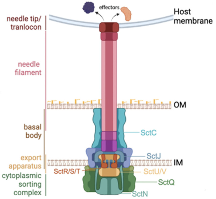
The injectisome is a macromolecule composed of more than 20 proteins [4,5] and consists of four components (Figure 1): (1) an extracellular part comprising a needle and tip/translocon complex, (2) a basal body of the syringe, (3) an export apparatus formed by inner membrane proteins, and (4) a cytoplasmic complex. Among these, the basal body and export apparatus are well conserved across a wide range of Gram-negative bacteria and homologous to bacterial flagellar components [6]. To refer to these injectisome proteins, we use the unified nomenclature of Sct for “Secretion and cellular translocation” [2]. Among around 20 proteins, SctN is an ATPase that works as the secretion pump and is usually most conserved. SctQ (cytoplasmic ring), SctR-S-T-U-V (export apparatus), SctJ (inner membrane ring) and SctC (outer membrane ring) are also well conserved proteins.
Figure 1.
An overview of T3SS core proteins and the bacterial membranes.
Based on the sequence similarity of these proteins, T3SS is classified into nine families across several bacterial phyla [6,7]: Ysc, Inv/Mxi-Spa, Ssa-Esc, Hrp/Hrc1, Hrp/Hrc2, Rhizobiales, Chlamydiales, Myxococcales, and Desulfovibrionales (Table 1). These family names originate in species names or secreted protein names required for infection.
Table 1.
| T3SS Family | Known Members | Known Relevant Hosts |
|---|---|---|
| Ysc |
Yersinia enterocolitica
Yersinia pestis Yersinia pseudotuberculosis |
Humans, rodents, insects |
|
Pseudomonas aeruginosa
Pseudomonas mosselii Pseudomonas otitidis |
Humans, plants, insects | |
| Aeromonas spp. | Humans, fish | |
|
Vibrio alginolyticus
Vibrio harveyi Vibrio parahaemolyticus |
Humans, fish, mollusks | |
| Inv/Mxi-Spa | Yersinia ruckeri | Salmonid fish |
|
Salmonella enterica
Salmonella bongori |
Humans | |
| Shigella boydii | Humans | |
| Burkholderia spp. | Mammals | |
| Ssa-Esc | Salmonella enterica | Humans |
|
Yersinia pseudotuberculosis
Yersinia enterocolitica Yersinia kristensenii |
Humans, rodents | |
| Edwardsiella ictaluri | Humans, fish | |
|
Vibrio aquimaris
Vibrio coralliilyticus Vibrio pectenicida |
Fish, mollusks | |
| Hrp/Hrc1 | Erwinia amylovora | Plants |
| Pantoea agglomerans | Plants | |
| Pseudomonas syringae | Plants | |
|
Vibrio cholerae
Vibrio parahaemolyticus Vibrio pectenicida |
Humans, fish, mollusks | |
| Hrp/Hrc2 | Ralstonia solanacearum | Plants |
| Xanthomonas spp. | Plants | |
| Burkholderia spp. | Plants | |
| Chlamydiales |
Chlamydia muridarum
Chlamydophila pneumoniae |
Mammals, birds, protists |
| Rhizobiales | Rhizobium spp. | Leguminous plants |
| Myxococcales | Myxococcus spp. | |
| Desulfovibrionales | Desulfovibrio vulgaris | Humans |
The first family Ysc comes from secreted Yersinia outer-membrane proteins. In Yersinia, T3SS is often encoded in a plasmid, and similar T3SSs in the Ysc family have been observed in two bacterial phyla, Alphaproteobacteria (e.g., Bordetella) and Gammaproteobacteria (Yersinia, Pseudomonas, Vibrio and others). Previously, T3SS of Desulfovibrio vulgaris was classified in this Ysc family but now it has become independent [8]. Upon infection, Yersinia pseudotuberculosis and Yersinia enterocolitica are known to cross the epithelium lining and stay extracellular in the mesenteric lymph nodes. T3SS effector proteins are used to avoid phagocytosis [9]. The second family, Inv/Mxi-Spa, comes from Inv-Spa and Mxi-Spa secreted protein complexes of Salmonella enterica and Shigella flexneri, respectively. This family is found in Betaproteobacteria (e.g. Burkholderia) and Gammaproteobacteria (Enterobacteriaceae). A well-known T3SS is encoded in Salmonella pathogenicity island 1 (SPI-1) of its chromosome and is associated with the invasion to host epithelial cells. The T3SS in Shigella is encoded in a large virulence plasmid of 230 kb. The third family, Ssa-Esc, indicates T3SSs in the SPI-2 region of Salmonella, in the locus of enterocyte effacement (LEE) regions of enteropathogenic/enterohemorrhagic Escherichia coli (EPEC/EHEC). In Salmonella, this T3SS is associated with later stages of infection to survive inside host cells and its mechanism is well investigated. After cell invasion, Salmonella forms a SCV (Salmonella-containing vacuole) and starts replication inside this vacuole by modulating the host cell using T3SS effectors [9]. On the other hand, EPEC/EHEC uses a single T3SS for infection and remains extracellular as Yersinia does [9].
The fourth and fifth families, Hrp/Hrc1 and Hrp/Hrc2, are found in plant pathogens such as Pseudomonas syringae and Burkholderia pseudomallei. The family names come from gene function as “Hypersensitive response and pathogenicity/conserved”. T3SSs in these families possess a longer injectisome for penetrating thick plant cell walls. Interestingly, some clinically isolated Vibrio species, including V. cholerae and V. parahaemolyticus, possess the second T3SS in this Hrp/Hrc1 family. Their length of injectisome has not been well documented.
The remaining four T3SS families (Rhizobiales, Chlamydiales, Myxococcales, and Desulfovibrionales) are order names identified through phylogenetic analyses. Among them, Rhizobium and Myxococcus are Alpha- and Deltaproteobacteria, respectively, while Chlamydia and Desulfovibrio belong to different phyla, i.e., Chlamydiota and Thermodesulfobacteria, respectively. They are outside of Pseudomonadota (formerly Proteobacteria).
The presence of T3SS in Vibrio was first revealed in V. parahaemolyticus by whole genome sequencing [10] and later its distribution in some other Vibrio species was also confirmed. Most Vibrio species possess only one T3SS gene cluster, but V. parahaemolyticus as well as some others possess two [10,11]. Within V. parahaemolyticus, the ubiquitous T3SS, referred to as T3SS1, is most similar to the Yersinia Ysc family and is encoded in its major chromosome. The other one, called T3SS2, is similar to the Hrp/Hrc1 family. The latter is non-essential and is encoded in a pathogenicity island called VpaI-7 in V. parahaemolyticus. The island is 81 kb in length and on the auxiliary chromosome, and is characterized by the presence of multiple transposase genes in contrast to the integrase genes found in the remaining 6 pathogenicity islands (VpaI-1~6) [12]. Both T3SSs of V. parahaemolyticus are associated with pathogenicity: T3SS1 is responsible for the cytotoxicity in cultured human cells, while T3SS2 is known to induce enterotoxicity in animal infection models and cytotoxic activity in intestinal cell lines [13,14]. So far, all Vibrio T3SSs have been phylogenetically classified into the above two types: one is similar to T3SS1 in the Ysc family and the other is similar to T3SS2 in the Hrp/Hrc1 family [7].
Here, we report the third type of Vibrio T3SS, named T3SS3, which belongs to the Ssa-Esc family and is most similar to the T3SSs in Salmonella, Sodalis and Shewanella. Among the T3SSs in these bacteria, the most well studied is the one located in the Salmonella SPI-2 pathogenicity island. The SPI-2 T3SS translocates its effectors across the membrane of SCVs after internalization, and they link the vacuole to the Golgi network of the host cell [15]. The effectors used in this process were also found in strains with T3SS3 investigated in this study.
2. Materials and Methods
2.1. Conservation Analysis
The initial dataset of T3SS core proteins was obtained from the work of Hu et al. [16]. All protein sequences were scanned for common domains using Pfam [17] and those without the common domain or those on plasmids were removed manually. To obtain T3SS protein sequences in Vibrio species comprehensively, the obtained T3SS core sequences were searched against Vibrio genomes in GenBank/ENA/DDBJ and sequences with >80% identity were selected. At this stage the sequence coverage was not considered. Then, the sequences were aligned using MAFFT [18], trimmed using trimAl [19]. The conservation rate of aligned sequences was calculated using MstatX [20].
2.2. Phylogenetic Analysis
Sequences of the most conserved core proteins (SctN, SctS, SctU and SctV) were aligned using MAFFT and concatenated using catfasta2phyml [21]. A phylogenetic tree of the concatenated core sequences was created using RAxML with a bootstrap value of 1000 [22]. The result was visualized using iTOL [23]. Branches of less than 70 bootstrap scores were deleted.
2.3. Prediction of Putative Type III Secretion Effectors (T3SEs)
Protein sequences in the twelve T3SS3 clusters were binned into homologous groups using SonicParanoid [24]. The sequences of each homologous group were aligned using MAFFT and their HMMER profile was generated. Experimentally validated T3SEs were collected from previous studies [7,15] and from the Virulence Factor Database (VFDB: http://www.mgc.ac.cn/VFs/main.htm accessed on 17 January 2023) online [25]. Protein sequences of Aliivibrio fischeri ES114 and Escherichia coli K-12 were used as the negative control set as they lack functional T3SS but possess flagella. Then, the HMMER profiles were scanned against the validated T3SEs and the negative control. Homologous groups that had no positive hit against the T3SEs dataset (E-value > 1 × 10⁻³) were dismissed from further analysis. Protein sequences in the homologous groups, whose HMMER profiles had at least one positive hit in the T3SEs dataset, were again searched in the T3SEs dataset using BLASTp. The Python script for HMMER and BLASTp evaluation was adopted from a previous study [26].
Homologous sequences that had no BLASTp hit against the negative control dataset were classified as true positives and those that had no hit against the T3SEs dataset were classified as false positives. The sequences with hits in both datasets were further classified as follows. If a sequence had three or more hits in both datasets, a t-test was performed to evaluate the significance of sequence similarity to the T3SEs dataset in comparison with the negative control dataset (p < 0.05). If the number of hits was less than three in either of the datasets, the bitscore of BLASTp was used. If the lowest bitscore against theT3SE alignment was at least 1.5-fold larger than the maximum bitscore against the negative-control alignment, the sequence was considered true positive, and otherwise, false positive. The parameters of this pipeline were determined so that known effectors in T3SS1 and T3SS2 clusters were accurately classified as true positives.
2.4. Genomic Island Prediction and Gene Order Analysis
A dataset of 12 different Vibrio T3SS clusters (4 each from T3SS1, T3SS2, and T3SS3) was used. Genomic islands in the Vibrio genomes were predicted using IslandViewer 4 [27] and their T3SS gene clusters were assigned to their corresponding positions within the genomes. Then, T3SS gene clusters in all genomes were reannotated using Prokka with default parameters [28] and the annotations were manually revised to fit the unified nomenclature of T3SS proteins and some of them were abbreviated to fit in the figures. In addition, transposase, integrase, and phage-related genes were manually searched within 50-kb regions. The order and orthology of genes in each T3SS gene cluster was visualized using Clinker [29].
3. Results
3.1. Identification of T3SS3 Clusters from Protein Similarity
We first searched the set of 10 conserved proteins (SctC, D, J, N, Q, R, S, T, U, V) against the publicly available genomes, and identified 208 bacterial strains that possess all of them (Supplementary Table S1). Among the 10 proteins, SctD was excluded from further analyses due to the lack of its complete sequence in many bacterial strains. T3SS clusters that were confirmed to be located on a plasmid were also excluded to keep the phylogenetic analysis evolutionarily clearer. T3SS-possessing strains including Yersinia spp., D. vulgaris, Grimontia hollisae and Vibrio mimicus were excluded from our analysis at this stage.
From the remaining strain set, only one strain was kept for each species to eliminate duplicate information and to reduce the volume of phylogeny. Thus, we obtained a dataset of 9 core proteins from 12 Vibrio T3SS clusters (two sets from V. pectenicida 99-46-Y, V. aquimaris strain THAF100, V. coralliilyticus OCN008, two sets from V. parahaemolyticus RIMD 2210633, V. cholerae strain SA3G, V. tubiashii ATCC19109, V. harveyi E385, V. alginolyticus NBRC 15630, undefined Vibrio spp. THAF191c and 2521-89) and the other bacteria (Supplementary Table S2). The amino acid sequence conservation rates of the four core proteins (SctN, SctS, SctU and SctV) were > 35% in this dataset, whereas the conservation rate of three proteins (SctC, SctJ and SctQ) were lower.
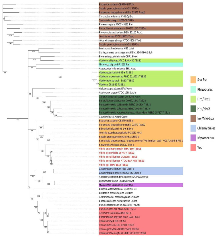
To create a reliable phylogenetic tree that represents the evolutionary picture of T3SS clusters, concatenated sequences of highly conserved proteins (SctN, SctS, SctU and SctV of >35% identity) were used to classify T3SS families for the 48 bacterial species. The resulting tree was confirmed to be compatible with previous studies (Figure 2) [6,8].
Figure 2.
Phylogenetic tree of concatenated SctN-SctS-SctU-SctV proteins for bacteria with T3SS. Vibrio strains possessing a T3SS3 cluster are marked in red. V. coralliilyticus ATCC BAA-450 was added, in addition to the well-studied OCN008 strain, as the type strain possessing T3SS. Background colors show 8 T3SS families previously proposed for a subset of species analyzed [6,8].
In the phylogenetic tree, upstream branches were not stable, but the Vib-T3SS1 and Vib-T3SS2 were found within the Ysc and Hrp/Hrc1 families, respectively, as previously reported. In addition, sequences from 4 Vibrio strains (V. pectenicida 99-46-Y, V. aquimaris strain THAF100, V. coralliilyticus OCN008, unidentified strain THAF191c) were located within the Ssa-Esc family. These four strains were all isolated from seashells or corals and not from human patients. This new T3SS family is called T3SS3 in this report. To explicitly specify the source organism of these clusters, we also use the notation Vib-T3SS1/2/3.
3.2. Genomic Location and Gene Order of T3SS3 Gene Clusters
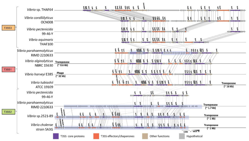
Within Vibrio genomes, T3SS1 and T3SS3 clusters were located outside of genomic islands on the larger chromosome (chromosome I) according to the prediction by IslandViewer 4 (see Methods). In contrast, the T3SS2 clusters were mostly located within a genomic island, probably on chromosome II. For example, V. parahaemolyticus possesses its T3SS1 cluster on chromosome I, and its T3SS2 cluster in a genomic island on chromosome II (Figure 3). A possible exception was V. pectenicida 99-46-Y, whose T3SS2 cluster was not associated with transposase and was not located within the genomic island, although it should be noted that the V. pectenicida genome was not closed.
Figure 3.
Gene orders of the T3SS1/2/3 clusters. Arrow direction and color correspond to respective coding strand and gene function, respectively. Arrow size reflects amino acid length, and the lines between arrows represent orthologs as computed by the Clinker software (see Methods). Genomic island is visualized as a blue shade. Genomic positions are shown in Supplementary Table S3.
Within each of the T3SS1/2/3 clusters, the injectisome genes and secretory genes exhibited well conserved ordering, with only a handful of insertions or deletions (Figure 3). In particular, the genes encoding SctC-SctD (basal body) and SctR-SctS-SctT-SctU (export apparatus) were found in exactly the same consecutive order in all the investigated T3SS1 and T3SS3 sequences, but not in the T3SS2 sequences. The gene order of T3SS2 clusters was shuffled entirely when compared with that of T3SS1 or T3SS3 clusters. Three out of four T3SS2 sequences were computationally predicted to be on genomic islands, but the gene order and composition were very well conserved within the T3SS2 group.
In contrast to the different ordering and less sequence similarity of core injectisome proteins between T3SS1 and T3SS2, they shared a similar repertoire for effector proteins. Vop (Vibrio outer membrane) proteins dominate among the predicted gene functions in T3SS1 and T3SS2 clusters. On the other hand, effectors in T3SS3 were mostly similar to Sse proteins for Salmonella secretory effectors and no Vop annotation was assigned in the neighborhood of injectisome core proteins.
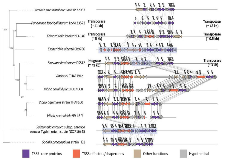
The gene order in Vib-T3SS3 was roughly conserved in several other bacteria of different orders in the Ssa-Esc family: Shewanella violacea DSS12 in Shewanellaceae (Alteromonadales), Salmonella enterica subsp. enterica serovar Typhimurium strain NCCP16345 in Enterobacteriaceae (Enterobacterales), and Sodalis praecaptivus strain HS1 in Pectobacteriaceae (Enterobacterales) (Figure 4). In these strains, the conservation of gene order spanned not only for core injectisome proteins (SctC-SctD and SctR-SctS-SctT-SctU), but also for translocons (SseB-SseC-SseD) and chaperones (CesD-IpgC). However, the clusters of Escherichia albertii CB9786, Edwardsiella icutaluri 93-146 in Enterobacteriaceae, and Pandoraea faecigallinarum DSM 23572 in Burkholderiaceae exhibited different gene ordering. They also did not share Salmonella effectors or translocons.
Figure 4.
Phylogenetic tree of concatenated SctN-SctS-SctU-SctV proteins and the gene order of the Ssa-Esc family. Graphical notation is the same as Figure 3.
Regarding genomic location, the T3SS genes in S. violacea DSS12, S. Typhimurium strain NCCP16345 and E. albertii CB9786 were found on a genomic island. In S. violacea DSS12, the cluster was sided by integrase and transposase genes. For the investigated Vib-T3SS3 clusters, no transposase/integrate genes were found in their 50 kb neighborhood.
3.3. Prediction of Putative T3SS3 Effectors, Translocons, and Chaperones
The gene order analysis focused on the neighborhood of injectisome genes, but effectors are often encoded outside of the cluster. In order to confirm the similarity of T3SS effectors (T3SEs) between Vib-T3SS3 and those in Salmonella spp., the sequences of experimentally validated effectors, translocons and chaperones were collected from previous studies [7,15,25] (see Methods). A dataset of 205 experimentally validated T3SEs and related proteins (Supplementary Table S4) was obtained.
Two hundred and five proteins were searched against the HMMER profiles of 3673 homologous gene groups created from protein sequences in the four Vibrio genomes with T3SS3 clusters, and 35 groups showed significant hits (E-value: <1 ×10⁻³). In order to minimize the number of false positives, the protein sequences of Aliivibrio fischeri and Escherichia coli K-12 were used as the negative control (see Methods). The combination of BLASTp searches of the 35 homologous groups against both 205 positive- and the negative-control datasets identified 14 true-positive protein groups (Table 2 and Supplementary Table S5). Eleven out of the 14 groups were similar to effectors, translocons, or chaperones in the Ssa-Esc family, just like the core T3SS proteins of T3SS3. The remaining few sequences were common with proteins in the Ysc or Inv/Mxi-Spa families. Salmonella translocons (SseB, SseC, SseD), EPEC/EHEC chaperone (CesD), and some core effectors in Salmonella (SseF, PipB) were conserved across Vib-T3SS3 strains. Not all the core Salmonella effectors, however, were detected in Vib-T3SS3 clusters in our analysis. For example, Jennings et al. defined seven core Salmonella effectors shared by all serovars (SseF, SseG, PipB, SteA, SteD and PipB2) [3], but the majority were completely absent from these Vibrio species (Table 2).
Table 2.
Putative T3SS3 effectors, translocons, and chaperones. The ‘Average identity’ column shows amino acid conservation rates in the Vib-T3SS3 strains. The letters in the ‘Strains’ column indicate the presence of a homolog (A: V. aquimaris strain THAF100; C: V. coralliilyticus OCN008; P: V. pectenicida 99-46-Y; and T: Vibrio sp. THAF191c).
| Protein Names | T3SS Family | Average Identity % | Strains | Biochemical and Cellular Functions |
|---|---|---|---|---|
| SseF | Ssa-Esc | 29.5 | A, C, P | Core effector in all pathogenic Salmonella serovars, tethering Salmonella-containing vacuoles (SCVs) to the Golgi network. SseF functions with another core effector, SseG protein [3]. |
| SspH2 | Ssa-Esc | 32.5 | T | Effector (E3 ubiquitin ligase) in pathogenic Salmonella, interfering with host immune signaling. SspH2 is associated with SspH1 and SlrP, which also show E3 ligase activity [3]. |
| SseJ | Ssa-Esc | 33.4 | A, P | Effector (acytransferase) in pathogenic Salmonella, preventing collapse of microtubules to provide a solid network around SCVs [3,30]. |
| PipB | Ssa-Esc | 32.4 | A, C, P, T | Core effector of unknown function in all pathogenic Salmonella serovars, localizing to SCVs and SIFs. Its associated protein PipB2 controls the kinesin-1 motor protein of host cells [3]. |
| SopD2 | Ssa-Esc | 33.3 | A, C | Effector that prevents from directing SCVs into late endosomes and lysosomes [3,30] |
| SopD | Ssa-Esc Inv/Mxi-Spa |
18.9 | A | Effector that promotes plasma membrane scission and the generation of SCVs [30] |
| CesD | Ssa-Esc | 39.5 25.4 |
A, C, P, T | T3SS chaperone in enteropathogenic E. coli strains for more efficient secretion [31,32] |
| CesT | C, T | |||
| SseB | Ssa-Esc | 37.7 30.3 28.4 |
A, C, P, T | Translocon proteins in Salmonella strains to transfer T3SS effectors [33] |
| SseC | ||||
| SseD | ||||
| BopA/IcsB | Inv/Mxi-Spa | 23.4 | C, T | Effector in Shigella or Burkholderia strains helping to evade the host autophagy defense system [34] |
| BopC | Inv/Mxi-Spa | 44.5 | A, P | Effector in Bordetella strains contributing to the necrotic cell death of mammalian host cells [35] |
| ExoY | Ysc | 31.2 | A, P | Common effector (adenylate cyclase) in clinically isolated Pseudomonas aeruginosa, delaying the inflammatory pathways in mammalian host cells [36] |
| Scc2 | Chlamydiales | 27.5 | C | T3SS chaperone specific to Chlamydia species and is similar to SycD of Yersinia, SicA of Salmonella, and IpgC of Shigella [37] |
4. Discussion
4.1. Evolutionary Distance of T3SS3 from T3SS1 and T3SS2
Our bioinformatics analysis described the third variation of T3SS in Vibrio, here called T3SS3. From the sequence similarity of injectisome proteins, the new variant belongs to the Ssa-Esc family, and its T3SS effectors are also more similar to those in Salmonella spp. than those in previously identified T3SS1 and T3SS2 effectors (Table 2). Additionally, from the gene ordering, T3SS3 resembled the Ssa-Esc family. Between Vibrio species, T3SS3 and T3SS1 are more closely related in terms of sequence similarity and gene order in comparison with T3SS2, which resides in genomic islands (Figure 3). The T3SS2 cluster in V. pectenicida 99-46-Y was not predicted within a genomic island in our analysis, but this is probably a consequence of incomplete genome information.
Within the Ssa-Esc family, the T3SS3 clusters showed a gene composition and ordering similar to intestinal pathogens such as Salmonella and Sodalis (Figure 4, Table 2). In addition to SPI-2 T3SS in Salmonella, one representative T3SS cluster in the Ssa-Esc family is that of enteropathogenic E. albertii CB9786 [38]. This cluster resides within a LEE pathogenicity island in the chromosome and contributes to its pathogenicity. Its T3SS gene order is very different from those of Vib-T3SS3 clusters as well as Salmonella, Sodalis, and Shewanella strains in the Ssa-Esc family. This cannot be easily explained as the result of shuffling in the genomic island, because T3SS clusters in Salmonella and Shewanella are also within genomic islands. This highlights an interesting similarity between these strains and T3SS3-possessing Vibrio strains across different bacterial phyla.
Our analysis relies on the phylogenetic tree created from the four most conserved proteins (SctN, SctS, SctU and SctV) of the injectisome across bacteria with T3SS. The SctN is the ATPase to translocate effector proteins through the narrow T3SS syringe and is the most conserved component of the entire system (Figure 1). The remaining three proteins compose the export apparatus and their amino acid conservation rate across strains was >35%. The phylogenetic analysis supported the previously proposed classification of T3SS families, with Vib-T3SS1 and Vib-T3SS2 clusters being clustered within the Ysc and Hrp/Hrc1 families, respectively (Figure 2). Hu et al. [16] presented a different classification based on the phylogeny of a set of core T3SS proteins and their conserved syntenic orders. The resulting 13 classes were not compatible with the traditional T3SS families, but also exhibited a unique position of Vibrio coralliilyticus RE98 within the Escherichia LEE and Salmonella SPI-2 group, corresponding to our T3SS3. We created our phylogeny to keep its consistency with known T3SS families for easier functional studies. Our tree shape kept a similar classification, even if we added a few more conserved proteins (e.g., SctR), but the clean clustering was lost when we used all 10 conserved proteins.
4.2. Effect of Mobile Elements and Horizontal Gene Transfer
Gene order and location are important indicators of genetic exchange in bacteria. Because the computational prediction of genomic islands might not always be accurate, we manually searched mobile element-related genes within ~50-kb regions around each T3SS gene cluster in this study. Within this range, all T3SS3 clusters were free from transposes or integrases, whereas the cluster in S. violacea DSS12 was associated with mobile genes. The cluster in S. Typhimurium strain NCCP16345 was also mobile gene-free, but the entire cluster was known to be within the SPI2 pathogenicity island. Although our detailed analysis surveyed only a handful of genomes, this suggests that the T3SS3 clusters may be horizontally transferred between Vibrio and other bacterial species such as Salmonella and Shewanella sharing a common habitat. Indeed, the food poisoning pathogen S. Typhimurium is occasionally found in molluscan shellfish such as mussels and clams [39]. Similarly, Shewanella is found in marine environments as a symbiont of squid and corals [40]. Vibrio species also live in marine and freshwater environments, and Vibrio strains with T3SS3 clusters have been isolated from mollusks and corals. The direction of the horizontal gene transfer, however, is difficult to clarify only from the current bacterial genomes.
4.3. Repertoire of Effectors, Translocons, and Chaperones in T3SS3
To gain more understanding of the infection mechanism of strains with T3SS3, we selected candidate T3SS3 effectors in a rather strict way (Table 2). Most of the predicted T3SS3 proteins were homologous to those in Salmonella. Of note were the core effectors shared by multiple strains with T3SS3, e.g., the orthologs of SseF and PipB proteins in Salmonella. These effectors are associated with Salmonella-containing vacuoles (SCVs) and Salmonella-induced filaments (SIFs); they contribute to the intracellular life of Salmonella in host cells. This was unexpected because well-known Vibrio species such as V. parahaemolyticus and V. cholerae are historically considered extracellular pathogens [7,41]. There has been no report for species with T3SS3 that suggests they internalize into host cells.
The list of candidate effectors alone, however, is insufficient to suggest the infection pathway of the strains with T3SS3. The selected 15 putative protein groups (effectors, translocons and chaperones) are not the full set but rather a partial list. To clarify the true function of each protein, experimental evidence is essential. The number and type of effectors are known to differ even among the same species [3], and a recent report also indicates diversity of effectors in T3SS2 clusters [42]. Bioinformatics analysis can find many putative effectors as in our study, and their functional characterization through experiments is awaited [43].
In summary, we described a new variation of the T3SS gene cluster in Vibrio species. This group, T3SS3, shares core and related proteins with Salmonella and other enteropathogenic species that infect a wide range of marine and terrestrial hosts. Of note is the similarity of translocons, chaperones, and effectors to those in Salmonella, although the mode of infection is considered very different between Vibrio and Salmonella. Further experimental analyses on its effectors will provide more information about their functions and mechanisms of action in comparison with their Ssa-Esc counterpart in other bacterial species.
Acknowledgments
Computational work was performed on the NIG supercomputer at the Research Organization of Information and Systems.
Supplementary Materials
The following supporting information can be downloaded at: https://www.mdpi.com/article/10.3390/microorganisms11020290/s1. Supplementary Table S1: Genomes containing T3SS; Table S2: T3SS core proteins; Table S3: genomic positions of T3SS; Table S4: validated effectors; Table S5: putative T3SS3 effectors.
Author Contributions
D.Z., M.A., S.M. and T.I. conceived the research outline. D.Z. conducted the data acquisition and initial analysis and wrote the first version of the manuscript. T.H. reviewed the initial results and contributed critical information. All authors have read and agreed to the published version of the manuscript.
Institutional Review Board Statement
Not applicable.
Informed Consent Statement
Not applicable.
Data Availability Statement
All genetic data are available from the GenBank/ENA/DDBJ repository. Accession numbers of all strains are summarized in Supplementary Table S1.
Conflicts of Interest
The authors declare no conflict of interest.
Funding Statement
This research was funded by KAKENHI, Japan (AMED 17940819; NBDC Togo Database).
Footnotes
Disclaimer/Publisher’s Note: The statements, opinions and data contained in all publications are solely those of the individual author(s) and contributor(s) and not of MDPI and/or the editor(s). MDPI and/or the editor(s) disclaim responsibility for any injury to people or property resulting from any ideas, methods, instructions or products referred to in the content.
References
- 1.Baker-Austin C., Trinanes J., Gonzalez-Escalona N., Martinez-Urtaza J. Non-cholera vibrios: The microbial barometer of climate change. Trends Microbiol. 2017;25:76–84. doi: 10.1016/j.tim.2016.09.008. [DOI] [PubMed] [Google Scholar]
- 2.Galán J.E., Lara-Tejero M., Marlovits T.C., Wagner S. Bacterial type III secretion systems: Specialized nanomachines for protein delivery into target cells. Ann. Rev. Microbiol. 2014;68:415–438. doi: 10.1146/annurev-micro-092412-155725. [DOI] [PMC free article] [PubMed] [Google Scholar]
- 3.Jennings E., Thurston TL M., Holden D.W. Salmonella SPI-2 type III secretion system effectors: Molecular mechanisms and physiological consequences. Cell Host Microbe. 2017;22:217–231. doi: 10.1016/j.chom.2017.07.009. [DOI] [PubMed] [Google Scholar]
- 4.Deng W., Marshall N.C., Rowland J.L., McCoy J.M., Worrall L.J., Santos A.S., Strynadka N.C., Finlay B.B. Assembly, structure, function and regulation of type III secretion systems. Nat. Rev. Microbiol. 2017;15:323–337. doi: 10.1038/nrmicro.2017.20. [DOI] [PubMed] [Google Scholar]
- 5.Wagner S., Grin I., Malmsheimer S., Singh N., Torres-Vargas C.E., Westerhausen S. Bacterial type III secretion systems: A complex device for the delivery of bacterial effector proteins into eukaryotic host cells. FEMS Microbiol. Lett. 2018;365:fny201. doi: 10.1093/femsle/fny201. [DOI] [PMC free article] [PubMed] [Google Scholar]
- 6.Abby S.S., Rocha EP C. The non-flagellar type III secretion system evolved from the bacterial flagellum and diversified into host-cell adapted systems. PLoS Genet. 2012;8:e1002983. doi: 10.1371/journal.pgen.1002983. [DOI] [PMC free article] [PubMed] [Google Scholar]
- 7.Matsuda S., Hiyoshi H., Tandhavanant S., Kodama T. Advances on Vibrio parahaemolyticus research in the postgenomic era. Microbiol. Immunol. 2020;64:167–181. doi: 10.1111/1348-0421.12767. [DOI] [PubMed] [Google Scholar]
- 8.Troisfontaines P., Cornelis G.R. Type III secretion: More systems than you think. Physiology. 2005;20:326–339. doi: 10.1152/physiol.00011.2005. [DOI] [PubMed] [Google Scholar]
- 9.Pinaud L., Sansonetti P.J., Phalipon A. Host cell targeting by enteropathogenic bacteria T3SS effectors. Trends Microbiol. 2018;26:266–283. doi: 10.1016/j.tim.2018.01.010. [DOI] [PubMed] [Google Scholar]
- 10.Makino K., Oshima K., Kurokawa K., Yokoyama K., Uda T., Tagomori K., Iijima Y., Najima M., Nakano M., Yamashita A., et al. Genome sequence of Vibrio parahaemolyticus: A pathogenic mechanism distinct from that of V. cholerae. Lancet. 2003;361:743–749. doi: 10.1016/S0140-6736(03)12659-1. [DOI] [PubMed] [Google Scholar]
- 11.Miller K.A., Tomberlin K.F., Dziejman M. Vibrio variations on a type three theme. Curr. Opin. Microbiol. 2019;47:66–73. doi: 10.1016/j.mib.2018.12.001. [DOI] [PMC free article] [PubMed] [Google Scholar]
- 12.Hurley C.C., Quirke A., Reen F.J., Boyd E.F. Four genomic islands that mark post-1995 pandemic Vibrio parahaemolyticus isolates. BMC Genom. 2006;7:104. doi: 10.1186/1471-2164-7-104. [DOI] [PMC free article] [PubMed] [Google Scholar]
- 13.Ham H., Orth K. The role of type III secretion system 2 in Vibrio parahaemolyticus pathogenicity. J. Microbiol. 2012;50:719–725. doi: 10.1007/s12275-012-2550-2. [DOI] [PubMed] [Google Scholar]
- 14.Yang H., de Santos M.S., Lee J., Law H.T., Chimalapati S., Verdu E.F., Orth K., Vallance B.A. A novel mouse model of enteric Vibrio parahaemolyticus infection reveals that the type III secretion system 2 effector VopC plays a key role in tissue invasion and gastroenteritis. mBio. 2019;10:e02608-19. doi: 10.1128/mBio.02608-19. [DOI] [PMC free article] [PubMed] [Google Scholar]
- 15.Figueira R., Holden D.W. Functions of the Salmonella pathogenicity island 2 (SPI-2) type III secretion system effectors. Microbiology. 2012;158:1147–1161. doi: 10.1099/mic.0.058115-0. [DOI] [PubMed] [Google Scholar]
- 16.Hu Y., Huang H., Cheng X., Shu X., White A.P., Stavrinides J., Köster W., Zhu G., Zhao Z., Wang Y. A global survey of bacterial type III secretion systems and their effectors. Environ. Microbiol. 2017;19:3879–3895. doi: 10.1111/1462-2920.13755. [DOI] [PubMed] [Google Scholar]
- 17.Mistry J., Chuguransky S., Williams L., Qureshi M., Salazar G.A., Sonnhammer E.L., Tosatto S.C., Paladin L., Raj S., Richardson L.J., et al. Pfam: The protein families database in 2021. Nucleic Acids Res. 2021;49:D412–D419. doi: 10.1093/nar/gkaa913. [DOI] [PMC free article] [PubMed] [Google Scholar]
- 18.Katoh K., Rozewicki J., Yamada K.D. MAFFT online service: Multiple sequence alignment, interactive sequence choice and visualization. Briefings Bioinform. 2019;20:1160–1166. doi: 10.1093/bib/bbx108. [DOI] [PMC free article] [PubMed] [Google Scholar]
- 19.Capella-Gutierrez S., Silla-Martinez J.M., Gabaldon T. Trimal: A tool for automated alignment trimming in large-scale phylogenetic analyses. Bioinformatics. 2009;25:1972–1973. doi: 10.1093/bioinformatics/btp348. [DOI] [PMC free article] [PubMed] [Google Scholar]
- 20.Collet G. Gcollet/Mstatx: A multiple alignment analyser. GitHub. Dec 3, 2017. [(accessed on 16 November 2021)]. Available online: https://github.com/gcollet/MstatX.
- 21.Nylander J.A.A. Nylander/Catfasta2phyml: Concatenates FASTA formatted files to “Phyml” (PHYLIP) format. GitHub. Sep 28, 2018. [(accessed on 16 November 2021)]. Available online: https://github.com/nylander/catfasta2phyml.
- 22.Stamatakis A. RAxML version 8: A tool for phylogenetic analysis and post-analysis of large phylogenies. Bioinformatics. 2014;30:1312–1313. doi: 10.1093/bioinformatics/btu033. [DOI] [PMC free article] [PubMed] [Google Scholar]
- 23.Letunic I., Bork P. Interactive tree of life (iTol) V4: Recent updates and new developments. Nucleic Acids Res. 2019;47:W256–W259. doi: 10.1093/nar/gkz239. [DOI] [PMC free article] [PubMed] [Google Scholar]
- 24.Cosentino S., Iwasaki W. SonicParanoid: Fast, accurate and easy orthology inference. Bioinformatics. 2019;35:149–151. doi: 10.1093/bioinformatics/bty631. [DOI] [PMC free article] [PubMed] [Google Scholar]
- 25.Liu B., Zheng D., Jin Q., Chen L., Yang J. VFDB 2019: A comparative pathogenomic platform with an interactive web interface. Nucleic Acids Res. 2019;47:D687–D692. doi: 10.1093/nar/gky1080. [DOI] [PMC free article] [PubMed] [Google Scholar]
- 26.Rangel L.T., Marden J., Colston S., Setubal J.C., Graf J., Gogarten J.P. Identification and characterization of putative Aeromonas spp. T3SS effectors. PLoS ONE. 2019;14:e0214035. doi: 10.1371/journal.pone.0214035. [DOI] [PMC free article] [PubMed] [Google Scholar]
- 27.Bertelli C., Laird M.R., Williams K.P., Lau B.Y., Hoad G., Winsor G.L., Brinkman F.S.L. IslandViewer 4: Expanded prediction of genomic islands for larger-scale datasets. Nucleic Acids Res. 2017;45:W30–W35. doi: 10.1093/nar/gkx343. [DOI] [PMC free article] [PubMed] [Google Scholar]
- 28.Seemann T. Prokka: Rapid prokaryotic genome annotation. Bioinformatics. 2014;30:2068–2069. doi: 10.1093/bioinformatics/btu153. [DOI] [PubMed] [Google Scholar]
- 29.Gilchrist C.L., Chooi Y.-H. Clinker Clustermap.js: Automatic generation of gene cluster comparison figures. Bioinformatics. 2021;37:2473–2475. doi: 10.1093/bioinformatics/btab007. [DOI] [PubMed] [Google Scholar]
- 30.Boddy K.C., Zhu H., D’Costa V.M., Xu C., Beyrakhova K., Cygler M., Grinstein S., Coyaud E., Laurent E.M., St-Germain J., et al. Salmonella effector SopD promotes plasma membrane scission by inhibiting Rab10. Nat. Commun. 2021;12:4047. doi: 10.1038/s41467-021-24983-z. [DOI] [PMC free article] [PubMed] [Google Scholar]
- 31.Thomas N.A., Deng W., Puente J.L., Frey E.A., Yip C.K., Strynadka N.C., Finlay B.B. CesT is a multi-effector chaperone and recruitment factor required for the efficient type III secretion of both Lee- and non-Lee-encoded effectors of enteropathogenic Escherichia coli. Mol. Microbiol. 2005;57:1762–1779. doi: 10.1111/j.1365-2958.2005.04802.x. [DOI] [PubMed] [Google Scholar]
- 32.MacDonald J., Miletic S., Gaildry T., Chin-Fatt A., Menassa R. Co-expression with the type 3 secretion chaperone CesT from enterohemorrhagic E. coli increases accumulation of recombinant Tir in plant chloroplasts. Front. Plant Sci. 2017;8:283. doi: 10.3389/fpls.2017.00283. [DOI] [PMC free article] [PubMed] [Google Scholar]
- 33.Hölzer S.U., Hensel M. Functional dissection of translocon proteins of the Salmonella pathogenicity island 2-encoded type III secretion system. BMC Microbiol. 2010;10:104. doi: 10.1186/1471-2180-10-104. [DOI] [PMC free article] [PubMed] [Google Scholar]
- 34.Kayath C.A., Hussey S., El Hajjami N., Nagra K., Philpott D., Allaoui A. Escape of intracellular Shigella from autophagy requires binding to cholesterol through the type III effector, IcsB. Microb. Infect. 2010;12:956–966. doi: 10.1016/j.micinf.2010.06.006. [DOI] [PubMed] [Google Scholar]
- 35.Kuwae A., Matsuzawa T., Ishikawa N., Abe H., Nonaka T., Fukuda H., Imajoh-Ohmi S., Abe A. BopC is a novel type III effector secreted by Bordetella bronchiseptica and has a critical role in type III-dependent necrotic cell death. J. Biol. Chem. 2006;281:6589–6600. doi: 10.1074/jbc.M512711200. [DOI] [PubMed] [Google Scholar]
- 36.Jeon J., Kim Y.-J., Shin H., Ha U.-H. T3SS effector ExoY reduces inflammasome-related responses by suppressing bacterial motility and delaying activation of NF-ΚB and caspase-1. FEBS J. 2017;284:3392–3403. doi: 10.1111/febs.14199. [DOI] [PubMed] [Google Scholar]
- 37.Fields K.A., Fischer E.R., Mead D.J., Hackstadt T. Analysis of putative Chlamydia trachomatis chaperones Scc2 and Scc3 and their use in the identification of type III secretion substrates. J. Bacteriol. 2005;187:6466–6478. doi: 10.1128/JB.187.18.6466-6478.2005. [DOI] [PMC free article] [PubMed] [Google Scholar]
- 38.Ooka T., Ogura Y., Katsura K., Seto K., Kobayashi H., Kawano K., Tokuoka E., Furukawa M., Harada S., Yoshino S., et al. Defining the genome features of Escherichia albertii, an emerging enteropathogen closely related to Escherichia coli. Genome Biol. Evol. 2015;7:3170–3179. doi: 10.1093/gbe/evv211. [DOI] [PMC free article] [PubMed] [Google Scholar]
- 39.Martinez-Urtaza J., Saco M., Hernandez-Cordova G., Lozano A., Garcia-Martin O., Espinosa J. Identification of Salmonella serovars isolated from live molluscan shellfish and their significance in the marine environment. J. Food Protect. 2003;66:226–232. doi: 10.4315/0362-028X-66.2.226. [DOI] [PubMed] [Google Scholar]
- 40.Alex A., Antunes A. Whole-genome comparisons among the genus Shewanella reveal the enrichment of genes encoding ankyrin-repeats containing proteins in sponge-associated bacteria. Front. Microbiol. 2019;10:5. doi: 10.3389/fmicb.2019.00005. [DOI] [PMC free article] [PubMed] [Google Scholar]
- 41.Ritchie J.M., Rui H., Zhou X., Iida T., Kodoma T., Ito S., Davis B.M., Bronson R.T., Waldor M.K. Inflammation and disintegration of intestinal villi in an experimental model for Vibrio parahaemolyticus-induced diarrhea. PLoS Pathog. 2012;8:e1002593. doi: 10.1371/journal.ppat.1002593. [DOI] [PMC free article] [PubMed] [Google Scholar]
- 42.Jerez S.A., Plaza N., Bravo V., Urrutia I.M., Blondel C.J. The Vibrio Type III secretion system 2 is not restricted to the Vibrionaceae and encodes differently distributed repertoires of effector proteins. bioRxiv. 2022 doi: 10.1101/2022.08.23.504659v3. [DOI] [PMC free article] [PubMed] [Google Scholar]
- 43.Hubbard T.P., Chao M.C., Abel S., Blondel C.J., Wiesch PA Z., Zhou X., Davis B.M., Waldor M.K. Genetic analysis of Vibrio parahaemolyticus intestinal colonization. Proc. Natl. Acad. Sci. USA. 2016;113:6283–6288. doi: 10.1073/pnas.1601718113. [DOI] [PMC free article] [PubMed] [Google Scholar]
Associated Data
This section collects any data citations, data availability statements, or supplementary materials included in this article.
Supplementary Materials
Data Availability Statement
All genetic data are available from the GenBank/ENA/DDBJ repository. Accession numbers of all strains are summarized in Supplementary Table S1.


