Abstract
A novel, in vitro bioassay for detection of the botulinum type B neurotoxin in a range of media was developed. The assay is amplified by the enzymic activity of the neurotoxin’s light chain and includes the following three stages: first, a small, monoclonal antibody-based immunoaffinity column captures the toxin; second, a peptide substrate is cleaved by using the endopeptidase activity of the type B neurotoxin; and finally, a modified enzyme-linked immunoassay system detects the peptide cleavage products. The assay is highly specific for type B neurotoxin and is capable of detecting type B toxin at a concentration of 5 pg ml−1 (0.5 mouse 50% lethal dose ml−1) in approximately 5 h. The format of the test was found to be suitable for detecting botulinum type B toxin in a range of foodstuffs with a sensitivity that exceeds the sensitivity of the mouse assay. Using highly specific monoclonal antibodies as the capture phase, we found that the endopeptidase assay was capable of differentiating between the type B neurotoxins produced by proteolytic and nonproteolytic strains of Clostridium botulinum type B.
Various strains of the bacterium Clostridium botulinum produce seven structurally related but antigenically different protein neurotoxins (botulinum neurotoxin type A [BoNT/A] to BoNT/G) which cause the syndrome botulism (8). The symptoms of this syndrome include widespread flaccid paralysis, which often results in death if the individual is not treated rapidly with antitoxin. There has been much effort by the food industry to ensure that food treatment processes prevent the growth of C. botulinum and toxin production by C. botulinum, and there is a need for rapid, sensitive, and specific assays for the C. botulinum toxins. At present, the only method which can be used with confidence to detect the toxins is the acute toxicity test performed with mice (9). Although this test is exquisitely sensitive, with a detection limit of 1 mouse 50% lethal dose (MLD50), which is equivalent to 10 to 20 pg of neurotoxin/ml, it has a number of drawbacks; it is expensive to perform, requires a large number of animals, and is not specific for the neurotoxin unless neutralization tests with a specific antiserum are carried out in parallel. In addition, the test takes up to 4 days to complete. The increasing resistance to animal tests has resulted in the development of alternative rapid in vitro assays that have the sensitivity and reliability of the mouse bioassay. A number of immunoassay systems with sensitivities comparable to the sensitivity of the mouse bioassay have been described (2, 16). These methods, however, require complicated, expensive amplification systems which have not become widely available. In addition, these immunoassays do not measure the biological activity of the neurotoxin and can lead to false-positive results.
Over the past 5 years significant progress has been made in deciphering the mode of action of the clostridial neurotoxins. It has been demonstrated that these toxins act at the cellular level as highly specific zinc endoproteases that cleave various isoforms of three small proteins which control the docking of the synaptic vesicles with the synaptic membrane. BoNT/A and BoNT/E specifically cleave the 25-kDa synaptosome-associated protein (SNAP-25) (1, 10, 13). BoNT/C cleaves the membrane protein syntaxin and SNAP-25 (3, 11). BoNT/B, BoNT/D, BoNT/F, and BoNT/G act on a different intracellular target, vesicle-associated membrane protein (VAMP) or synaptobrevin (10, 12, 13). BoNT/B cleaves VAMP at a single peptide bond between Gln-76 and Phe-77. Recent studies have shown that synthetic peptides of VAMP isoform 2 are also cleaved by BoNT/B (14, 15). These peptides have been exploited in the development of in vitro assays based on the cleavage of solid-phase immobilized peptide substrates by BoNT/B (6). While such assays are rapid and specific and include a measurement of the biological activity of the neurotoxin, they do not match the sensitivity of the mouse bioassay and are not realistic replacements. In addition, the stringent conditions required to support the endopeptidase activity of the neurotoxins is unlikely to be supported in matrices as diverse as food, sera, and feces (14). Here we describe an assay with a sensitivity that exceeds the sensitivity of the mouse bioassay, and the new bioassay is sufficiently robust to detect BoNT/B in a range of foodstuffs.
MATERIALS AND METHODS
Purification of BoNT/B.
C. botulinum Okra BoNT/B was purified from 200 liters of culture by ion-exchange chromatography as described previously (15). The toxin was dialyzed against 50 mM HEPES–0.15 M NaCl (pH 7.4) and stored at −80°C. The biological activities of toxins were assessed by the mouse bioassay as described previously (5, 9).
Production of hybridoma cell lines.
Hybridoma cell lines that secreted antibody specific for BoNT/B were generated by using purified strain Okra and the procedure described previously for BoNT/A (6).
Test cultures.
The strains used and their origins are shown in Table 1. Proteolytic and nonproteolytic type B cultures were grown in cooked meat carbohydrate medium (Oxoid, Basingstoke, United Kingdom) for 48 h at 37 and 30°C, respectively, before we assayed for the presence of BoNT/B.
TABLE 1.
Evaluation of monoclonal antibodies for detecting C. botulinum type B toxin with the ELISA
| Strain | Sourcea | Reactions with monoclonal antibodiesb
|
||||
|---|---|---|---|---|---|---|
| 5BB/25.3 | 5BB/21.3 | 5BB/9.3 | 5BB/4.3 | 3BB/110.3 | ||
| Proteolytic strains | ||||||
| Okrac | CAMR | 3.1 ± 0.9d | 7.4 ± 2.5 | 20.5 ± 11.8 | 104 ± 82 | 6.4 ± 3.1 |
| NCTC 7273 | NCTC | + | + | + | +/− | ND |
| Danish | DSI | + | + | + | + | ND |
| UN180284/1 | CAMR | + | + | + | +/− | ND |
| UN180284/2 | CAMR | + | + | + | + | ND |
| 53B | NFPA | + | + | + | + | ND |
| 1356/B | WB | + | + | + | + | ND |
| NCTC 751 | NCTC | + | + | + | + | ND |
| ATCC 439 | ATCC | + | + | +/− | + | ND |
| NCTC 3807 | NCTC | + | + | + | +/− | ND |
| 115B/1 | NFPA | +/− | + | − | +/− | ND |
| 5009 | WB | + | + | + | +/− | ND |
| 657 | CAMR | + | + | +/− | + | ND |
| B6 | UR | + | + | − | + | ND |
| BL 143 | UR | + | + | + | + | ND |
| BL 86 | UR | + | + | +/− | + | ND |
| BL 150 | UR | + | + | +/− | + | ND |
| BL 152 | UR | + | + | + | + | ND |
| BL 146 | UR | + | + | + | + | ND |
| BL 147 | UR | + | + | + | + | ND |
| NCTC 438 | NCTC | + | + | + | + | ND |
| BL 264 | UR | + | + | + | + | ND |
| BL | UR | + | + | + | + | ND |
| Nonproteolytic strains | ||||||
| ATCC 17844 | ATCC | − | − | − | − | +/− |
| 2B | UR | − | − | − | − | +/− |
| BL 151 | UR | +/− | − | − | − | +/− |
| ATCC 17B | ATCC | − | − | − | − | +/− |
| FT50 | UR | − | − | − | − | +/− |
| CDC 3875 | CDC | +/− | − | − | − | +/− |
| CDC S900A-T-A-3 | CDC | +/− | − | − | − | +/− |
| 2129B | NFPA | +/− | − | − | − | +/− |
| CDC 5900 | CDC | − | − | − | − | +/− |
| KapchunkaB2 | NFPA | − | − | − | − | +/− |
| KapchunkaB5 | NKPA | − | − | − | − | +/− |
CAMR, Centre for Applied Microbiology and Research, Salisbury, United Kingdom; NCTC, National Collection of Type Cultures, London, United Kingdom; DSI, Danish Serum Institute; NFPA, National Food Processors Association, Berkeley, Calif.; WB, Wellcome Biotech, Beckenham, United Kingdom; ATCC, American Type Culture Collection, Rockville, Md.; UR, Unilever Research, Bedford, United Kingdom; CDC, Centers for Disease Control, Atlanta, Ga.
+, toxin detected with sensitivity similar to the sensitivity of control strain detection; +/−, toxin detected with sensitivity lower than the sensitivity of control strain detection; −, toxin not detected; ND, not determined. The values are the amounts of toxin (expressed as nanograms per milliliter) which resulted in optical density values which were 0.3 U greater than the background value.
Control strain.
Mean ± standard deviation (n − 1; n = 11).
ELISA for C. botulinum BoNT/B.
Antibody enzyme-linked immunoassays (ELISA) were performed essentially as described previously (16); 3,3,5,5-tetramethylbenzidine was used as the peroxidase substrate.
Synthesis of BP.
The type B peptide substrate (BP) was synthesized on preloaded Wang resin (Calbiochem-Novabiochem UK, Nottingham, United Kingdom) with an automated solid-phase peptide synthesizer (model 413A; Applied Biosystems, Warrington, United Kingdom) by using Perkin-Elmer FastMoc chemistry. A peptide substrate representing residues 60 to 94 of human VAMP isoform 1 [VAMP(60-94)] was used in the BoNT/B assay. A C-terminal cysteine was added to the peptide, which was postsynthetically modified to contain a biotin moiety as follows. First, 100 mg of crude peptide (2 mg/ml in water) was added to an equal volume of 50 mM sodium phosphate–2 mM EDTA (pH 6.5). A two-fold molar excess (compared to the amount of peptide) of biotin maleimide [N-biotinyl-N′-(6-maleimidohexanoyl)-hydrazide] was then added in dimethyl sulfoxide, and the reaction mixture was stirred overnight at room temperature. The trifluoroacetic acid (0.1%, vol/vol) and acetonitrile (12%, vol/vol) were added, and the derivative was purified by reverse-phase high-performance liquid chromatography on a C8 column as described previously (15). The purified peptide was dried in vacuo and added to distilled water at a concentration of approximately 1 mg/ml. The concentration of the peptide was estimated by determining the absorbance at 280 nm with a molar extinction coefficient of 12,315 M−1 cm−1. Incorporation of the biotin was monitored by measuring the loss of the free thiol, as determined by reaction with 3 mM dithionitrobenzoic acid and by mass spectrometry (15, 17). The peptide was stored at −20°C.
Production of antibodies to peptides.
Antisera were raised against the peptide FESSAAKC, which represents the C-terminal side of the cleavage site of VAMP. To produce antisera, the peptide was coupled to maleimide-activated keyhole limpet hemocyanin (Pierce and Warriner UK Ltd., Chester, United Kingdom) by following the manufacturers instructions. Guinea pigs were immunized by intraperitoneally injecting peptide coupled to keyhole limpet hemocyanin (50 μg) at zero time and on days 14, 28, and 42. Serum collected 10 days after the last immunization was dialyzed against 20 mM sodium phosphate (pH 7.0) and was purified on a protein G-Sepharose Fast Flow column (Pharmacia Biotech, Uppsala, Sweden). The immunoglobulin G (IgG) fraction was eluted with 0.1 M citric acid (pH 2.7) and dialyzed against 0.1 M Tris-HCl (pH 8.0). The IgG concentration was determined by using an absorption coefficient of 1.4 ml mg−1 cm−1, and the samples were stored at −20°C.
Preparation of immunoaffinity columns.
The immunoaffinity columns used to extract BoNT/B were prepared as follows. One gram of cyanogen bromide-activated Sepharose 4B (Pharmacia Biotech) was swollen to a volume of approximately 3.5 ml in 30 ml of distilled water, and the gel was recovered by gentle centrifugation with a bench top centrifuge. The gel was washed four times with 15 ml of ice-cold 1 mM HCl and then with ice-cold distilled water to ensure that all residual acid was removed. Equimolar amounts of monoclonal antibodies 5BB/21.3 and 5BB/9.3 (final amount, 175 μg) that previously had been dialyzed against coupling buffer (0.2 M Na2HCO3, 0.5 M NaCl; pH 8.75) were added in 10 ml (total volume) of the same buffer. The gel was rocked gently at room temperature for 2 h. The gel was recovered, and reactive sites were blocked by incubation with 10 ml of 0.1 M Tris–0.5 M NaCl (pH 8.0) for 2 h. The gel was washed with coupling buffer and then with acetate buffer (0.1 M sodium acetate, 0.5 M NaCl; pH 4.0). This cycle was repeated five more times, after which the gel was washed twice with 5 mM glycine (pH 2.7) and equilibrated in 10 gel volumes of 0.1 M Tris–0.5 M NaCl–0.02% thimerosal (pH 8.0). One milliliter of the gel slurry was then added to a disposable plastic column (65 by 10 mm; Rhône Diagnostics Technologies, Glasgow, United Kingdom), which resulted in approximately 200 μl of packed gel containing 10 μg of immobilized monoclonal antibody. The columns were stored at 4°C.
Preparation of streptavidin-coated microtiter plates.
Immulon 2 microtiter plates were coated with streptavidin (5 μg/ml) in 50 mM sodium hydrogen carbonate (pH 9.6) for 1 h at 37°C. Each plate was washed once with phosphate-buffered saline (PBS) containing 0.1% Tween 20 (PBS-Tw), and the remaining sites on the plastic were blocked for 1 h at 37°C by using PBS-Tw containing 5% fetal calf serum (FCS). Then the plates were washed with 100 mM Tris-HCl (pH 8.0), air dried, and stored at 4°C in the presence of desiccant.
C. botulinum BoNT/B endopeptidase assay.
A test sample was mixed with an equal volume of HEPES-buffered saline and centrifuged with a bench top centrifuge (5 min, 11,600 × g). The supernatant was filtered through a 0.45-μm-pore-size disposable filter. Storage buffer was drained from the immunoaffinity columns, and 2-ml samples were added. Unless indicated otherwise, the positive control samples contained 2 ml of purified BoNT/B, which was equivalent to 1 MLD50 in 50 mM HEPES–20 μM ZnCl2 (pH 7.4) (HZ buffer) containing 5% FCS. The columns were then sealed and shaken horizontally at 37°C for 15 min, which ensured that there was adequate movement of the gel matrix. Then the columns were washed three times with 2.5 ml of HZ buffer and drained. Peptide substrate (BP) (100 μl of a 25 μM solution) was added in HZ buffer containing 10 mM dithiothreitol. Each column was shaken at 37°C for 2.5 h in an upright position. Four hundred microliters of PBS-Tw was added to the column, the contents were mixed, and 100 μl of the eluate was added to four wells of a streptavidin-coated microtiter plate (Immulon 2). The plate was shaken for 5 min at 37°C, and unbound material was removed by washing with PBS-Tw. Antibody specific to the cleaved peptide was then added (1.2 μg/ml in PBS-Tw containing FCS), and the plate was incubated for 1 h at 37°C. Unbound material was removed by washing the plate three times with PBS-Tw. Rabbit anti-guinea pig IgG-horseradish peroxidase conjugate was added, and the plate was incubated for 1 h at 37°C. After washing, a 3,3,5,5-tetramethylbenzidine substrate solution was added.
Preparation of food extracts.
Food samples (processed cheese, meat pate, and cod) were stomached with an equal volume of gelatin-phosphate buffer and stored at 4°C for 18 h. Each sample was centrifuged at 13,000 × g for 20 min at 4°C, and then the supernatant was removed and spiked with strain Okra BoNT/B. Extracts were then assayed by using both the endopeptidase assay and the mouse bioassay. In order to generate toxin in situ, food samples (20 g) were autoclaved in universal bottles and allowed to cool. C. botulinum Okra spores were injected into the center of the food in 100 μl of distilled water (4.3 × 10² spores g of food−1). The inoculated food samples were incubated anaerobically at 30°C for 4 days, after which extracts were prepared as described above.
RESULTS
Production, characterization, and selection of BoNT/B-specific monoclonal antibodies for use in in vitro assays.
Five monoclonal antibodies specific to BoNT/B toxin were generated and designated 5BB/4.3, 5BB/9.3, 5BB/21.3, 5BB/25.3, and 3BB/110.3. In studies of binding to solid-phase BoNT/B, antibodies 5BB/21.3 and 5BB/25.3 were found to be competitive, suggesting that they recognized either the same epitope or two epitopes that are located close to each other on BoNT/B. Antibodies 5BB/9.3 and 5BB/4.3 were not competitive with 5BB/21.3 for binding to BoNT/B, suggesting that different epitopes were recognized. To select the monoclonal antibodies that were best suited for use in the initial capture phase of the BoNT/B assay, the abilities of several monoclonal antibodies to detect BoNT/B produced by a variety of proteolytic and nonproteolytic C. botulinum type B strains were assessed. A number of type B strains were cultured, and a single sandwich, polyclonal ELISA system, which was calibrated by using a purified BoNT/B standard, was used to assess the levels of toxin produced by each of the type B strains. For each of the monoclonal antibodies, the level of toxin detected in the control strain (C. botulinum type B strain Okra) was then compared with the level detected in each of the test strains. Table 1 summarizes the data obtained from an assessment of 34 C. botulinum type B strains. Antibodies secreted by hybridoma cell lines 5BB/21.3 and 5BB/25.3 were the most efficient capture antibodies, and 5BB/21.3 recognized the BoNT/B produced by all of the proteolytic stains tested. A total of 11 nonproteolytic type B strains were examined, and only one antibody, 3BB/110.3, recognized the BoNT/B produced by all of these strains, albeit with a sensitivity that was lower than the sensitivity obtained with the control strain. Antibodies 5BB/21.3, 5BB/9.3, and 5BB/4.3 did not recognize the neurotoxin produced by any of the nonproteolytic type B strains.
In order to develop an in vitro assay for BoNT/B produced by proteolytic strains of C. botulinum type B, antibodies 5BB/21.3 and 5BB/9.3 were combined and used as the capture phase antibodies. This antibody combination was chosen because BoNT/B from all proteolytic strains should be detected and because separate epitopes on BoNT/B are recognized by the two antibodies. Antibody 3BB/110.3 was used to develop an assay for BoNT/B produced by nonproteolytic type B strains.
Development of an endopeptidase assay for BoNT/B.
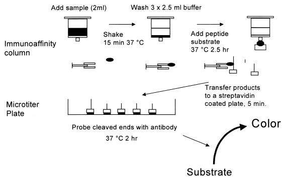
The assay system developed for detection of BoNT/B consists of the following three stages (Fig. 1): capture of BoNT/B on an immunoaffinity column, cleavage of a peptide substrate by the neurotoxin, and detection of the peptide cleavage products by a modified immunoassay procedure.
FIG. 1.
Assay format for detection of C. botulinum BoNT/B.  , immobilized monoclonal antibody;
  , neurotoxin;
  , biotinylated substrate; ▄, streptavidin.
(i) Step 1: capture of the toxin on the immunoaffinity column.
During step 1 BoNT/B is captured from the sample by immobilized monoclonal antibodies, and sample medium components which could potentially interfere with subsequent assay steps are eluted from the column. Assay performance was assessed by using various amounts of monoclonal antibody immobilized on the Sepharose gel. Increases in assay sensitivity were observed as the antibody load was increased to 10 μg per 200 μl of gel, but greater antibody loads resulted in no further improvements. In similar experiments to determine the optimum time of incubation of samples with the immobilized antibodies, little improvement in assay sensitivity was observed when the incubation time was extended beyond 15 min at 37°C.
(ii) Step 2: cleavage of the VAMP peptide substrate.
During step 2 biotinylated VAMP(60-94) peptide is cleaved by the immobilized BoNT/B under buffer conditions optimized previously (14). Incubation for 2.5 h at 37°C in the presence of 25 μM peptide was determined to give the desired assay sensitivity. Assays in which higher concentrations (50 and 100 μM) of peptide were used under similar incubation conditions did not improve the overall sensitivity of the assay.
(iii) Step 3: detection of the peptide cleavage products by immunoassay.
In the final stage of the assay the specific VAMP peptide cleavage products generated by the endopeptidase activity of BoNT/B are detected by using cleavage product-specific antibodies (6). In the assay format used for the BoNT/B assay, the mixture of cleaved and uncleaved peptides is first immobilized onto streptavidin-coated microtiter plates, and the cleaved peptide is detected by using the cleavage product-specific antibody (Fig. 1).
An alternative step 3 assay format in which the cleavage product-specific antibody was used as the solid-phase capture antibody was also examined. In this format, the solid-phase antibody selectively bound the cleaved peptide from the mixture, which was then detected by using a streptavidin-labelled horseradish peroxidase conjugate. Attempts to use this assay format, however, were unsuccessful due to strong nonspecific binding of the uncleaved VAMP peptide to the solid phase, which resulted in high assay blank values.
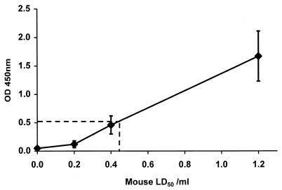
Using monoclonal antibodies 5BB/9.3 and 5BB/21.3 as capture antibodies in the optimized BoNT/B assay, we found that the detection limit for purified neurotoxin derived from a proteolytic strain (strain Okra) was approximately 0.5 MLD50/ml when an arbitrary cutoff of 0.5 absorbance unit above the background value was used. BoNT/B assayed at a concentration of 1 MLD50/ml was easily detected by eye; this concentration resulted in an absorbance that was >1.0 U greater than the background value of <0.1 U. Figure 2 shows the data obtained in 33 toxin assays.
FIG. 2.
Assay for detection of C. botulinum BoNT/B. Data were obtained from 33 assays in which we used HZ buffer containing 5% FCS spiked with purified BoNT/B. The error bars indicate standard deviations (n − 1). The dotted line shows the detection limit at 0.5 absorbance unit above the background value. OD 450nm, optical density at 450 nm.
We developed a similar assay system for detection of BoNT/B from nonproteolytic C. botulinum type B strains. To assess the endopeptidase activity of BoNT/B derived from a nonproteolytic type B strain, the abilities of neurotoxin produced by type B strain ATCC 17844 and BoNT/B produced by strain Okra to cleave solid-phase VAMP(60-94) peptide were compared (6). We found that after treatment with trypsin, the two neurotoxins cleaved the VAMP substrate at similar rates (data not shown). An assay for nonproteolytic BoNT/B was developed by using antibody 3BB/110.3 as the capture antibody in step 1. The assay which we developed was capable of detecting BoNT/B from a C. botulinum type B strain ATCC 17844 culture at a concentration of 1 MLD50/ml. At this concentration of BoNT/B, the assay mixture gave an absorbance reading of 0.66 ± 0.04 U (n = 4), compared with the blank value of 0.203 U.
Detection of BoNT/B in foods.
The results obtained with the BoNT/B endopeptidase assay when several types of food were examined are shown in Table 2. The BoNT/B endopeptidase assay was found to be capable of detecting 1 MLD50 in a variety of foods. In additional experiments in which we compared the BoNT/B endopeptidase assay with the mouse lethality test, assays were carried out with food extracts which were spiked with a range of BoNT/B concentrations (Table 3). Data obtained in these experiments showed that the endopeptidase assay was approximately fourfold more sensitive than the mouse bioassay. In addition, the presence of food extract (pate and sausage) had little affect on the performance of the endopeptidase assay.
TABLE 2.
Detection of BoNT/B in food extracts and neurotoxin generated in situ by the BoNT/B endopeptidase assaya
| Prepn | Mean optical density at 450 nm | Coefficient of variance | No. of tests | MLD50/ml |
|---|---|---|---|---|
| Spiked food extracts | ||||
| Pate | 2.26 | 25.5 | 50 | 1 |
| Cheese | 1.83 | 39.4 | 49 | 1 |
| Cod | 2.58 | 11.2 | 54 | 1 |
| Minced beef | 1.22 | 64.8 | 36 | 1 |
| Yogurt | 2.04 | 11.3 | 6 | 1 |
| In situ-generated toxin | ||||
| Pate | 0.56 | 8.9 | 3 | Negb |
| Cheese | 0.42 | 9.5 | 3 | Neg |
| Cod | 2.16 | 14.8 | 3 | >1,000 |
| Minced beef | 2.66 | 3.4 | 3 | 10-100 |
| Sausage | 2.66 | 0.8 | 3 | 1-10 |
The optical densities of negative controls consisting of unspiked food extracts were typically 0.02 to 0.04 U.
Neg, sample was negative in the mouse lethality test.
TABLE 3.
Comparison of the abilities of the BoNT/B endopeptidase assay and the mouse lethality test to detect crude neurotoxin present in spiked food samples
| BoNT/B concn (pg/ml) | Pate
|
Sausage
|
Buffer
|
|||
|---|---|---|---|---|---|---|
| Endo-peptidase assaya | Mouse test | Endo-peptidase assaya | Mouse test | Endo-peptidase assaya | Mouse test | |
| 50 | 2.78 | + | 2.02 | + | 2.57 | + |
| 25 | 2.80 | + | 1.90 | + | 2.54 | + |
| 12.5 | 2.81 | + | 2.09 | + | 2.69 | + |
| 6.3 | 2.15 | + | 1.82 | − | 1.80 | + |
| 3.1 | 1.24 | − | 1.19 | − | 1.09 | − |
| 1.5 | 0.78 | − | 1.05 | − | 1.02 | − |
| 0 | 0.02 | − | 0.004 | − | 0.04 | − |
The endopeptidase assay values are mean absorbance values (n = 4).
DISCUSSION
In the present study we developed a novel, rapid, in vitro assay for detection of BoNT/B in food products; the sensitivity of this assay is greater than the sensitivity of the mouse bioassay. The new assay is effectively a modified immunoassay amplified by the endopeptidase activity in the light chain of BoNT/B. Since the assay format relies on a key biological activity of the neurotoxin, this assay more closely resembles the mouse bioassay than a conventional immunoassay, since denatured enzymically inactive BoNT/B cannot be detected. In contrast to previously described assays for botulinum neurotoxins, the present assay requires only a few specialized reagents and is sufficiently sensitive to give a visual reading for the presence or absence of toxin in a sample within 5 to 6 h. The format of the assay also makes false-positive results highly unlikely, and no false-positive results were observed during the study. For an agent to give a false-positive reaction it would have to be retained by the antibody solid phase, which consists of monoclonal antibodies specific for BoNT/B, and it would also have to cleave specifically the VAMP(60-94) peptide substrate between the Gln-76–Phe-77 bond, an endopeptidase activity which has been described only for BoNT/B and tetanus toxin (12). Of more concern in the development of an assay to replace the existing mouse bioassay is the possibility of false-negative results in the test. The present format relies on immobilized antibody to capture BoNT/B from the sample media and therefore depends on epitopes that are conserved in the toxin structure. Previously developed immunoassays for BoNT/A and BoNT/B in which a single monoclonal antibody was used as the capture antibody proved to be unreliable because one or more toxin strains containing each neurotoxin type may go undetected (4, 5). For this reason extensive screening of BoNT/B strains was performed with a panel of monoclonal antibodies. While we found that antibody 5BB/21.3 recognizes BoNT/B produced by all of the proteolytic strains assessed, an additional antibody, 5BB/9.3, which recognizes a different epitope, was added to the assay solid phase, which lessened the possibility of false-negative results in future applications.
A surprising result obtained in this study is the finding that only one of the five antibodies screened, 3BB/110.3, recognized BoNT/B produced by all of the nonproteolytic strains tested. Thus, at least two antigenic determinants present on BoNT/B from proteolytic strains are not present on the neurotoxin from nonproteolytic strains. A comparison of the primary sequences obtained for these two BoNT/B subclasses (7) revealed that there was only 7% sequence heterogeneity. The observations described above suggest that a number of the epitopes on BoNT/B are determined by the regions that are dissimilar in the toxin subclasses. Despite significant differences in antigenic structure, a comparison of the endopeptidase activities of BoNT/B purified from proteolytic and nonproteolytic strains showed that the two types of toxin cleave the VAMP(60-94) peptide at the same site and at similar rates. However, while the endopeptidase-based assay developed for nonproteolytic BoNT/B was as sensitive as the mouse bioassay, it was less sensitive than the assay for proteolytic BoNT/B. Given the similarity in enzymic activities, this probably means that the affinity of monoclonal antibody 3BB/110.3 for nonproteolytic BoNT/B is lower than the affinities of monoclonal antibodies 5BB/21.3 and 5BB/9.3 for proteolytic BoNT/B. Production of additional antibody reagents for BoNT/B purified from nonproteolytic type B strains would therefore improve the assay further.
First-generation endopeptidase assays for botulinum neurotoxin based on toxin-dependent cleavage of solid-phase immobilized VAMP peptide substrates have been described previously (6). While such assays have a use as research tools, they cannot be realistic replacements for the mouse assay due to their low levels of sensitivity (approximately 1 ng of BoNT/B ml−1). Our principal modification to the previously described assay format is that the VAMP peptide substrate is presented to the BoNT/B in solution rather than as a solid-phase substrate. This significantly increases the peptide cleavage rate and, hence, increases the assay sensitivity. The sensitivity of the assay described here is largely dependent on the proportion of peptide captured on the streptavidin-coated plate that is cleaved. Increases in the free peptide concentration during the cleavage step, therefore, do not lead to further increases in assay sensitivity, as the increase in the rate of endopeptidase activity is compensated for by the amount of competing uncleaved peptide added to the second solid phase. The optimum free concentration of peptide was found to be 25 μM, a value far below the Km reported previously for the endopeptidase activity (14). At this peptide concentration an incubation time of 2.5 h was required to give the desired sensitivity (10 pg ml−1). Assays in which the sensitivity is increased or reduced may be obtained by extending or reducing the peptide cleavage time.
The dual solid phase of the BoNT/B assay described here provides a particularly robust system for detecting toxin in difficult media, such as fatty foods, since the second solid-phase ELISA preparation is not exposed to the medium components. We found that the assay can detect purified, crude, and in situ-derived BoNT/B in food samples and that food has little effect on the sensitivity of the assay. The detection of crude BoNT/B in culture supernatants also shows that the nontoxic protein components associated with BoNT/B in its complex state do not interfere with any of the assay steps.
A major use of the mouse bioassay is safety validation of foodstuffs which have been challenged with known strain types, and it is hoped that the assay described here will be a realistic replacement in such situations. While extensive validation of the assay format in a wide range of food types will be required before our assay can replace the mouse bioassay, we hope that this assay will greatly reduce the number of mice used for detection of BoNT/B.
ACKNOWLEDGMENT
This work was supported by the Ministry of Agriculture, Fisheries and Foods, United Kingdom.
REFERENCES
- 1.Blasi J, Chapman E R, Link E, Binz T, Yamasaki S, DeCamilli P, Südhof T C, Niemann H, Jahn R. Botulinum neurotoxin A selectively cleaves the synaptic protein SNAP-25. Nature (London) 1993;365:160–163. doi: 10.1038/365160a0. [DOI] [PubMed] [Google Scholar]
- 2.Doellgast G J, Triscott M X, Beard G A, Bottoms J D, Cheng T, Roh B H, Roman M G, Hall P A, Brown J E. Sensitive enzyme-linked immunosorbent assay for the detection of Clostridium botulinum neurotoxins A, B, and E using signal amplification via enzyme-linked coagulation assay. J Clin Microbiol. 1993;31:2402–2409. doi: 10.1128/jcm.31.9.2402-2409.1993. [DOI] [PMC free article] [PubMed] [Google Scholar]
- 3.Foran P, Lawrence G W, Shone C C, Foster K A, Dolly J O. Botulinum neurotoxin C1 cleaves both syntaxin and SNAP-25 in intact and permeabilised chromatin cells: correlation with its blockade of catecholamine release. Biochemistry. 1996;35:2630–2636. doi: 10.1021/bi9519009. [DOI] [PubMed] [Google Scholar]
- 4.Gibson A M, Modi N K, Roberts T A, Shone C C, Hambleton P, Melling J. Evaluation of a monoclonal antibody-based immunoassay for detecting type A Clostridium botulinum toxin produced in pure culture and an inoculated model cured meat system. J Appl Bacteriol. 1987;63:217–226. doi: 10.1111/j.1365-2672.1987.tb04939.x. [DOI] [PubMed] [Google Scholar]
- 5.Gibson A M, Modi N K, Roberts T A, Shone C C, Hambleton P, Melling J. Evaluation of a monoclonal antibody-based immunoassay for detecting type B Clostridium botulinum toxin produced in pure culture and an inoculated model cured meat system. J Appl Bacteriol. 1988;64:285–291. doi: 10.1111/j.1365-2672.1988.tb01873.x. [DOI] [PubMed] [Google Scholar]
- 6.Hallis B, James B A F, Shone C C. Development of novel assays for botulinum type A and B neurotoxins based on their endopeptidase activities. J Clin Microbiol. 1996;34:1934–1938. doi: 10.1128/jcm.34.8.1934-1938.1996. [DOI] [PMC free article] [PubMed] [Google Scholar]
- 7.Minton N P. Molecular genetics of clostridial neurotoxins. In: Montecucco C, editor. Clostridial neurotoxins: the molecular pathogenesis of tetanus and botulism. Berlin, Germany: Springer; 1995. pp. 161–194. [Google Scholar]
- 8.Niemann H. Molecular biology of the clostridial neurotoxins. In: Alouf J E, Freer J H, editors. A sourcebook on bacterial protein toxins. London, United Kingdom: Academic Press; 1991. pp. 303–348. [Google Scholar]
- 9.Reed L J, Muench H. A simple method of estimating fifty percent endpoints. Am J Hyg. 1938;27:493–497. [Google Scholar]
- 10.Schiavo G, Santucci A, DasGupta B R, Mehta P P, Jontes J, Benfenati F, Wilson M C, Montecucco C. Botulinum neurotoxins serotypes A and E cleave SNAP-25 at distinct COOH-terminal peptide bonds. FEBS Lett. 1993;335:99–103. doi: 10.1016/0014-5793(93)80448-4. [DOI] [PubMed] [Google Scholar]
- 11.Schiavo G, Shone C C, Bennett M K, Scheller R H, Montecucco C. Botulinum neurotoxin type C cleaves a single Lys-Ala bond within the carboxy terminal region of syntaxins. J Biol Chem. 1995;270:10566–10570. doi: 10.1074/jbc.270.18.10566. [DOI] [PubMed] [Google Scholar]
- 12.Schiavo G, Benefenati F, Poulain B, Rossetto O, Polverino de Laureto P, DasGupta B R, Montecucco C. Tetanus and botulinum-B neurotoxins block neurotransmitter release by proteolytic cleavage of synaptobrevin. Nature (London) 1992;359:832–835. doi: 10.1038/359832a0. [DOI] [PubMed] [Google Scholar]
- 13.Schiavo G, Rossetto O, Catsicas S, Polverino de Laureto P, DasGupta B R, Benfenati F, Montecucco C. Identification of the nerve terminal targets of botulinum nuerotoxin serotypes A, D and E. J Biol Chem. 1993;268:23784–23787. [PubMed] [Google Scholar]
- 14.Shone C C, Roberts A K. Peptide substrate specificity and properties of the zinc-endopeptidase activity of botulinum type B neurotoxin. Eur J Biochem. 1994;225:263–270. doi: 10.1111/j.1432-1033.1994.00263.x. [DOI] [PubMed] [Google Scholar]
- 15.Shone C C, Quinn C P, Wait R, Hallis B, Fooks S G, Hambleton P. Proteolytic cleavage of synthetic fragments of VAMP isoform-2 by botulinum type B neurotoxin. Eur J Biochem. 1993;217:965–971. doi: 10.1111/j.1432-1033.1993.tb18327.x. [DOI] [PubMed] [Google Scholar]
- 16.Shone C C, Wilton-Smith P, Appleton N, Hambleton P, Modi N K, Gatley S, Melling J. Monoclonal antibody-based immunoassay for type A Clostridium botulinum toxin is comparable to the mouse bioassay. Appl Environ Microbiol. 1985;50:63–67. doi: 10.1128/aem.50.1.63-67.1985. [DOI] [PMC free article] [PubMed] [Google Scholar]
- 17.Wictome M, Rossetto O, Montecucco C, Shone C C. Substrate residues N-terminal to the cleavage site of botulinum type B neurotoxin play a role in determining the specificity of its endopeptidase activity. FEBS Lett. 1996;386:133–136. doi: 10.1016/0014-5793(96)00431-0. [DOI] [PubMed] [Google Scholar]


