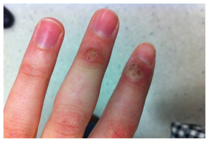
A 21-year-old woman presented to the emergency department with a 2-week history of lesions on her left hand. As a student in an agriculture program, she had had ungloved contact with goats and the mouth of a baby calf during a farm practicum 1 week before presentation. The animals had no evidence of disease. The patient reported no history of symptomatic herpes infections, recent travel or chronic illnesses. She had 2 discreet, nonpainful, well-circumscribed lesions on the dorsum of her left second and third fingers, each measuring less than 1 cm in diameter (Figure 1). She had a small, tender, left epitrochlear lymph node, and mild swelling and tenderness of the left wrist.
Figure 1:
Photograph of lesions on the left fingers of a 21-year-old woman with pseudocowpox. The lesion on her second finger had a slightly depressed ulcerative appearance; the lesion on her third finger was semi-firm and filled with fluid, in keeping with the exudative nodular phase. Both lesions contained purplish–red punctate areas and mild surrounding erythema.
The infectious differential diagnosis included herpetic whitlow (herpes simplex virus [HSV] 1 and 2); varicella zoster virus (VZV); Staphylococcus aureus; poxviruses, including orf virus (contagious ecthyma), pseudocowpox virus (milker’s nodule) and bovine papular stomatitis virus; cutaneous anthrax (Bacillus anthracis); and bovine tuberculosis (Mycobacterium bovis), given her exposures. This patient presented to us before the current epidemic of mpox, which should now be considered in the differential diagnosis of patients with similar lesions.
Lesion swabs were negative for HSV 1, HSV 2 and VZV via polymerase chain reaction (PCR) and bacterial culture. No virus was visualized on electron microscopy. Specialized PCR testing showed the presence of poxvirus DNA, and further sequencing confirmed the presence of pseudocowpox virus (paravaccinia virus). Results of PCR testing for orthopoxviruses were negative. Our patient’s lesions had resolved without scarring at follow-up 1 month later.
Because pseudocowpox is not a reportable disease, its exact incidence is unknown; however, it is commonly reported among farmworkers. The incubation period is 5–15 days.1 Humans are infected by direct contact with infected dairy cattle teats or udders, or with the mouths of nursing calves, via exposure to abraded skin on the hands and fingers. Infection is generally limited to the epithelium, and disseminated disease is uncommon.2,3
Lesions progress through maculopapular, exudative nodular, crusted and papillomatous stages, with complete healing without scarring within 4–6 weeks.1,2 The diagnosis can be clinical, but confirmitory testing with PCR or electron microscopy is helpful. Management is supportive.1 People with exposure risks, (e.g., agriculture students, farmers) should wear gloves and perform stringent hand hygiene to prevent infection.
Acknowledgements
The authors would like to acknowledge the contributions of the late Dr. Geoff Taylor (University of Alberta) who was involved in the clinical care of this patient and who reviewed content for a previous poster presentation at a conference of the Association of Medical Microbiology and Infectious Disease Canada.
Footnotes
Competing interests: None declared.
This article has been peer reviewed.
The authors have obtained patient consent.
References
- 1.Barraviera SRCS. Diseases caused by poxvirus — orf and milker’s nodules: a review. J Venom Anim Toxins Incl Trop Dis 2005;11:102–8. [Google Scholar]
- 2.Phillips ZC, Holfeld KI, Lang ALS, et al. A case of milker’s nodules in Saskatchewan, Canada. SAGE Open Med Case Rep 2020;8:2050313X20984118. doi: 10.1177/2050313X20984118. [DOI] [PMC free article] [PubMed] [Google Scholar]
- 3.Adriano AR, Quiroz CD, Acosta ML, et al. Milker’s nodule — case report. An Bras Dermatol 2015;90:407–10. [DOI] [PMC free article] [PubMed] [Google Scholar]


