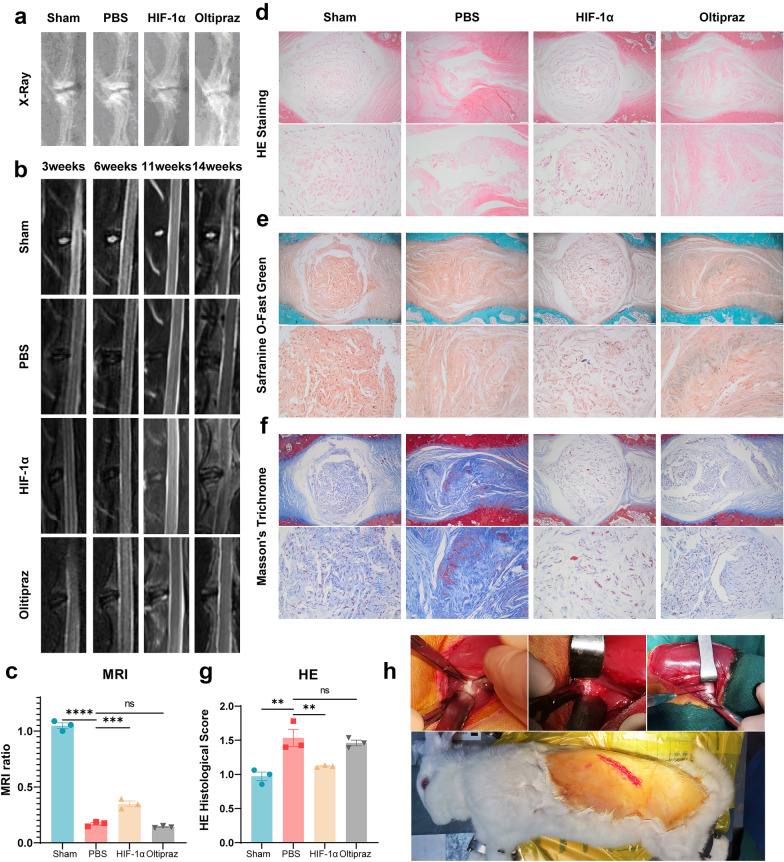
Fig. 5.
Exogenous supplementation of HIF-1α exhibited a protective effect on the regeneration of NP tissue in vivo. a X-ray and b, c MRI to assess the degree of IVDD in rabbits from the sham group, PBS group, HIF-1α group and Oltipraz group. d–g HE, Safranin O and Masson staining indicated morphological degeneration score such as the height of IVD in this IVDD model was alleviated by supplementation of HIF-1α in comparison with the PBS group and Oltipraz group. Safranin O and Masson staining showed that HIF-1α reduced proteoglycan and collagen loss during the process of IVDD, but suppression of HIF-1α expression aggravated that. The histological scores of each indicated group were calculated according to the grading scale previously published. h Photos of the surgical procedure in vivo. The scale bar is 500 or 100 μm. The values are the mean of at least three independent experiments. *P < 0.05, **P < 0.01, ***P < 0.001 and ****P < 0.0001.

