ABSTRACT
There are several Entamoeba species that colonize humans, but only Entamoeba histolytica causes severe disease. E. histolytica is transmitted through the fecal-oral route to colonize the intestinal tract of 50 million people worldwide. The current mouse model to study E. histolytica intestinal infection directly delivers the parasite into the surgically exposed cecum, which circumvents the natural route of infection. To develop a fecal-oral mouse model, we screened our vivarium for a natural murine Entamoeba colonizer via a pan-Entamoeba PCR targeting the 18S ribosomal gene. We determined that C57BL/6 mice were chronically colonized by Entamoeba muris. This amoeba is closely related to E. histolytica, as determined by 18S sequencing and cross-reactivity with an E. histolytica-specific antibody. In contrast, outbred Swiss Webster (SW) mice were not chronically colonized by E. muris. We orally challenged SW mice with 1 × 10⁵ E. muris cysts and discovered they were susceptible to infection, with peak cyst shedding occurring between 5 and 7 days postinfection. Most infected SW mice did not lose weight significantly but trended toward decreased weight gain throughout the experiment compared to mock-infected controls. Infected mice treated with paromomycin, an antibiotic used against noninvasive intestinal disease, do not become colonized by E. muris. Within the intestinal tract, E. muris localizes exclusively to the cecum and colon. Purified E. muris cysts treated with bovine bile in vitro excyst into mobile, pretrophozoite stages. Overall, this work describes a novel fecal-oral mouse model for the important global pathogen E. histolytica.
KEYWORDS: Entamoeba, parasitology, fecal-oral, intestinal infection, excystation
INTRODUCTION
Parasitic diseases are underappreciated causes of morbidity and mortality because disease outcomes are variable (1), cases are often underreported (2), and disease disproportionately impacts geographical locations experiencing poverty (1–3). Diarrheal diseases are a significant and underreported cause of child mortality in tropical regions (4, 5). Such infections are exacerbated by factors such as resource availability, lack of sanitation infrastructure, and malnutrition (6). For these reasons, diarrheal diseases represent a long-standing and significant burden, particularly in Latin America, Southeast Asia, and sub-Saharan Africa (6). Diarrheal diseases are caused by a range of pathogens, such as bacteria, viruses, and parasites, including Entamoeba histolytica. E. histolytica is an extracellular parasite that causes human infection with variable outcomes ranging from asymptomatic colonization to diarrhea, invasive colitis, liver abscesses, and metastatic infection. E. histolytica ranks among the top 15 causes of diarrhea in children under the age of 2 in developing countries (7, 8). Prevalence rates vary significantly due to earlier misdiagnoses resulting from use of microscopy as a diagnostic tool, which is unable to differentiate between E. histolytica and the nonpathogenic Entamoeba dispar.
Entamoeba infection starts with the ingestion of the cyst stage from contaminated food or water. Presumably in the small intestine, the Entamoeba cyst molts from its chitinous shell and differentiates into the metabolically active trophozoite form through a process known as excystation. Trophozoites then attach to the intestinal epithelium where they undergo asexual reproduction and encystation. The infectious fecal cysts are shed and contaminate the environment to complete the parasite life cycle. Trophozoites can either stay contained within the intestinal tract or may disseminate to soft tissue organs like the liver, the lungs, or the brain (9–12), although in ~90% of the cases, the infection remains in the intestinal lumen (13, 14). The current murine intestinal infection model surgically delivers trophozoites into the cecum of animals and has provided immense insight into host-pathogen interactions but produces no cysts (15). To date, attempts at generating cysts in vitro using the E. histolytica reference strain, HM1:IMSS, have successfully yielded cyst-like structures (CLS) resembling immature cysts (16–18). However, the infectivity of these CLS in an animal infection model has not yet been determined, and efficient production of mature cysts has not yet been achieved. Thus, modeling the critical developmental stage interconversion that E. histolytica undergoes between ingestion and colonization of the cecum is not yet possible (19). A model that includes the developmental changes of excystation and encystation would allow the field to understand the transmission of the pathogen and find targets for intervention (reviewed in reference 20), as only viable parasites completely encysted and shed via the fecal-oral route can contaminate food and water and infect a new host.
The parasitology field has used species that naturally colonize animals to expand the knowledge of infectious diseases that are fastidious to culture or model in the laboratory. Murine pathogens like Plasmodium chabaudi have been pivotal to studying the in vivo pathology of malaria. For the Entamoeba field, Entamoeba invadens, a reptile-specific pathogen, has provided critical insights related to developmental changes like encystation. Here, we screened for a natural murine Entamoeba colonizer and developed an oral infection model using Swiss Webster (SW) mice. SW mice treated with paromomycin showed no E. muris colonization. We further determine infection location within the intestinal tract and excystation cues for the purified fecal cysts.
RESULTS
C57BL/6 mice, but not Swiss Webster mice, are chronically colonized with a naturally occurring Entamoeba organism.
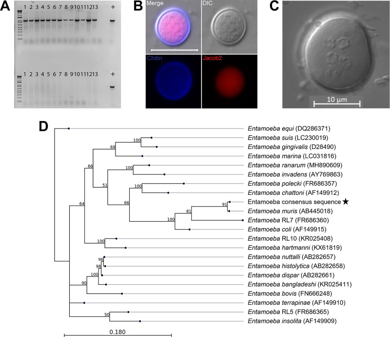
We screened transgenic and wild-type animals within our vivarium facility (Fig. 1A) using a Pan-Entamoeba PCR (Fig. S1). We screened fecal material from males and females with ages ranging from newly weaned to mice that spent up to a 1 year in our facility. Eighty percentage of the mice from the C57BL/6 background were colonized with an Entamoeba organism. To address if an Entamoeba is naturally occurring in other vivariums, we requested fecal samples from C57BL/6 mice from five collaborators around the country (n = 18) and ran both single-step pan-Entamoeba PCR and 2-step, nested PCR on all samples obtained. We were able to detect Entamoeba gDNA using single-step PCR in samples from one other institution, while for another institution, we were only able to detect the parasite using nested PCR (Table S1). Colonization was by no means ubiquitous, as the majority of these institutions were PCR-negative even after using the nested PCR approach (Table S1). In contrast, all the SW mice from our vivarium tested were PCR negative, regardless of age or sex (Fig. 1A).
FIG 1.
Identification of a murine Entamoeba species. (A) Representative Pan-Entamoeba PCR for C57BL/6 mice (top) and Swiss Webster mice (bottom) within our vivarium. The loading control was murine GAPDH (data not shown). Each lane number represents a mouse cage. Positive control (+) is isolated genomic DNA from axenic Entamoeba histolytica culture. (B) Immunofluorescence assay staining for chitin (Calcofluor White), Jacob2 (1A4 antibody), scale bar represents 20 μm. (C) Representative phenotypic characterization of the number of nuclei of E. muris cysts (>4 nuclei). Image was taken using DIC. Scale bar represents 10 μm. (D) 18S phylogeny is based on a 1,033 bp alignment, including 27 published Entamoeba sequences, labeled as species name (NCBI accession), plus the Entamoeba found in our vivarium, indicated by a star. Numbers on branches are bootstrap values (%) based on 1,000 replicates (values >50% are shown). The scale bar indicates nucleotide substitutions per site.
Pan-Entamoeba primer design. Full-length Entamoeba 18S rRNA sequences (n = 63, 25 Entamoeba species) were downloaded from NCBI GenBank, aligned in CLC Genomics Workbench v20.0.4 (Qiagen, Hilden, Germany), and conserved regions were the target for the placement of the forward and reverse primers, which together amplify a 1 kb product.Forward: 5′-AGATACCGTCGTAGTCCT-3′ Reverse: 5′-ACGACTTCTCCTTCCTCTAA-3′. Download FIG S1, TIF file, 2.9 MB (3MB, tif) .
Copyright © 2023 Mendoza Cavazos et al.
This content is distributed under the terms of the Creative Commons Attribution 4.0 International license.
Colonization rates at vivarium facilities around the United States. Both 40-cycle PCR and nested PCR targeting the Entamoeba 18S gene were run on all donated fecal samples. Reactions were run on agarose gels to determine presence or absence of Entamoeba gDNA, and percentages of positive samples out of total samples obtained from each institution are shown. Download Table S1, DOCX file, 0.01 MB (15.8KB, docx) .
Copyright © 2023 Mendoza Cavazos et al.
This content is distributed under the terms of the Creative Commons Attribution 4.0 International license.
To confirm the PCR findings in our animals, we developed a sucrose density gradient protocol based on fecal isolation methods from the Entamoeba literature (21) (Fig. S2) and conducted phenotypic characterization of the isolated cysts (Fig. 1B and C). We processed feces of PCR-positive mice within a sucrose gradient of 1.33 specific gravity and isolated structures of 15 to 20 μm in diameter. These structures were cyst-like and stained with Calcofluor White, indicating the presence of chitin (Fig. 1B). We further characterized these cysts by immunofluorescence detection of a previously published Entamoeba-specific antibody targeting the lectin Jacob2 (22). The sucrose gradient results for B6 and SW mice were 100% replicative of the Pan-Entamoeba PCR; SW mice did not display cyst-like structures in their fecal samples, while the B6 fecal samples contained 15 to 20 μm-diameter cysts that stained positive for both chitin and Jacob2 (Fig. 1B). We also observed the presence of multiple nuclei within individual cysts (Fig. 1C).
Sucrose gradient isolation protocol. (A) Fecal samples (0.25 to 5 g) that were collected overnight were processed as indicated above. Briefly, fecal samples were ground to a fine powder using a mortar and pestle then shortly homogenized with Nanopure water for 15 minutes using a Mini Rotator (Glas-Col) at 60 rpm. The resulting solution was filtered through four-ply cotton gauze, and samples were pelleted for 10 minutes at 2,500 × g. The resulting pellet was layered on top of 1.5 M sucrose solution. The mid-layer was washed with Nanopure water and pelleted again at the same speed. Pellet 3 was suspended in Nanopure water, and the isolated unfixed cysts were used as the input for oral infection. (B) While cysts can be found in the P2 pellet during the winter months, they have a dehydrated appearance compared to the cysts in the P3 pellet, likely due to the low humidity of the vivarium. Download FIG S2, TIF file, 2.9 MB (3MB, tif) .
Copyright © 2023 Mendoza Cavazos et al.
This content is distributed under the terms of the Creative Commons Attribution 4.0 International license.
To determine the species of Entamoeba present in our mice, we gel-purified and Sanger-sequenced the 1 kb pan-Entamoeba PCR product from cecum, colon, and fecal samples from 8 mice (n = 24 total). The resulting reads were 100% identical to each other and most closely matched Entamoeba muris (GenBank accession number AB445018) with 98% query coverage at 92% identity and an E value of 0.00. We then performed phylogenetic analysis to further confirm this preliminary species identification. Alignment of 21 published Entamoeba 18S sequences from NCBI GenBank along with our consensus Sanger sequence yielded a final alignment length of 1,033 bp. A maximum-likelihood phylogeny built from this alignment shows our organism to cluster with Entamoeba muris, as expected from the BLASTn results, and form a clade with Entamoeba RL7 and Entamoeba coli (Fig. 1D). Pairwise comparison of the E. muris/coli clade based on the 18S phylogenetic tree across the entire length of the alignment shows our organism to be 91.63% identical to E. muris with 9 total alignment gaps (0.87%) compared to 81.82% identity and 42 gaps (4.1%) with its next-closest relative, Entamoeba RL7 (Table S2). Thus, we will refer to this organism as E. muris here.
Pairwise comparison of Entamoeba muris/coli clade. Comparison is based on a 1,033 bp alignment. Percent nucleotide identity is shown above the diagonal and number of gaps are. Download Table S2, DOCX file, 0.01 MB (15.6KB, docx) .
Copyright © 2023 Mendoza Cavazos et al.
This content is distributed under the terms of the Creative Commons Attribution 4.0 International license.
Swiss Webster mice are susceptible to Entamoeba muris oral challenge.
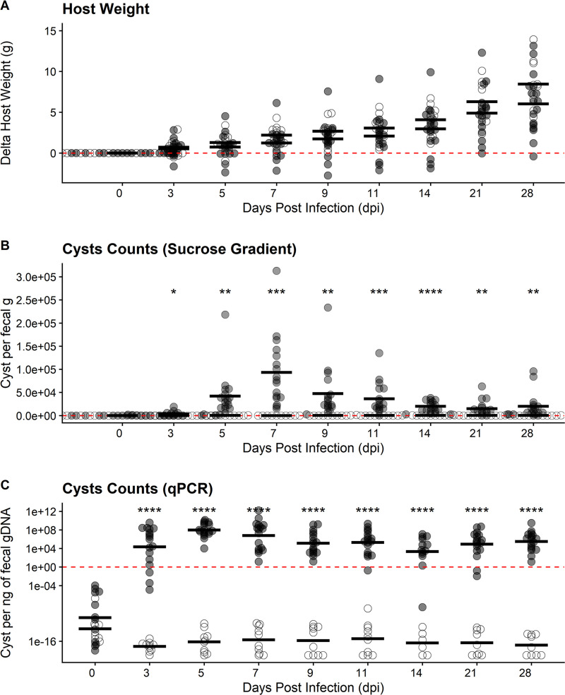
As SW mice were not currently colonized, we determined if these mice were suitable for E. muris infection and characterization using an oral challenge model. When orally challenged with a low cyst dosage (1 × 10⁵ isolated from B6 mice), SW mice were able to host a patent infection, as evidenced by fecal cyst shedding, but displayed asymptomatic infections in all cases. Specifically, we did not observe any hallmark signs or symptoms of distress among our infected SW mice during our daily health checks. We also did not see any notable changes in grooming, movement, food and water consumption, or overall activity that would indicate symptomatic disease caused by E. muris. We tracked changes in host weight during the infection and found that both uninfected and infected animals displayed a modest degree of variability in weight changes, with most mice gaining weight over time (Fig. 2A and S3A). No statistically significant differences in weight change were found between uninfected animals and animals orally challenged with E. muris cysts. Despite this, infected mice seemed to display an overall trend in slower weight gain compared to uninfected control mice (Fig. 2A). Weight loss was only observed for one of the biological replicates (Fig. S3A) and was not correlated with cyst shedding (Fig. S3B). Fecal samples showed cysts as early as 3 days postinfection (dpi) via sucrose gradient. When using this method, we determined the peak of infection to be 7 dpi based on average cysts counts across four independent infection replicates (n = 27 mice, 17 infected and 10 uninfected controls). Shedding per biological replicates does not show significant differences (Fig. S3B). By sucrose gradient, we detected a significant decrease in cyst shedding by 11 dpi, and very few E. muris cysts were detected using this method by 28 dpi, indicating a sharp decline in viable cyst shedding.
FIG 2.
Swiss Webster mice are susceptible to Entamoeba muris oral challenge. (A) Host weight was monitored through the course of infection. (B) Quantification of cysts isolated by sucrose gradient from Swiss Webster fecal samples (normalized by fecal mass). Peak of infection was determined to be 7 dpi. (C) Quantification of cysts in fecal samples via qPCR isolated from Swiss Webster’s fecal samples (normalized by gDNA per qPCR). Each dot represents a single mouse (n = 27 mice, 17 infected and 10 uninfected controls). Open circles represent uninfected mice while gray circles represent infected mice. Significance was determined using a two-tailed t test between the uninfected versus infected average per DPI. Data combines four independent biological replicates (see Fig. S3 for individual biological replicate [n = 4] plotting).
Swiss Webster mice are susceptible to Entamoeba muris oral challenge, per biological replicate. (A) Host weight was monitored through the course of infection. (B) Quantification of cysts isolated by sucrose gradient from Swiss Webster fecal samples (normalized by fecal mass). Each dot represents a single mouse. Open circles represent uninfected mice while gray circles represent infected mice. Significance was determined using a two-tailed t-test between the uninfected versus infected average per DPI. Download FIG S3, TIF file, 2.9 MB (2.9MB, tif) .
Copyright © 2023 Mendoza Cavazos et al.
This content is distributed under the terms of the Creative Commons Attribution 4.0 International license.
Using a similar approach to the Pan-Entamoeba screen, we designed qPCR detection primers that amplified a 200-bp amplicon to quantify E. muris shedding. We generated a standard curve using cyst samples of known concentrations based on counts, ranging from 1,562 cysts to 100,000 cysts on a 2-fold scale (Fig. S4A). Our qPCR results determined that E. muris was detectable as early as 3 dpi, in accordance with the sucrose gradient isolation. However, the peak of infection was at 5 dpi, and a significant reduction was evident by 9 dpi. In contrast to our sucrose gradient data (Fig. 2B), SW mice appear to maintain infection by E. muris up to 28 dpi when using qPCR as a detection method. Taken together, these results suggest that qPCR detection occurs prior to peak viable cyst isolation and that SW mice may remain colonized by E. muris even after viable cysts are no longer being shed.
Standard curves for qPCR cyst quantification. Primers were designed as described above for pan-Entamoeba PCR (Fig. S1), except here they were chosen to amplify a 200 bp product (see primers below). Two standard curves were generated for each real-time PCR system used in our studies using cyst samples of known concentrations, ranging from (A) 1,562 cysts to 100,000 cysts or (B) 1,562 cysts to 50,000 cysts expressed on a 2-fold scale. Concentrations for Fig. 2C and 3B were calculated using the cycle threshold (CT) values of the experimental samples and the linear trendline equations presented above.Forward: 5′-TCGAGATAAACGAGAGCGAAAG-3′Reverse: 5′-GTCAGGACTACGACGGTATCTA-3′. Download FIG S4, TIF file, 2.9 MB (2.9MB, tif) .
Copyright © 2023 Mendoza Cavazos et al.
This content is distributed under the terms of the Creative Commons Attribution 4.0 International license.
Paromomycin treatment prevents colonization of Entamoeba muris.
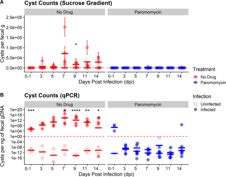
Patients infected with E. histolytica are prescribed antibiotics depending on the degree of pathogenicity. Two common drug treatments for patients infected with E. histolytica are metronidazole and paromomycin. For invasive disease, metronidazole is the gold standard for treatment, but some E. histolytica strains can develop resistance over time (23). Paromomycin is the treatment of choice for noninvasive E. histolytica infections (24) due to its antimicrobial activity against facultative anaerobes and lack of absorption in the gut. To determine if paromomycin recapitulates its amoebicidal effect against E. muris, we orally infected SW mice with 7 × 10⁴ cysts isolated from B6 mice and immediately began treatment with paromomycin (16 g/L) in their drinking water for the first 7 days postinfection before reverting to untreated drinking water. Using the approaches presented earlier, we monitored E. muris colonization and host weight for 2 weeks postinfection. Overall, SW mice treated with paromomycin showed higher variability in weight changes throughout the course of treatment compared to untreated mice (Fig. S5). Infected mice treated with paromomycin again trended toward slower weight gain than their uninfected treated counterparts, although this observation was not statistically significant (Fig. S5). In contrast, both uninfected and infected mice given untreated drinking water displayed similar weight changes over the course of 14 days (Fig. S5). As expected, SW mice that were challenged with E. muris but received no paromomycin treatment shed fecal cysts as early as 3 to 5 dpi via sucrose gradient (Fig. 3A), even at a lower infectious dose of 7 × 10⁴ cysts. In concordance with our previous results, cyst shedding peaked at 7 dpi and declined significantly by 9 dpi (Fig. 3A). In contrast, SW mice challenged with E. muris and treated with paromomycin never shed any viable E. muris throughout 14 dpi as determined by sucrose gradient isolation (Fig. 3A).
FIG 3.
Paromomycin effectively inhibits colonization of SW mice by Entamoeba muris. (A) Quantification of cysts isolated by sucrose gradient from (Swiss Webster fecal samples normalized by fecal mass). (B) Quantification of cysts in fecal samples via qPCR isolated from Swiss Webster fecal samples (normalized by gDNA per qPCR). Each circle represents a single mouse (n = 16 mice, 12 infected and 4 uninfected controls). Open circles represent uninfected mice while filled circles represent infected mice. Red circles represent untreated mice while blue circles represent mice treated with paromomycin. Bars indicate calculated mean values for each experimental group per DPI. Significance was determined using a two-tailed t test between the uninfected versus infected average per DPI. Data combines two independent biological replicates.
Paromomycin-treated SW mice display variable changes in host weight compared to untreated SW mice. (A) Host weight was monitored through the course of infection at 0, 3, 5, 7, 9, 11, and 14 dpi. Open circles represent uninfected mice while filled circles represent infected mice. Red circles represent untreated mice while blue circles represent mice treated with paromomycin. Bars indicate calculated mean values for each experimental group per DPI. Download FIG S5, TIF file, 0.6 MB (669.4KB, tif) .
Copyright © 2023 Mendoza Cavazos et al.
This content is distributed under the terms of the Creative Commons Attribution 4.0 International license.
We also monitored E. muris shedding via qPCR as a second method of detection. In agreement with our sucrose gradient data (Fig. 3A), the peak of infection in untreated mice challenged with E. muris was also found to be 7 dpi by qPCR (Fig. 3B). Unlike our sucrose gradient data, qPCR results indicated that all infected mice did have detectable E. muris between the day of infection (0 dpi) to 1 dpi (Fig. 3B). However, infected mice treated with paromomycin no longer shed detectable E. muris by 3 dpi (Fig. 3B). These mice continued to lack detectable E. muris up to 14 dpi, except for one mouse on day 14 (Fig. 3B). Taken together, these results suggest that paromomycin effectively inhibits E. muris colonization when administered as a prophylactic and that our model can be used for drug screening studies relevant to luminal Entamoeba infections.
Entamoeba muris resides in the large intestine at 5 days postinfection.
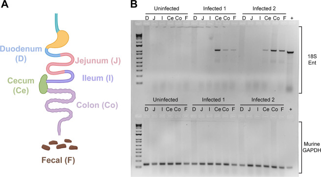
E. histolytica is thought to replicate in the colon and has been found during diagnostic colonoscopies (25). As our model uses the natural oral route of infection, we aimed to determine where E. muris is located during primary infection. We infected SW mice with 105 cysts by oral gavage and collected the intestinal content and mucus layer of murine gastrointestinal sections at 5 dpi, when most of the animals were shedding cysts by qPCR detection (Fig. 2C). The small intestine was sectioned into three parts: duodenum (D), jejunum (J), and ileum (I). The cecum (Ce) and the colon (Co) correspond to the large intestine (Fig. 4A). As a positive control for the presence of cysts, we included two fresh fecal pellets (F) and we used a standard loading control, murine GAPDH (Fig. 4B, lower panel). As expected from clinical data (reviewed in reference 26), Entamoeba localizes within the large intestine (Fig. 4B, upper panel). While we found mouse to mouse variability between levels of E. muris detection within the cecum, colon, and fecal samples, no Entamoeba muris gDNA was isolated from the small intestine (Fig. 4B, upper panel).
FIG 4.
Entamoeba muris localizes to the large intestine of infected animals. (A) Schematics of the murine intestine. (B) The Entamoeba 18S gene was amplified from gastrointestinal sections (gel top). The positive control is genomic DNA extracted from an Entamoeba histolytica axenic culture. As a loading control, the murine GAPDH gene was amplified from various gastrointestinal sections, with gDNA isolated from mice tail snips serving as a positive control (gel bottom). Gel image is a representative of two independent biological replicates (n = 6, 4 infected and 2 uninfected controls).
Bile extract triggers Entamoeba muris excystation in vitro.
To determine if the previously published E. invadens cues could trigger consistent E. muris excystation in vitro, we incubated the isolated cysts with Nanopure water, 80 mM sodium bicarbonate, 1% bovine bile, or a combination of both treatments for 24 h (27). We scored excystation efficiency based on the percentage of the parasite that was outside the chitin shell. An intact cyst was given a score of 0 (Fig. 1B). We scored an open cyst with less than 50% of the trophozoite-mass excysted as a 1, cysts where 50% or more of the parasite was outside the chitin shell as a 2, and empty chitin shells were given a score of 3 (Fig. 5A). We observed excystation to be an asynchronous process, as scores ranged within each condition (Fig. S6). Treatment of cysts with only 1% bovine bile resulted in greater than 70% excystation by 24 h, which was statistically higher than the excystation rate of the Nanopure water treatment (P = 0.0040). This excystation rate was not enhanced by the addition of the sodium bicarbonate, and sodium bicarbonate alone did not significantly enhance excystation compared to water only (Fig. 5B). These results strongly indicate that 1% bile is sufficient to trigger excystation, which implies that excystation of E. muris is occurring in the small intestinal tract as we would expect.
FIG 5.
Entamoeba muris shows reliable excystation in vitro when treated with upper gastrointestinal tract components. Fecal cysts were purified by sucrose density gradient and then acid washed (0.1 M HCl). Cysts were inoculated into excystation conditions (1% bovine bile, 80 mM sodium bicarbonate, or a combination of both), then incubated for 24 h at room temperature. (A) Cysts were scored from 0 to 3, where 0 represented an intact cyst and 3 is an empty chitin shell. Chitin (Calcofluor White), Jacob2 (1A4 antibody [17]), and nuclei (Syto11). Scale bar represents 10 μm. (B) Excystation rates (score ≥ 1) were quantified under these conditions. Significance was determined using a two-tailed t test. Only significant pairwise comparisons are shown; Bile (P = 0.004) and, NaHCO3 + Bile (P = 0.0064). Each dot represents a biological replicate (n = 3 independent experiments), black horizontal line is the average of the three biological replicates.
Entamoeba muris excystation is an asynchronous process. Fecal cysts were purified by sucrose density gradient and then acid washed (0.1 M HCl). Cysts were inoculated into excystation conditions (1% bile, 80 mM sodium bicarbonate, or a combination of both), then incubated for 24 h at room temperature. Cysts were scored from 0 to 3, where 0 represented an intact cyst and 3 is an empty chitin shell. Each panel (A–C) represents the individual biological replicates averaged in Fig. 5. Download FIG S6, TIF file, 0.9 MB (967KB, tif) .
Copyright © 2023 Mendoza Cavazos et al.
This content is distributed under the terms of the Creative Commons Attribution 4.0 International license.
DISCUSSION
To the best of our knowledge, this work is the first to demonstrate that C57BL/6 mice can be chronically colonized with E. muris. C57BL/6 mice have been previously described as naturally resistant to E. histolytica when injecting trophozoites directly into a surgically exposed cecum (28). Other protozoans, such as Tritrichomonas musculis, have been reported to chronically colonize many mouse colonies on the East Coast of the United States (29). T. musculis was found to change the immune response to protect against pathogens in mice with chronic colonization, and E. muris may also be changing the immune responses. Understudied intestinal protozoans may account for the variability of results between research institutions. Many of the fecal samples from C57BL/6 mice across the United States did not amplify Entamoeba, but the gDNA isolated from the feces was of high quality because we could detect host gDNA in the Entamoeba negative samples when using a primer set for murine GAPDH. One of the limitations of this study is that the Entamoeba identification is based on the 18S gene (Fig. 1D and Table S2) and phenotypic characterization of the number of nuclei (>4) via microscopy (Fig. 1C); thus, further characterization will be required.
One surprising result was the positive Jacob2 staining for the E. muris cysts (Fig. 1B) given the genetic divergence we observed in our phylogenetic analyses of the E. muris 18S ribosomal gene (Fig. 1D). The 1A4 antibody was previously described to distinguish E. histolytica cysts without cross-reacting either with E. dispar or E. bangladeshi cysts. The 1A4 antibody was generated against the flexible, serine-rich spacer of the Jacob2 lectin in E. histolytica (22), so perhaps this region is similar in E. muris. It is also interesting that the E. muris cyst Jacob2 staining is associated with the pretrophozoite during excystation and not the chitin-rich wall (Fig. 4), as previously shown in stool samples and xenic cultures with another anti-Jacob2 antibody (30). Thus, E. muris may be a good model organism for comparative studies examining Entamoeba species convergence and divergence at both the structural and genetic level.
We demonstrated that previously uninfected SW mice can be infected with E. muris via oral gavage, sustain replication of E. muris in the gut, and shed intact cysts, recapitulating the stage interconversion processes that constitute the Entamoeba life cycle. We were especially interested in the natural route of infection as it has been established that the route of infection impacts disease progression for other parasites like Toxoplasma gondii (31, 32). Because SW are outbred mice, they have been used for the evaluation of vaccines due to their unbiased immune response (33). In contrast, inbred mice of various genetic backgrounds exhibit different immune responses to an infectious challenge (34), specifically for parasitic infections that are intracellular (35) or extracellular (36). Inbreeding within human populations has been linked to protection against malaria (37), but inbreeding in wild European badgers intensified sex- and age-dependent tuberculosis disease (38). Considering these factors, our E. muris oral infection model may be useful for investigations into host variations in susceptibility and transmissibility of Entamoeba via use of collaborative cross mice (39). Indeed, future studies will examine E. muris oral infection in other inbred and outbred mice as well as immune deletion strains to determine the inflammatory responses necessary for Entamoeba control.
We also demonstrate that SW mice can be protected from E. muris infection oral infection using paromomycin. A surprising result from these experiments was that mice treated with paromomycin exhibited higher variability in weight changes compared to untreated mice (Fig. S5). This observation may hint at subtle differences in how a host responds to antibiotic treatment. In addition, paromomycin served as an effective prophylactic by inhibiting E. muris colonization after oral infection, demonstrated by a complete lack of cyst shedding as quantified by sucrose gradient (Fig. 3A) and no qPCR detection by 3 dpi (Fig. 3B). However, most patients do not take anti-parasitic drugs as prophylaxis, but rather as treatment for an already established infection. In our paromomycin treatment studies, we did observe early detection of E. muris in infected mice by qPCR at 1 dpi but not by 3 dpi. This early but transient detection could be attributed to E. muris cysts passively shed in the feces as a by-product of oral infection or early colonization of parasites that were later killed by paromomycin treatment. Although more studies are needed to demonstrate that paromomycin can directly kill an active E. muris infection in SW mice, these experiments are a proof of principle that our E. muris oral infection model can be applied to characterization of currently available anti-parasitic drugs. While drug development must be particularly targeted against E. histolytica, we foresee our E. muris oral infection model proving useful for the discovery and testing of novel anti-amebic compounds broadly effective against all Entamoeba species, not just E. histolytica.
Contrary to our expectations, about 20% of cysts isolated from unfixed fecal material via a sucrose gradient undergo asynchronous excystation when stored overnight at 4°C in Nanopure water. These results are surprising because, for axenic Entamoeba invadens, a combination of cues encountered in the upper gastrointestinal tract are required for comparable levels of excystation (27). Perhaps chemical signals present in the fecal samples, not eliminated during the sucrose gradient purification and not present in E. invadens literature, are triggering excystation in the isolated cysts. It may also be possible that exposure to sucrose during density gradient purification may serve as a nutritional cue for E. muris to excyst at low levels. The experimental induction of excystation with the bile treatment alone was also surprising because for E. invadens, bile alone yields less than 40% excystation (27). This may allude to differences between reptilian and murine hosts in gut physiology and metabolism. We did not perform the water pretreatment as described in the E. invadens protocol. Perhaps our isolation process (Fig. S2) might act as a water pretreatment, given the excystation yield of treatment with bile alone is comparable to the combination treatment previously reported (27).
There was a significant difference in the number of parasites quantified when using qPCR detection versus sucrose gradient isolation. This result is an important limitation that might be explained by the nature of the two selected assays. The sucrose isolation protocol selects for healthy cysts with a specific gravity of 1.33. A parasite that is in the trophozoite state, currently excysting, or that has a suboptimal cyst wall would be lost during the density gradient protocol. Meanwhile, the qPCR assay detects parasites in any state regardless of viability. In addition, each cyst can contain more than 4 nuclei, further increasing the amount of detectable E. muris gDNA in fecal samples.
During this project, we also discovered the importance of humidity on cyst viability, which has been characterized previously in other diarrhea causing parasites (40). Vivarium records indicate that there are drastic differences in humidity between the winter (20%) and summer (50%). The number of cysts isolated that were “healthy” and presumably viable at the specific gravity of 1.33 (Fig. S2B, pellet 3) was dramatically reduced during the winter months. When we examined the waste sections of the gradient, where the material of different density would be expected, we found many cysts with a desiccated appearance (Fig. S2B, pellet 2). Thus, room humidity will be important for researchers to monitor as they develop this model in their own facilities. Humidity may also play an important role in the seasonality that is seen with increases in human E. histolytica infections (41–43).
While the oral infection model presented herein does not recapitulate invasive disease associated with amebiasis caused by E. histolytica, we foresee its utility in investigating specific aspects of the Entamoeba life cycle that could not be achieved in vivo previously. It is important to note that only a small subset of cases involving E. histolytica infection become invasive, while the majority of E. histolytica infections remain confined to the large intestine and are either asymptomatic or mild to moderate in disease severity. Our model will open new avenues to study biological processes of Entamoeba, such as oral infection and excystation, persistence within the host, interactions with the immune system and the resident gut microbiome, and finally cyst formation and viability. The abundance of cysts produced in this model will also be useful for improving detection methods. Lastly, we have confidence that with further studies, by our and other groups, the establishment of a robust culturing protocol is attainable to study parasite-microbiome interactions in vitro. We are excited to present these results, which allow for a myriad of new research avenues focusing on parasite physiology, host-parasite interactions, and transmission.
MATERIALS AND METHODS
All mice were treated according to the guidelines established by the Institutional Animal Care and Use Committee (IACUC) of the University of Wisconsin School of Medicine and Public Health (protocol number M005217). The institution adheres to the regulations and guidelines set by the National Research Council.
Screen for colonized mice.
Fecal samples were collected from various institutions within the continental United States (Table S2), as well as our own vivarium facility. Genomic DNA (gDNA) was isolated following previously published protocols with the following modifications (44, 45): briefly, whole feces (~0.10 g) were placed in solvent-resistant screw-cap tubes containing 0.1 mm zirconia/silica beads (BioSpec Products 11079101z) and 1 large stainless steel bead (BioSpec Products 11079132ss) suspended in 20% SDS buffer (200 mM Tris·HCl, pH 8.0/200 mM NaCl/20 mM EDTA) and UltraPure Phenol/Chloroform/Isoamyl alcohol, pH 7.9, 25:24:1 (Invitrogen 15593-049). Samples were bead beat on high for 3 min at room temperature, and gDNA was precipitated with 3 M sodium acetate and isopropanol overnight. gDNA was cleaned using DNA Clean & Concentrator 5 (Zymo Research D4004). For identification of Entamoeba muris, a set of pan-Entamoeba primers were designed by downloading full-length Entamoeba 18S rRNA sequences (n = 63, 25 Entamoeba species) from NCBI GenBank, aligning them in CLC Genomics Workbench v20.0.4 (Qiagen, Hilden, Germany), and identifying conserved regions to target forward and reverse primers (Forward: 5′-AGATACCGTCGTAGTCCT-3′ and Reverse: 5′-ACGACTTCTCCTTCCTCTAA-3′) which together amplify a 1 kb product (Fig. S1, reaction 1). A total of 500 ng per PCR were the genetic template for reaction 1, while for some samples a 2-step, nested PCR, was performed using 5 μL of reaction 1 as genetic template, using the same primer set and thermocycler conditions.
Cyst purification.
Cyst counts: Fecal samples were processed used sucrose gradients as previously described with some modifications (46). Briefly, fecal samples (0.25 to 5 g) were ground to a fine powder using a mortar and pestle then shortly homogenized with Nanopure water for 15 min using a Mini Rotator (Glas-Col) at 60 rpm. The resulting solution was filtered through four-ply cotton gauze, and samples were pelleted for 10 min at 2500 × g. The resulting pellet layered on top of 1.5 M sucrose solution. The mid-layer was washed with Nanopure H2O and pelleted again at same speed. Isolated, unfixed cysts were used as the input for oral infection.
Immunofluorescence assays.
Fresh fecal sample was used to isolate cysts as described above then fixed in 10% formalin, washed twice, and resuspended. Isolated cysts were blocked for 5 min in 3% normal goat serum at room temperature with rotation. After washing, primary antibody 1A4 (22) was added at 1:1000 dilution (2.9 μg/mL concentration) and incubated for 2 h with rotation. As the secondary antibody, we utilized a goat anti-mouse IgG conjugated to Alexa Fluor 594 (Thermo Fischer Scientific) and incubated under the same conditions overnight. A set of washes in between antibodies was conducted. Lastly, the samples were stained with 0.1% Calcofluor White Stain (Sigma-Aldrich) according to the manufacturer’s instructions. To target nucleic acids, 0.025% Syto11 stain (Thermo Fisher Scientific) was used. Equal parts of sample and VECTASHIELD Mounting Media (Vector Laboratories) were utilized. Samples were visualized using an Axio Imager 2 microscope (Zeiss). Images were captured at 40× and ×100 magnification using the DAPI, DIC, GFP and TexRed channels.
Sanger sequencing and phylogenetic analysis.
Approximately 1 kb products from the Pan-Entamoeba PCR above were gel purified using a Zymoclean Gel DNA Recovery kit (Zymo Research) and submitted to the UW-Madison Biotechnology Center for Sanger Sequencing using the amplification primers described above. Sanger reads were manually inspected and edited using Sequencher v10.1 (Gene Codes Corporation) and queried against NCBI GenBank using Megablast (47) and default parameters. Twenty-seven full-length 18S Entamoeba sequences were downloaded from NCBI GenBank and aligned, along with our consensus Sanger sequence, using CLC Genomics workbench v20.0.4 (final length 1033 positions). A phylogenetic tree was inferred from the alignment with PhyML v.1.8.1 (48) using the general time reversible (GTR) substitution model and 1000 bootstrapped data sets were used to estimate statistical confidences of clades. To quantify nucleotide-level distances within the clade containing our organism, a pairwise distance matrix was constructed with the 4 clade members in CLC Genomics Workbench v20.0.4.
Mouse infections.
Characterization of E. muris oral infection: House-bred male and female Swiss Webster Outbred mice were used to characterize Entamoeba muris infection for biological replicate 1 (Fig. 2 and S3). Male and female Swiss Webster (CFW) Outbred mice, purchased from Charles River Laboratories, were used to characterize Entamoeba muris infection for biological replicates 2 to 4 (Fig. 2 and S3). Mice were 6 to 8 weeks of age at the time of oral challenge, individually caged, and provided enrichment for the duration of the experiments. All animals were gavage-fed either purified cysts (1 × 10⁵) or 1× PBS as a control.
Paromomycin treatment: Male and female Swiss Webster (CFW) Outbred mice were purchased from Charles River Laboratories and used to test paromomycin efficacy against Entamoeba muris oral challenge. Mice were 8 to 15 weeks of age at the time of oral challenge, individually caged, and provided enrichment for the duration of the experiments. All animals were gavage-fed either purified cysts (7 × 10⁴ due to limited input cyst amounts) or 1× PBS as a control. Treated mice were administered paromomycin sulfate (Research Products International) via drinking water (16 g/L) ad libitum for 7 days before switching to normal drinking water. Throughout the length of the experiments, mice consumed an average of 5 mL per day with no difference in water consumption between untreated and treated mice.
Localization: House-bred male and female Swiss Webster Outbred mice were infected and euthanized at 5 dpi. The entire murine intestine was isolated and placed in 1× PBS. The small intestine was divided into three sections. Starting from the stomach, the first third was determined to be the duodenum, the following section was labeled as the jejunum, and the most proximal to the cecum was labeled ileum. For the large intestine, the entire cecum pouch and colon were used as independent sections. Intestinal contents of each section and a generous scraping of the host epithelial layer were pelleted at 2500 × g at room temperature for 5 min. The pellet was then processed in the previously described gDNA extraction protocol.
Cyst quantification.
Sucrose Gradient: Cysts were purified as described above. For sucrose gradients, ~0.25 g fecal sample were used to isolate cysts. The cysts were then counted using a hemocytometer, and fecal mass was used to normalize counts.
qPCR: For quantification of Entamoeba muris by qPCR detection, standard curves were generated with known concentrations of cysts (Fig. S5) and intercalated dye (Bio-Rad SsoAdvanced Universal SYBR green Supermix) using either a QuantStudio 7 Flex real-time PCR system or a StepOnePlus real-time PCR system (Applied Biosystems). Individual standard curves were generated specifically for each system. The standard curve for the QuantStudio 7 Flex PCR system ranged from 1,562 to 100,000 cysts (Fig. S4A) and was used to calculate cyst concentrations in all biological replicates of Fig. 2C and biological replicate 1 of Fig. 3B. The standard curve for the StepOnePlus PCR system ranged from 1,562 to 50,000 cysts (Fig. S4B) and was used to calculate cyst concentrations for biological replicate 2 of Fig. 3B. Primers were designed as described above for pan-Entamoeba PCR (Fig. S1), except here they were chosen to amplify a 200 bp product. The following primers were used: Forward: 5′-TCGAGATAAACGAGAGCGAAAG-3′ and Reverse: 5′-GTCAGGACTACGACGGTATCTA-3′. Fecal samples were collected, and ~0.10 g of whole feces were used as the starting material for gDNA isolation as described above. Per qPCR well, 100 ng of sample gDNA were loaded and analyzed. The total number of E. muris cysts present in each sample per nanogram of gDNA was calculated using the CT values of the experimental samples and the linear trendline equations of their respective standard curves.
Excystation assay.
Assays were conducted as previously described for Entamoeba invadens (27). Briefly, isolated cysts were acid washed with 0.1 M HCl for 10 min, followed by a second wash with Nanopure water. Cysts were then inoculated into each excystation treatment condition at a final amount of 10,000 cysts per condition: Nanopure water, 1% bovine bile, 80 mM sodium bicarbonate, or a combination of both bovine bile and sodium bicarbonate. Samples were incubated for 24 h at room temperature, washed with Nanopure water, and fixed in 10% formalin. Fixed cysts were stained with 0.1% Calcofluor White. Cysts were mounted and visualized as described for immunofluorescence assays. A total of 50 cysts per biological replicate were scored per tested condition.
ACKNOWLEDGMENTS
We sincerely thank Upinder Singh and her lab for advice, and Jerry Cangelosi and his lab for the 1A4 anti-Jacob2 antibodies. We thank researchers from across the United States for providing mouse fecal samples. We also thank members of the Knoll lab (Nicole M. Davis, Billy J. Erazo Flores, Carlos J. Ramirez Flores, Katie M. Cataldo, and Jasmine N. Hughes) for assistance with experiments, software, as well as scientific advice. We thank Apoorva P. Maru and Sarah K. Wilson for their critical reading and editing of the manuscript.
This work was funded by the National Institutes of Health (R01-AI144016-01 [L.J.K.]; T32007215 [C.M.C.]; T32AI007414 [L.A.O.]), SciMed Graduate Research Scholars Fellowship from the University of Wisconsin-Madison (C.M.C. and M.Y.H.), E. Michael and Winona Foster Wisconsin Distinguished Graduate Fellowship from the Food Research Institute (C.M.C.), and the Robert H. and Carol L. Deibel Distinguished Graduate Fellowship in Probiotic Research from the Food Research Institute (M.Y.H.). Funding bodies had no role in the design of the study and collection, analysis, and interpretation of data, or in writing the manuscript.
Contributor Information
Laura J. Knoll, Email: ljknoll@wisc.edu.
Patricia J. Johnson, University of California Los Angeles
REFERENCES
- 1.Kappus K, Lundgren R, Juranek D, Roberts J, Spencer HC. 1994. Intestinal parasitism in the United States: update on a continuing problem. American J Tropical Medicine and Hygiene 50:705–713. doi: 10.4269/ajtmh.1994.50.705. [DOI] [PubMed] [Google Scholar]
- 2.Singer R, Xu TH, Herrera LNS, Villar MJ, Faust KM, Hotez PJ, et al. 2020. Prevalence of intestinal parasites in a low-income Texas community. The American J Tropical Medicine and Hygiene 102(6):1386–1395. doi: 10.4269/ajtmh.19-0915. [DOI] [PMC free article] [PubMed] [Google Scholar]
- 3.Hotez PJ. 2014. Neglected parasitic infections and poverty in the United States. PLoS Negl Trop Dis 8(9):e3012. doi: 10.1371/journal.pntd.0003012. [DOI] [PMC free article] [PubMed] [Google Scholar]
- 4.Liu L, Johnson HL, Cousens S, Perin J, Scott S, Lawn JE, Rudan I, Campbell H, Cibulskis R, Li M, Mathers C, Black RE, Child Health Epidemiology Reference Group of WHO and UNICEF . 2012. Global, regional, and national causes of child mortality: an updated systematic analysis for 2010 with time trends since 2000. Lancet 379:2151–2161. doi: 10.1016/S0140-6736(12)60560-1. [DOI] [PubMed] [Google Scholar]
- 5.Kotloff KL, Platts-Mills JA, Nasrin D, Roose A, Blackwelder WC, Levine MM. 2017. Global burden of diarrheal diseases among children in developing countries: incidence, etiology, and insights from new molecular diagnostic techniques. Vaccine 35:6783–6789. doi: 10.1016/j.vaccine.2017.07.036. [DOI] [PubMed] [Google Scholar]
- 6.Collaborators GDD. 2018. Estimates of the global, regional, and national morbidity, mortality, and aetiologies of diarrhoea in 195 countries: a systematic analysis for the Global Burden of Disease Study 2016. Lancet Infect Dis 18:1211–1228. doi: 10.1016/S1473-3099(18)30362-1. [DOI] [PMC free article] [PubMed] [Google Scholar]
- 7.Shirley DT, Watanabe K, Moonah S. 2019. Significance of amebiasis: 10 reasons why neglecting amebiasis might come back to bite us in the gut. PLoS Negl Trop Dis 13:e0007744. doi: 10.1371/journal.pntd.0007744. [DOI] [PMC free article] [PubMed] [Google Scholar]
- 8.Shirley D-AT, Farr L, Watanabe K, Moonah S. 2018. A Review of the Global Burden, New Diagnostics, and Current Therapeutics for Amebiasis. Open Forum Infect Dis 5:ofy161. doi: 10.1093/ofid/ofy161. [DOI] [PMC free article] [PubMed] [Google Scholar]
- 9.Skappak C, Akierman S, Belga S, Novak K, Chadee K, Urbanski SJ, Church D, Beck PL. 2014. Invasive amoebiasis: a review of Entamoeba infections highlighted with case reports. Can J Gastroenterol Hepatol 28:355–359. doi: 10.1155/2014/745130. [DOI] [PMC free article] [PubMed] [Google Scholar]
- 10.Lichtenstein A, Kondo AT, Visvesvara GS, Fernandez A, Paiva EF, Mauad T, Dolhnikoff M, Martins MA. 2005. Pulmonary amoebiasis presenting as superior vena cava syndrome. Thorax 60:350–352. doi: 10.1136/thx.2004.021014. [DOI] [PMC free article] [PubMed] [Google Scholar]
- 11.Wuerz T, Kane JB, Boggild AK, Krajden S, Keystone JS, Fuksa M, Kain KC, Warren R, Kempston J, Anderson J. 2012. A review of amoebic liver abscess for clinicians in a nonendemic setting. Can J Gastroenterol 26:729–733. doi: 10.1155/2012/852835. [DOI] [PMC free article] [PubMed] [Google Scholar]
- 12.Petri WA, Haque R. 2013. Entamoeba histolytica brain abscess. Handb Clin Neurol 114:147–152. doi: 10.1016/B978-0-444-53490-3.00009-1. [DOI] [PubMed] [Google Scholar]
- 13.Stanley SL. 2003. Amoebiasis. Lancet 361:1025–1034. doi: 10.1016/S0140-6736(03)12830-9. [DOI] [PubMed] [Google Scholar]
- 14.Haque R, Huston CD, Hughes M, Houpt E, Petri WA. 2003. Amebiasis. N Engl J Med 348:1565–1573. doi: 10.1056/NEJMra022710. [DOI] [PubMed] [Google Scholar]
- 15.Houpt ER, Glembocki DJ, Obrig TG, Moskaluk CA, Lockhart LA, Wright RL, Seaner RM, Keepers TR, Wilkins TD, Petri WA. 2002. The mouse model of amebic colitis reveals mouse strain susceptibility to infection and exacerbation of disease by CD4+ T cells. J Immunol 169:4496–4503. doi: 10.4049/jimmunol.169.8.4496. [DOI] [PubMed] [Google Scholar]
- 16.Wesel J, Shuman J, Bastuzel I, Dickerson J, Ingram-Smith C. 2021. Encystation of Entamoeba histolytica in Axenic Culture. Microorganisms 9:873. doi: 10.3390/microorganisms9040873. [DOI] [PMC free article] [PubMed] [Google Scholar]
- 17.Aguilar-Díaz H, Díaz-Gallardo M, Laclette JP, Carrero JC. 2010. In vitro induction of entamoeba histolytica cyst-like structures from trophozoites. PLoS Negl Trop Dis 4:e607. doi: 10.1371/journal.pntd.0000607. [DOI] [PMC free article] [PubMed] [Google Scholar]
- 18.Barrón-González MP, Villarreal-Treviño L, Reséndez-Pérez D, Mata-Cárdenas BD, Morales-Vallarta MR. 2008. Entamoeba histolytica: cyst-like structures in vitro induction. Exp Parasitol 118:600–603. doi: 10.1016/j.exppara.2007.11.002. [DOI] [PubMed] [Google Scholar]
- 19.Debnath A, Rodriguez MA, Ankri S. 2019. Editorial: recent progresses in amebiasis. Front Cell Infect Microbiol 9:247. doi: 10.3389/fcimb.2019.00247. [DOI] [PMC free article] [PubMed] [Google Scholar]
- 20.Mendoza Cavazos C, Knoll LJ. 2020. Entamoeba histolytica: five facts about modeling a complex human disease in rodents. PLoS Pathog 16:e1008950. doi: 10.1371/journal.ppat.1008950. [DOI] [PMC free article] [PubMed] [Google Scholar]
- 21.Jyothi R, Foerster B, Hamelmann C, Shetty NP. 1993. Improved method for the concentration and purification of faecal cysts of Entamoeba histolytica for use as antigen. J Trop Med Hyg 96:249–250. [PubMed] [Google Scholar]
- 22.Spadafora LJ, Kearney MR, Siddique A, Ali IK, Gilchrist CA, Arju T, Hoffstrom B, Nguyen FK, Petri WA, Haque R, Cangelosi GA. 2016. Species-specific immunodetection of an entamoeba histolytica cyst wall protein. PLoS Negl Trop Dis 10:e0004697. doi: 10.1371/journal.pntd.0004697. [DOI] [PMC free article] [PubMed] [Google Scholar]
- 23.Ehrenkaufer GM, Suresh S, Solow-Cordero D, Singh U. 2018. High-throughput screening of entamoeba identifies compounds which target both life cycle stages and which are effective against metronidazole resistant parasites. Front Cell Infect Microbiol 8:276. doi: 10.3389/fcimb.2018.00276. [DOI] [PMC free article] [PubMed] [Google Scholar]
- 24.Centers for Disease Control and Prevention. Amebiasis. 2013. https://www.cdc.gov/dpdx/amebiasis/tx.html.
- 25.Lee KC, Lu CC, Hu WH, Lin SE, Chen HH. 2015. Colonoscopic diagnosis of amebiasis: a case series and systematic review. Int J Colorectal Dis 30:31–41. doi: 10.1007/s00384-014-2040-6. [DOI] [PubMed] [Google Scholar]
- 26.Kantor M, Abrantes A, Estevez A, Schiller A, Torrent J, Gascon J, Hernandez R, Ochner C. 2018. Entamoeba histolytica: updates in clinical manifestation, pathogenesis, and vaccine development. Can J Gastroenterol Hepatol 2018:4601420. [DOI] [PMC free article] [PubMed] [Google Scholar]
- 27.Mitra BN, Pradel G, Frevert U, Eichinger D. 2010. Compounds of the upper gastrointestinal tract induce rapid and efficient excystation of Entamoeba invadens. Int J Parasitol 40:751–760. doi: 10.1016/j.ijpara.2009.11.012. [DOI] [PMC free article] [PubMed] [Google Scholar]
- 28.Hamano S, Asgharpour A, Stroup SE, Wynn TA, Leiter EH, Houpt E. 2006. Resistance of C57BL/6 mice to amoebiasis is mediated by nonhemopoietic cells but requires hemopoietic IL-10 production. J Immunol 177:1208–1213. doi: 10.4049/jimmunol.177.2.1208. [DOI] [PubMed] [Google Scholar]
- 29.Chudnovskiy A, Mortha A, Kana V, Kennard A, Ramirez JD, Rahman A, Remark R, Mogno I, Ng R, Gnjatic S, Amir E-AD, Solovyov A, Greenbaum B, Clemente J, Faith J, Belkaid Y, Grigg ME, Merad M. 2016. Host-protozoan interactions protect from mucosal infections through activation of the inflammasome. Cell 167:444–456.e14. doi: 10.1016/j.cell.2016.08.076. [DOI] [PMC free article] [PubMed] [Google Scholar]
- 30.Ghosh SK, Van Dellen KL, Chatterjee A, Dey T, Haque R, Robbins PW, Samuelson J. 2010. The Jacob2 lectin of the Entamoeba histolytica cyst wall binds chitin and is polymorphic. PLoS Negl Trop Dis 4:e750. doi: 10.1371/journal.pntd.0000750. [DOI] [PMC free article] [PubMed] [Google Scholar]
- 31.Pittman KJ, Cervantes PW, Knoll LJ. 2016. Z-DNA binding protein mediates host control of Toxoplasma gondii infection. Infect Immun 84:3063–3070. doi: 10.1128/IAI.00511-16. [DOI] [PMC free article] [PubMed] [Google Scholar]
- 32.Cervantes PW, Di Genova BM, Erazo FB, Knoll LJ. 2021. RIPK3 facilitates host resistance to oral Toxoplasma gondii infection. Infect Immun 89:e00021-21. doi: 10.1128/IAI.00021-21. [DOI] [PMC free article] [PubMed] [Google Scholar]
- 33.Sunagar R, Kumar S, Namjoshi P, Rosa SJ, Hazlett KRO, Gosselin EJ. 2018. Evaluation of an outbred mouse model for Francisella tularensis vaccine development and testing. PLoS One 13:e0207587. doi: 10.1371/journal.pone.0207587. [DOI] [PMC free article] [PubMed] [Google Scholar]
- 34.Watanabe H, Numata K, Ito T, Takagi K, Matsukawa A. 2004. Innate immune response in Th1- and Th2-dominant mouse strains. Shock 22:460–466. doi: 10.1097/01.shk.0000142249.08135.e9. [DOI] [PubMed] [Google Scholar]
- 35.Ferreira BL, Ferreira ÉR, de Brito MV, Salu BR, Oliva MLV, Mortara RA, Orikaza CM. 2018. BALB/c and C57BL/6 Mice cytokine responses to. Front Microbiol 9:553. doi: 10.3389/fmicb.2018.00553. [DOI] [PMC free article] [PubMed] [Google Scholar]
- 36.Hartmann W, Blankenhaus B, Brunn ML, Meiners J, Breloer M. 2021. Elucidating different pattern of immunoregulation in BALB/c and C57BL/6 mice and their F1 progeny. Sci Rep 11:1536. doi: 10.1038/s41598-020-79477-7. [DOI] [PMC free article] [PubMed] [Google Scholar]
- 37.Denic S, Nicholls MG. 2007. Genetic benefits of consanguinity through selection of genotypes protective against malaria. Hum Biol 79:145–158. doi: 10.1353/hub.2007.0030. [DOI] [PubMed] [Google Scholar]
- 38.Benton CH, Delahay RJ, Smith FAP, Robertson A, McDonald RA, Young AJ, Burke TA, Hodgson D. 2018. Inbreeding intensifies sex- and age-dependent disease in a wild mammal. J Anim Ecol 87:1500–1511. doi: 10.1111/1365-2656.12878. [DOI] [PubMed] [Google Scholar]
- 39.Churchill GA, Airey DC, Allayee H, Angel JM, Attie AD, Beatty J, et al. 2004. The Collaborative Cross, a community resource for the genetic analysis of complex traits. Nature Genetics 36:1133–1137. doi: 10.1155/2014/210385. [DOI] [PubMed] [Google Scholar]
- 40.Alum A, Absar IM, Asaad H, Rubino JR, Ijaz MK. 2014. Impact of environmental conditions on the survival of cryptosporidium and giardia on environmental surfaces. Interdisciplinary perspectives on Infectious Diseases 2014. [DOI] [PMC free article] [PubMed] [Google Scholar]
- 41.Pawestri AR, Thima K, Leetachewa S, Maneekan P, Deesitthivech O, Pinna C, Yingtaweesak T, Moonsom S. 2021. Seasonal prevalence, risk factors, and One Health intervention for prevention of intestinal parasitic infection in underprivileged communities on the Thai-Myanmar border. Int J Infect Dis 105:152–160. doi: 10.1016/j.ijid.2021.02.015. [DOI] [PubMed] [Google Scholar]
- 42.Jaran AS. 2017. Prevalence and seasonal variation of human intestinal parasites in patients attending hospital with abdominal symptoms in northern Jordan. East Mediterr Health J 22:756–760. doi: 10.26719/2016.22.10.756. [DOI] [PubMed] [Google Scholar]
- 43.Chowdhury FR, Ibrahim QSU, Bari MS, Alam MMJ, Dunachie SJ, Rodriguez-Morales AJ, Patwary MI. 2020. Correction: the association between temperature, rainfall and humidity with common climate-sensitive infectious diseases in Bangladesh. PLoS One 15:e0232285. doi: 10.1371/journal.pone.0232285. [DOI] [PMC free article] [PubMed] [Google Scholar]
- 44.Turnbaugh PJ, Hamady M, Yatsunenko T, Cantarel BL, Duncan A, Ley RE, Sogin ML, Jones WJ, Roe BA, Affourtit JP, Egholm M, Henrissat B, Heath AC, Knight R, Gordon JI. 2009. A core gut microbiome in obese and lean twins. Nature 457:480–484. doi: 10.1038/nature07540. [DOI] [PMC free article] [PubMed] [Google Scholar]
- 45.McNulty NP, Yatsunenko T, Hsiao A, Faith JJ, Muegge BD, Goodman AL, et al. 2011. The impact of a consortium of fermented milk strains on the gut microbiome of gnotobiotic mice and monozygotic twins. Sci Transl Med 3:106ra. [DOI] [PMC free article] [PubMed] [Google Scholar]
- 46.Walderich B, Müller L, Bracha R, Knobloch J, Burchard GD. 1997. A new method for isolation and differentiation of native Entamoeba histolytica and E. dispar cysts from fecal samples. Parasitol Res 83:719–721. doi: 10.1007/s004360050326. [DOI] [PubMed] [Google Scholar]
- 47.Morgulis A, Coulouris G, Raytselis Y, Madden TL, Agarwala R, Schäffer AA. 2008. Database indexing for production MegaBLAST searches. Bioinformatics 24:1757–1764. doi: 10.1093/bioinformatics/btn322. [DOI] [PMC free article] [PubMed] [Google Scholar]
- 48.Guindon S, Dufayard JF, Lefort V, Anisimova M, Hordijk W, Gascuel O. 2010. New algorithms and methods to estimate maximum-likelihood phylogenies: assessing the performance of PhyML 3.0. Syst Biol 59:307–321. doi: 10.1093/sysbio/syq010. [DOI] [PubMed] [Google Scholar]
Associated Data
This section collects any data citations, data availability statements, or supplementary materials included in this article.
Supplementary Materials
Pan-Entamoeba primer design. Full-length Entamoeba 18S rRNA sequences (n = 63, 25 Entamoeba species) were downloaded from NCBI GenBank, aligned in CLC Genomics Workbench v20.0.4 (Qiagen, Hilden, Germany), and conserved regions were the target for the placement of the forward and reverse primers, which together amplify a 1 kb product.Forward: 5′-AGATACCGTCGTAGTCCT-3′ Reverse: 5′-ACGACTTCTCCTTCCTCTAA-3′. Download FIG S1, TIF file, 2.9 MB (3MB, tif) .
Copyright © 2023 Mendoza Cavazos et al.
This content is distributed under the terms of the Creative Commons Attribution 4.0 International license.
Colonization rates at vivarium facilities around the United States. Both 40-cycle PCR and nested PCR targeting the Entamoeba 18S gene were run on all donated fecal samples. Reactions were run on agarose gels to determine presence or absence of Entamoeba gDNA, and percentages of positive samples out of total samples obtained from each institution are shown. Download Table S1, DOCX file, 0.01 MB (15.8KB, docx) .
Copyright © 2023 Mendoza Cavazos et al.
This content is distributed under the terms of the Creative Commons Attribution 4.0 International license.
Sucrose gradient isolation protocol. (A) Fecal samples (0.25 to 5 g) that were collected overnight were processed as indicated above. Briefly, fecal samples were ground to a fine powder using a mortar and pestle then shortly homogenized with Nanopure water for 15 minutes using a Mini Rotator (Glas-Col) at 60 rpm. The resulting solution was filtered through four-ply cotton gauze, and samples were pelleted for 10 minutes at 2,500 × g. The resulting pellet was layered on top of 1.5 M sucrose solution. The mid-layer was washed with Nanopure water and pelleted again at the same speed. Pellet 3 was suspended in Nanopure water, and the isolated unfixed cysts were used as the input for oral infection. (B) While cysts can be found in the P2 pellet during the winter months, they have a dehydrated appearance compared to the cysts in the P3 pellet, likely due to the low humidity of the vivarium. Download FIG S2, TIF file, 2.9 MB (3MB, tif) .
Copyright © 2023 Mendoza Cavazos et al.
This content is distributed under the terms of the Creative Commons Attribution 4.0 International license.
Pairwise comparison of Entamoeba muris/coli clade. Comparison is based on a 1,033 bp alignment. Percent nucleotide identity is shown above the diagonal and number of gaps are. Download Table S2, DOCX file, 0.01 MB (15.6KB, docx) .
Copyright © 2023 Mendoza Cavazos et al.
This content is distributed under the terms of the Creative Commons Attribution 4.0 International license.
Swiss Webster mice are susceptible to Entamoeba muris oral challenge, per biological replicate. (A) Host weight was monitored through the course of infection. (B) Quantification of cysts isolated by sucrose gradient from Swiss Webster fecal samples (normalized by fecal mass). Each dot represents a single mouse. Open circles represent uninfected mice while gray circles represent infected mice. Significance was determined using a two-tailed t-test between the uninfected versus infected average per DPI. Download FIG S3, TIF file, 2.9 MB (2.9MB, tif) .
Copyright © 2023 Mendoza Cavazos et al.
This content is distributed under the terms of the Creative Commons Attribution 4.0 International license.
Standard curves for qPCR cyst quantification. Primers were designed as described above for pan-Entamoeba PCR (Fig. S1), except here they were chosen to amplify a 200 bp product (see primers below). Two standard curves were generated for each real-time PCR system used in our studies using cyst samples of known concentrations, ranging from (A) 1,562 cysts to 100,000 cysts or (B) 1,562 cysts to 50,000 cysts expressed on a 2-fold scale. Concentrations for Fig. 2C and 3B were calculated using the cycle threshold (CT) values of the experimental samples and the linear trendline equations presented above.Forward: 5′-TCGAGATAAACGAGAGCGAAAG-3′Reverse: 5′-GTCAGGACTACGACGGTATCTA-3′. Download FIG S4, TIF file, 2.9 MB (2.9MB, tif) .
Copyright © 2023 Mendoza Cavazos et al.
This content is distributed under the terms of the Creative Commons Attribution 4.0 International license.
Paromomycin-treated SW mice display variable changes in host weight compared to untreated SW mice. (A) Host weight was monitored through the course of infection at 0, 3, 5, 7, 9, 11, and 14 dpi. Open circles represent uninfected mice while filled circles represent infected mice. Red circles represent untreated mice while blue circles represent mice treated with paromomycin. Bars indicate calculated mean values for each experimental group per DPI. Download FIG S5, TIF file, 0.6 MB (669.4KB, tif) .
Copyright © 2023 Mendoza Cavazos et al.
This content is distributed under the terms of the Creative Commons Attribution 4.0 International license.
Entamoeba muris excystation is an asynchronous process. Fecal cysts were purified by sucrose density gradient and then acid washed (0.1 M HCl). Cysts were inoculated into excystation conditions (1% bile, 80 mM sodium bicarbonate, or a combination of both), then incubated for 24 h at room temperature. Cysts were scored from 0 to 3, where 0 represented an intact cyst and 3 is an empty chitin shell. Each panel (A–C) represents the individual biological replicates averaged in Fig. 5. Download FIG S6, TIF file, 0.9 MB (967KB, tif) .
Copyright © 2023 Mendoza Cavazos et al.
This content is distributed under the terms of the Creative Commons Attribution 4.0 International license.


