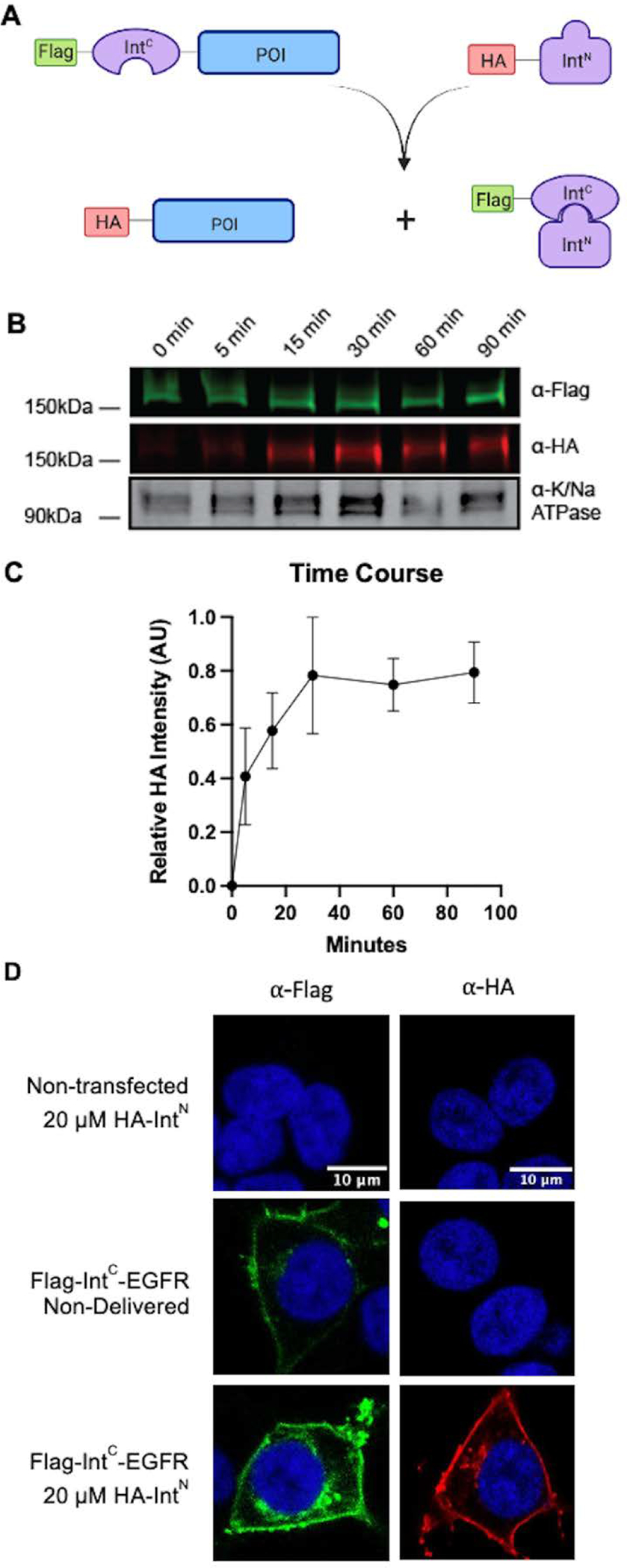
Figure 1. EGFR is site-specifically modified using a split intein system in live cells.

A) Scheme illustrating epitope tag exchange with split-intein splicing. B) Time course of splicing following the formation of HA-EGFR product. HEK293T cells were transfected with Flag-NpuC-EGFR and then treated with 20 μM of HA-AvaN over the course of 90 minutes. The membrane fraction was then extracted and analyzed by western blot (anti-Flag – green, anti-HA – red, anti-Na/K ATPase – gray scale). C. Quantification of B (n=3, error bars represent SEM) D) Immunofluorescence HEK293T cells transfected with Flag-NpuC-EGFR and incubated with HA-AvaN. Cells were fixed with paraformaldehyde and then stained with either anti-Flag (green) or anti-HA (red) and DAPI (blue) followed by imaging.
