Abstract
Background:
CXCR3 is a chemokine receptor and is expressed on innate and adaptive immune cells. It promotes the recruitment of T-lymphocytes and other immune cells to the inflammatory site in response to the binding of cognate chemokines. Upregulation of CXCR3 and its chemokines has been found during atherosclerotic lesion formation. Therefore, the detection of CXCR3 by positron emission tomography (PET) radiotracer may be a useful tool to detect atherosclerosis development noninvasively. Herein, we report the synthesis, radiosynthesis, and characterization of a novel fluorine-18 (F-18, 18F) labeled small-molecule radiotracer for the imaging of the CXCR3 receptor in mouse models of atherosclerosis.
Methods:
The reference standard (S)-2-(5-chloro-6-(4-(1-(4-chloro-2-fluorobenzyl)piperidin-4-yl)-3-ethylpiperazin-1-yl)pyridin-3-yl)-1,3,4-oxadiazole (1) and its corresponding precursor 9 were synthesized using organic syntheses. The radiotracer [18F]1 was prepared in one-pot, two-step synthesis via aromatic 18F-substitution followed by reductive amination. Cell binding assays were conducted using 1, [125I]CXCL10, and CXCR3A- and CXCR3B-transfected human embryonic kidney (HEK) 293 cells. Dynamic PET imaging studies over 90 min were performed on C57BL/6 and apolipoprotein E (ApoE) knockout (KO) mice that were subjected to a normal and high-fat diet for 12 weeks, respectively. Blocking studies were conducted with preadministration of the hydrochloride salt of 1 (5 mg/kg) to assess the binding specificity. Time-activity curves (TACs) for [18F]1 in both mice were used to extract standard uptake values (SUVs). Biodistribution studies were performed on C57BL/6 mice, and the distribution of CXCR3 in the abdominal aorta of ApoE KO mice was assessed by immunohistochemistry (IHC).
Results:
The reference standard 1 and its precursor 9 were synthesized over 5 steps from starting materials in good to moderate yields. The measured Ki values of CXCR3A and CXCR3B were 0.81 ± 0.02 nM and 0.31 ± 0.02 nM, respectively. [18F]1 was prepared with decay-corrected radiochemical yield (RCY) of 13 ± 2%, radiochemical purity (RCP) >99%, and specific activity of 44.4 ± 3.7 GBq/μmol at the end of synthesis (EOS) (n=6). The baseline studies showed that [18F]1 displayed high uptake in the atherosclerotic aorta and brown adipose tissue (BAT) in ApoE KO mice. The uptake of [18F]1 in these regions was reduced significantly in self-blocking studies, demonstrating CXCR3 binding specificity. Contrary to this, no significant differences in uptake of [18F]1 in the abdominal aorta of C57BL/6 mice were observed in both baseline and blocking studies, indicating increased CXCR3 expression in atherosclerotic lesions. IHC studies demonstrated that [18F]1-positive regions were correlated with CXCR3 expression, but some atherosclerotic plaques with significant size were not detected by [18F]1, and their CXCR3 expressions were minimal.
Conclusion:
The novel radiotracer, [18F]1 was synthesized with good RCY and high RCP. In PET imaging studies, [18F]1 displayed CXCR3-specific uptake in the atherosclerotic aorta in ApoE KO mice. [18F]1 visualized CXCR3 expression in different regions in mice is in line with the tissue histology studies. Taken together, [18F]1 is a potential PET radiotracer for the imaging of CXCR3 in atherosclerosis.
Keywords: Atherosclerosis, CXCR3 chemokine receptor, PET imaging, Inflammation, Small-molecule radiotracer
Introduction
Atherosclerosis is a chronic inflammatory disease of the artery wall and is characterized by ectopic fat deposition within the arterial wall, and the lesion development is mediated by a vicious cycle of inflammatory response and cell death.[1] Chemokines and chemokine receptors are key signal axis that mediates the leukocyte migration into the artery wall, thereby promoting the progression of atherosclerotic lesions.[2] Chemokines are a large family of small peptides that signal through cell surface receptors and induce a chemotactic response in the recipient cells.[3, 4] The two major subfamilies of chemokines are CC chemokines and CXC chemokines, named after their primary structures, wherein CC chemokines have two N-terminal cysteines next to each other and CXC chemokines have two N-terminal cysteines separated by one amino acid. Each chemokine has single or multiple chemokine receptors as its partners and vice versa. CXCR3 is one of the six CXC chemokine receptors and is highly expressed on different types of leukocytes, including CD4+ type-1 helper T cells (Th1), natural killer (Nk) cells, and monocytes under inflammation and infection conditions.[4] In fact, the upregulation of CXCR3 and its chemokines has been found in a variety of inflammatory diseases such as multiple sclerosis, arthritis, and atherosclerosis.[5] CXCR3 inhibition is emerging as a promising therapeutic strategy for inflammatory disorders.[5]
CXCR3 binds with three CXC chemokines, CXCL9, CXCL10, and CXCL11, and mediates intracellular signaling.[3, 4] These chemokines are secreted by macrophages and endothelial cells during the formation of atherosclerotic lesions,[6] thereby playing integral roles in the recruitment of CXCR3-expressing proinflammatory leukocytes to the lesion and thus promoting inflammation and, ultimately, atherosclerosis.[6–8] It has been reported that the atherosclerotic lesion formation was shown to decrease with the deletion of either CXCR3 or its ligand CXCL10 in apolipoprotein E (ApoE) knockout (KO) mice, a widely used rodent model of atherosclerosis.[9, 10]
Over the last two decades, an extensive array of small-molecule CXCR3 inverse agonists have been identified from high-throughput, virtual screening approaches.[14] Initially, Amgen and Neurocrine Biosciences reported 8-azaquinazolinone-based molecules as CXCR3 antagonists with nanomolar affinities [14, 15], then pyridyl-piperazinyl-based molecules with the highest affinities and selectivity were disclosed by Merck Research Laboratories.[14] The latter is taken into consideration in this study. Scheme 1 depicts the strategic and consecutive development of various antagonists based on a pyridyl-piperazinyl-piperidine scaffold flanked by a benzyl unit and a polar group such as amide or substituted amide. A representative compound of this class is VUF11211 (Kd = 0.65 nM) which antagonizes CXCR3-chemokines action.[16, 17] Despite the availability of such highly potent CXCR3 antagonists, the development of small-molecule radioligands is limited. Recently, tritium (H-3, 3H) labeled VUF11211 ([3H]VUF11211) was assessed in vitro, and the results suggested that it has a high affinity for human CXCR3 and has fast binding kinetics.[17] The positron emission tomography (PET) imaging probes of such compounds can provide sensitive Information that enables the investigation of pathophysiological processes of various inflammatory disorders, including atherosclerosis.
Various radiotracers for chemokine receptors, mostly labeled with radiometal, have previously been developed to visualize the inflammation mechanism involved in atherosclerosis.[18] [68Ga]Ga-Pentixafor and [64Cu]Cu-DOTA-DAPTA have successfully imaged inflammatory atherosclerosis via CXCR4 and CCR5 chemokine receptors, respectively.[19, 20] [18F]2-Fluorodeoxyglucose ([18F]FDG) has shown promise in atherosclerosis imaging, but its use is limited for such imaging because of its non-specific uptake by metabolically active myocardium.[21] To our knowledge, there is no reported PET tracer specific to CXCR3 receptor though its role in atherosclerosis is evident. In this study, we chose compound 1 from the reported CXCR3-specific compound libraries. VUF11211 may not be suitable for PET studies as it lacks a proper labeling site with carbon-11 or F-18, and the ethylamide part of VUF11211 is metabolically labile. The target compound 1 overcomes these disadvantages, and, in addition, its high potent binding affinity (IC50 = 0.8 nM) to CXCR3 encouraged us to develop and validate its isotopologue [18F]1.[13] Herein, we report the synthesis of [18F]1 and its PET validation in ApoE KO mice in comparison to control C57BL/6 mice as a proof-of-principle.
Results
Computer-Aided Modeling (CAM) Study
Figure 1 shows the results of the CAM experiment. The CAM work shows that 1 has interactions with 14 amino acid residues. It has total 6 p-p interactions with Phe 47, Tyr 60, Trp 109, Phe 131, His 202, and Tyr 205 and 3 hydrophobic interactions with Leu 56, Leu 190, and Leu 215. It also has 5 hydrogen bonds with Glu 204, Arg 212, Asp 297, Lys 300, and Ser 301. As a result, the docking score of 1 was −5.124. Additional hydrophobic interactions contribute to the binding of 1 with CXCR3. The residues involved in hydrophobic interactions include W109, A113, L190, and the carbon chain of R212. The primary stability comes from above mentioned hydrophobic and p-p interactions.
Figure 1.
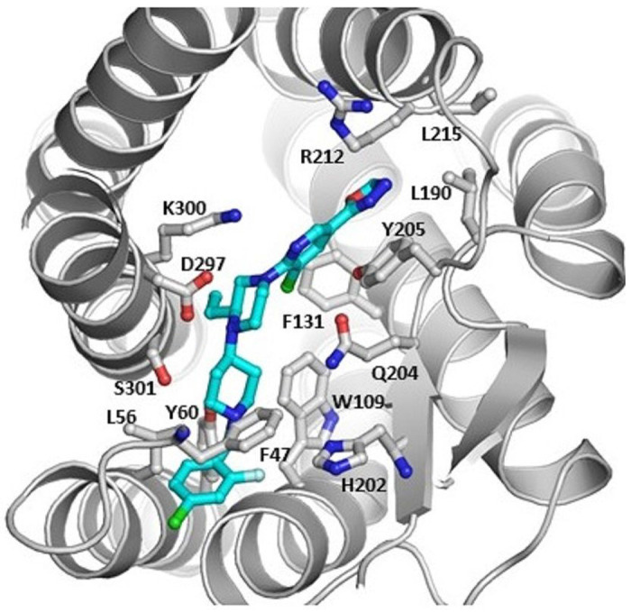
The cartoon of the docking model for compound 1 that has 14 interactions with the amino acids of CXCR3.
Chemistry
Reagents and conditions: a. DIPEA, DMF, 90°C, 18h, b. DCM, TFA, 0°C – rt, 2 h, 97%, c. NaBH(OAc)3, MgSO4 DCE, rt, 36 h, d. 65% NH2NH2, EtOH, 50°C, 12 h, e. CH(OEt)3, CHCl3, reflux, 16 h, then Pyridinium p-toluenesulfonate, reflux, 4 h, f. K2CO3, MeCN, reflux, overnight.
Synthesis of 1 and its radiolabelling precursor 9 are shown in Scheme 2. The syntheses were carried out using reported methods with modifications.15 At first, compound 5 was synthesized by heating 2 and (S)-N-Boc-3-ethylpiperazine (3) under basic conditions, followed by -Boc removal using trifluoroacetic acid (TFA). Compound 5 was used as a common intermediate to make 1 and its precursor 9. Reductive amination of 5 with 1-Boc-piperidin-4-one (6) afforded 7 and similarly with 12 obtained by benzylation of piperidin-4-one (10) gave 13. The carboxylic ester derivatives 7 and 13 were then treated with hydrazine followed by triethyl orthoformate and pyridinium p-toluenesulfonate to obtain the intermediate compound 8 and reference standard 1, respectively. Finally, precursor 9 was obtained from 8 upon Boc deprotection. Reference standard 1 was prepared as a hydrochloride (HCl) salt. After 1 was dissolved in the ethanol (EtOH), an excess amount of conc. HCl was added to the solution. The whole solution was further dried in a stream of nitrogen until a white solid was left behind. The residual solvent was further removed in vacuo and used as it was as a blocking agent. 1·HCl salt was easily dissolved in water.
Radiochemistry
Initially, literature method[22] for the automated preparation of F-18 benzylhalides has tempted us to exploit the strategy to prepare [18F]1. Scheme 3A depicts an attempted 4-step radiosynthesis approach that involves i) nucleophilic substitution of 4-chloro-2-nitrobenzaldehyde (14) with [18F]fluoride for [18F]15 ii) reduction to alcohol [18F]16, iii) bromination to [18F]17, iv) heating bromide with 9 under basic conditions to make [18F]1. We have used a semi-automated setup to prepare the desired radiolabelled intermediates and finally [18F]1. The intermediate [18F]15 was trapped on a C18 solid phase extraction (SPE) cartridge (1 g), and the subsequent reduction and halogenation were performed on SPE support. At first, an aqueous solution of sodium borohydride (NaBH4, 3 mL, 8 – 10 mg/mL) was passed slowly through support over 10 min for converting [18F]15 to [18F]16, and >85% radiochemical conversion (RCC) based on radio-thin layer chromatography (TLC) was noticed (n ≥ 10). After a rinse with water (5 mL) and a flush with nitrogen, 48% bromic acid (HBr) or 33% HBr/acetic acid (AcOH, 3 mL) was passed through the SPE support over 15 to 20 min for converting [18F]16 to [18F]17 and moderate RCC (35±10%, radio-TLC) was noticed at this step (n ≥ 8). Neither an increase in reaction time nor the pre-heated (50°C, 5 – 10 min) HBr was not useful in enhancing RCC to [18F]17. Considerable damage to C18 support was observed when warm HBr was used. At this stage, after a rinse with water (5 mL) and a flush with nitrogen, the radiolabelled intermediates mixture trapped in the C18 SPE cartridge was eluted with N,N-dimethylformamide (DMF) or acetonitrile (MeCN, 2 mL) and passed through an anhydrous sodium sulfate (Na2SO4) cartridge into a second reaction vial that already contained 9 (~3 mg) and potassium carbonate (K2CO3, ~10 mg) or diisopropylethylamine (DIPEA, ~10 mL). The vial was then stirred at 100°C for 20 – 30 min. However, no desired RCC was noticed during different time intervals and temperatures.
Reagents and Conditions a. K222/K2CO3/[18F]F (0.74 – 3.7 GBq), 14 (4 mg), DMSO (0.5 mL), 140°C, 15 min, 80±5% b. 9 (5±0.5 mg), NaBH3CN (11±1 mg), CH3COOH (10mL), DMSO (0.5 mL), 110°C, 20 min, 36±4% c. aq. NaBH4 (3 mL, 8 – 10 mg/mL), rt, 10 min, 85±5% d. 48% HBr (3 mL), rt, 20 min, 35±15%
Having seen the difficulties associated with this four-step radiosynthesis method, we decided to put efforts towards a one-pot, two-step reductive amination strategy (Scheme 3B) that involved 9 and [18F]15. [23] This approach rewarded us not only with reduced radiosynthesis time but also with good RCYs and reproducibility. Precursor 9 is mixed with sodium cyanoborohydride (NaBH3CN) and AcOH and transferred into the reaction vial that already has [18F]15 and heated at 110°C for 20 min (RCC 36 ± 4%, n ≥ 10).
Table 1 shows the optimization of step 2 towards good RCC of [18F]15 to [18F]1. Dimethylsulfoxide (DMSO) was found to be the optimal solvent compared to DMF or methanol (MeOH). The addition of AcOH and the high temperature was necessary for an improved RCC. A usual C18 SPE workup followed by semi-preparative reverse phase HPLC purification afforded [18F]1 in decay-corrected radiochemical yield (RCY) of 13 ± 2% (n = 10) at the end of synthesis (EOS). Figure 2(a) shows the semi-preparative HPLC chromatograms of [18F]1. The HPLC purification was performed using a Phenomenex Gemini C18 Semi-preparative column (5 μm, 250 X 10 mm) equipped with UV (254 nm) and radioactivity detectors by eluting with an isocratic mobile phase composed of 65% MeCN, 5% MeOH, 30% water, and 0.1% triethylamine (NEt3) at a flow rate of 5 mL/min. The radioactive [18F]1 was collected between 16 - 17 min and was well-separated from the rest of the UV and radioactivity peaks. After the purification, the collected product was formulated in 10% EtOH/0.9% saline containing 0.5% sodium ascorbate. The radiosynthesis took 100 min. The final reformulated aliquot was further characterized by HPLC analysis using a Phenomenex Gemini C18 analytical column (5 μm, 250 X 4.6 mm) equipped with UV and radioactivity detectors by eluting with the same HPLC solvent at a flow rate of 1 mL/min. The radioactive peak was eluted between 15-17 min. The single injection of the final aliquot confirmed that the obtained radioactive product was >99% radiochemical purity (RCP) with a specific activity of 44.4 ± 3.7 GBq/μmol at the EOS (n=6). The radiotracer was also shown to be stable for more than 10 hours. Then, the aliquot was mixed with nonradioactive 1 standard solution and injected into the same HPLC system. Figure 2(b) shows the result of the co-injection, confirming that the radioactive peak was eluted together with reference standard 1.
Table 1:
The table of optimization for step 2 from [18F]15 to [18F]1
| Entry | Solvent | Additive | Temp | RCC (%)a |
|---|---|---|---|---|
| 1 | MeOH | HOAc | 50 | <3 |
| 2 | MeOH | None | 50 | - |
| 3 | DMF | HOAc | 50 | 5±3 |
| 4 | DMF | HOAc | 110 | 12±5 |
| 5 | DMF | None | 110 | <5 |
| 6 | DMSO | None | 110 | 11±5 |
| 7 | DMSO | HOAc | 110 | 36±4 |
Conditions: [18F]15(740 – 2220 MBq), 9 (5±0.5 mg), NaBH3CN (11±1 mg), HOAc (10mL), 20 min
Based on radio-TLC (0.1% Et3N, 10% MeOH in DCM, Rf 0.3)
Figure 2.
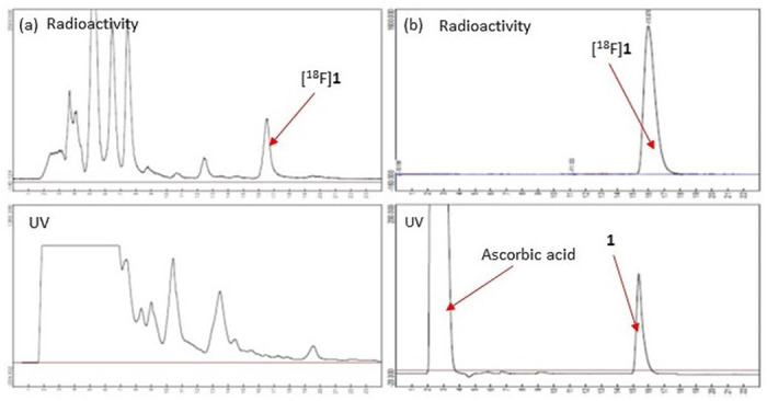
(a) The semi-preparative HPLC of [18F]1 and (b) The analytical HPLC of [18F]1. Co-injection of nonradioactive standard 1 confirmed that the collected product was an authentic product.
Cell-Binding Assay
The equilibrium inhibition constants (Ki) value of 1 at CXCR3 receptor was measured through competition binding assay using NBI74330 (Tocris Bioscience, Bristol, UK), an antagonist of CXCR3 receptor, [125I]CXCL10 (ViTrax Inc., Placentia, CA) and CXCR3 transfected human embryonic kidney (HEK) 293 cells. CXCR3 has two major variants, CXCR3A and CXCR3B, with distinctive biological roles,[24] so two types of HEK293 cells containing each CXCR3 variant were prepared. Before the cell-binding assay, two cDNA constructs containing CXCR3 transcript variant A (CXCR3A, GenBank accession No. NM_001504; GenScript) and CXCR3 transcript variant B (CXCR3B, GenBank accession No. NM_001142797; GenScript) and HEK293 cells were purchased from GenScript Biotech (Piscataway, NJ) and ATCC (Manassas, VA), respectively. Then the cDNA constructs were transfected to HEK293 cells, and the transfected cells were cultured to get enough amount for the cell-binding assay. After the CXCR3-transfected cell membranes were harvested, competitive binding assays were performed by measuring the concentration of free [125I]CXCL10 at different concentrations of 1 and NBI74330. The IC50 value of 1 was measured based on the regression plot between the free radioligand and the different concentrations of 1. The Ki values of 1 were calculated by the Cheng-Prusoff equation, indicating that 0.81 ± 0.02 nM and 0.31 ± 0.02 nM in CXCR3A and CXCR3B, respectively. The regression plots of the cell-binding assays can be found in Supplementary Information.
PET imaging Studies
[18F]1 was further evaluated for reliability as a CXCR3 radiotracer using mouse models of atherosclerosis. For this purpose, we tested [18F]1 in ApoE KO mice, a widely used murine model of atherosclerosis.[25] Six-week-old ApoE KO male mice (N=6) and C57BL/6 male control mice (i.e., a background strain of ApoE KO mouse, ApoE-wildtype control; N=6) were purchased from Jackson Laboratory (Bar Harbor, ME). The ApoE KO mice were placed on a high-fat diet over 12 weeks to accelerate atherosclerosis development. Meanwhile, C57BL/6 mice were fed a normal diet for the same period, and they did not produce atherosclerosis in the aorta. At 12 weeks, PET/computed tomography (CT) studies were performed using both mice groups. Dynamic PET scans (90 min acquisition) were performed beginning 5 min after the intravenous injection of [18F]1 followed by a ~10 min CT scan. Usually, one week after the first baseline PET/CT scan, the second blocking PET/CT scan was performed. This time, 1·HCl salt (5 mg/kg) was used as a blocking agent and intravenously injected 5 min before the radiotracer injection.
Figure 3 shows the representative PET images of two ApoE KO mice and one C57BL/6 mouse, and the blocking images of the same mouse is shown next to the baseline images. The PET images of the ApoE KO mice showed high uptake of [18F]1 in the atherosclerotic abdominal aorta (red arrow), brown adipose tissue (BAT, blue arrow), liver, kidney, and bladder. The PET images of the C57BL/6 mouse displayed a similar uptake except for the atherosclerotic abdominal aorta. Their blocking PET images indicated that both BAT and atherosclerotic abdominal aorta were missing in the ApoE KO mice, and the BAT was missing in the C57BL/6 control mice, demonstrating that [18F]1 is a CXCR3-specific radiotracer. The high uptake of the liver and bladder reflects its metabolism. The atherosclerotic plaques were observed at multiple sites in the abdominal aorta.
Figure 3.
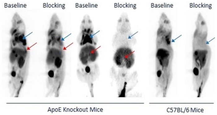
Representative CXCR3 PET images of [18F]1 in two ApoE KO mice and one C57BL/6 control mouse. The specific uptake of [18F]1 was observed in the atherosclerotic aorta (red arrow) and BAT (blue arrow) of ApoE KO mice and the BAT of C57BL/6 control mice.
Figure 4 indicates the average time-activity curves (TACs) of the BAT of both ApoE KO mice (N=3) and C57BL/6 control mice (N=5). Preadministration of 1.HCl (5 mg/kg) in blocking studies reduced the uptake of [18F]1 in the atherosclerotic abdominal aorta (standard uptake value (SUV) 1.1) in ApoE KO mice and reduced the uptake in BAT region (SUV 1.2 – 1.5) in both groups. A comparison of TACs from all the PET scans showed that the binding of [18F]1 is specific to CXCR3, especially in these regions of interest (ROIs). The uptake levels in all the scans in ROI were retained at the more or less same level throughout the 90 min scan, indicating a slower washout of the tracer from these regions. Figure 5 displays the area under the curves (AUCs) obtained based on the TACs. The blocking effects in the BAT of ApoE KO mice and C57BL/6 control mice were 23.4% and 45.9%, and those in the atherosclerotic abdominal aorta were 16.9% and 4.7%, respectively.
Figure 4.
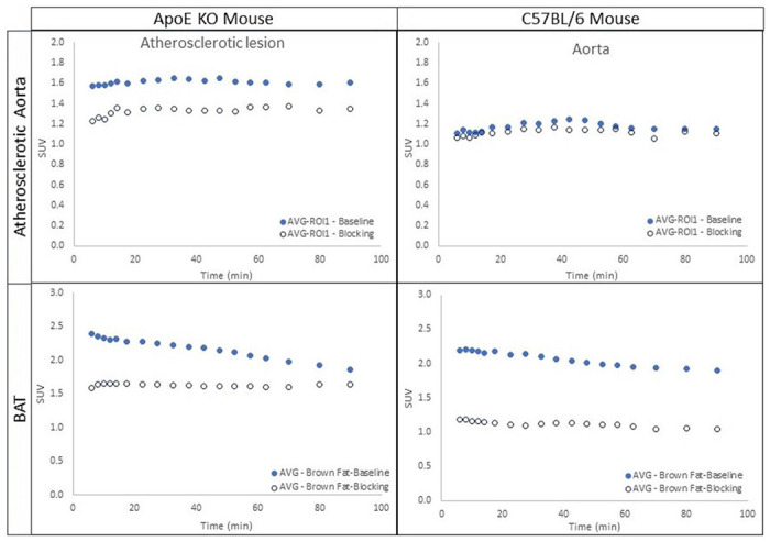
The average time-activity curves (TACs) of ApoE KO mice (N=3) and C57BL/6 control mice (N=5). The TACs indicate the blocking effects of [18F]1, demonstrating the specificity of [18F]1.
Figure 5.
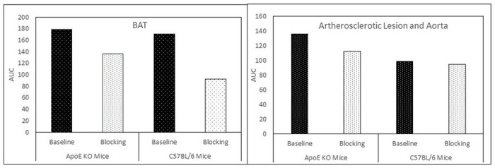
The areas under the curve (AUCs) of the BAT and atherosclerotic aorta of ApoE KO mice and the BAT and corresponding aorta of C57BL/6 mice.
Biodistribution Studies
Biodistribution studies were also performed to discover the drug distribution within normal mice. 17-week-old male and female C57BL/6 mice were purchased to match the age of the imaging study group. [18F]1 was intravenously injected into these mice (N=4/time point), then they were sacrificed at 10, 30, and 60 min after the injection. Their major organs, including the brain, blood, lung, liver, stomach, heart, spleen, kidney, muscle, bone, large intestine, small intestine, aorta, liquinal lymph nodes (LN), interscapular BAT, and tail, were harvested. Their radioactivity was counted by an automated g-counter. The biodistribution was calculated as an average of the percent injected dose per gram tissue (%ID/g tissue) where the counted radioactivity of each organ was divided by the weight of each tissue and the total radioactivity that circulated in the mouse body at the time of the euthanasia.
Figure 6 shows the results of the biodistribution study. The results show that the uptake properties of [18F]1 were similar between males and females. [18F]1 displayed high uptake in the lung (27% ID/g) and kidney (22% ID/g) followed by the BAT (15% ID/g), aorta (13% ID/g), and heart (12% ID/g) at 10 min postinjection. At 30 min postinjection., the uptake was reduced to ~60% and then to ~40% at 60 min postinjection in these regions. However, the radiotracer uptake is relatively low (12% ID/g) at 10 min postinjection but increased to 22% ID/g at 30 min postinjection in the liver. The uptake properties of [18F]1 in the liver were due to its metabolism. The blocking experiments would clarify the binding specificity in these regions.
Figure 6.
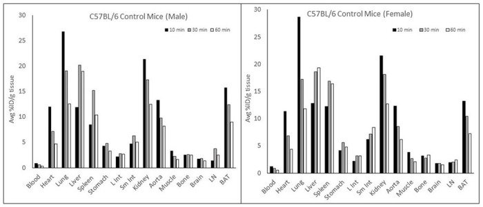
The results of biodistribution using male and female control C57BL/6 mice. The uptake of [18F]1 in each organ was measured at 10, 30, and 60 min after its intravenous injection.
Tissue Analysis and Immunohistochemistry (IHC) Analysis
Figure 7 indicated that [18F]1 could detect atherosclerotic plaques in the aorta where high levels of CXCR3 expression (green) were evident (Region I in Figure 7(a) and Region III in Figure 7(b). It is noteworthy that not all atherosclerotic plaques were detected by PET via [18F]1. For instance, Region II (Figure 7(a)) has a plaque, and its size is equal to the one in Region I (Figure 7(a), hematoxylin and eosin (H&E) staining images). Nevertheless, Region II was not detected by PET (Figure 7(a), PET image; blue arrow). Interestingly, the plaque at Region II has notably lower expression levels of CXCR3 compared to the plaque at Region I. These results further support CXCR3-specific radiotracer-dependent detection.
Figure 7.
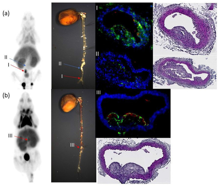
CXCR3 PET imaging using [18F]1 and their corresponding cardiovascular tissues obtained from the ApoE KO mice. (a) Region I (red arrow) is a [18F]1-positive region, and the corresponding aorta tissue was highly expressing CXCR3 (Green color); meanwhile, region II (blue arrow) has a significant atherosclerotic plaque. Nevertheless, it is a [18F]1-negative region of PET imaging and indicated low CXCR3 expression levels. These results suggest CXCR3 expression-dependent PET detection by [18F]1. (b) Region III also indicated [18F]1-positivity, and subsequent histologic analysis revealed a significant atherosclerotic plaque with high CXCR3 expression levels (green detection in the fluorescence image), consistent with CXCR3 expression-dependent detection by the radiotracer.
Discussion
We chose 1 from the reported CXCR3 ligand libraries because of its subnanomolar affinity and feasibility of labeling with fluorine-18 and decided to develop it as an F-18 CXCR3 radiotracer. We performed a CAM study to determine the CXCR3 binding pockets and the ligand binding interactions (hydrophobic and π-π in this case) that lead to a stable receptor-ligand complex. Reference standard 1 and its precursor 9 were prepared over five steps from commercially available starting materials. Although copper or palladium-catalyzed methods are available for aromatic 18F-substitution using arylboronate or aryltin precursors, we presumed that benzylamines are not appropriate precursors for such labeling reactions. In addition, the occurrence of protodestannylation and protodeboronylation side reactions during 18F-labeling and the presence of two chlorine atoms on two different aromatic positions in 1 made us use a non-catalyzed multi-step radiosynthesis.[26, 27] The first radiosynthesis that rapidly converted benzaldehyde derivative [18F]15 to benzyl bromide derivative [18F]17 via benzyl alcohol derivative [18F]16 failed in the final step that coupled [18F]17 with 9. We presumed the failed final step might be due to the presence of residual water and the decomposition of impure [18F]17 during the reaction.[22] In addition, technical differences in synthesis setup compared to the literature method amounted to moderate conversion of [18F]16 to [18F]17. Then, we tried two-step radiosynthesis that coupled [18F]15 with 9 with reductive amination, which turned out to be a more efficient method.[23] The result also indicated that the acidic catalyst facilitated reductive amination. The activity of NaBH3CN was also very critical for the success of the second step. When the storage of NaBH3CN was not strictly guaranteed under inert gas, and its freshness was in doubt, a new fresh bottle was purchased and used. When an opened bottle was routinely used for radiosynthesis every week or more, we slowly increased its amount to compensate for its lost freshness.
Cell-binding assays indicated that the Ki values of 1 were 0.81 ± 0.02 nM and 0.31 ± 0.02 nM in CXCR3A and CXCR3B, respectively. Therefore, 1 binds to two CXCR3 variants with the same affinity. CXCR3A mediates the chemotaxis of immune cells. [24] CXCR3A and CXCR3B co-express in the immune cells, but CXCR3A expresses almost 1000 times more abundant than CXCR3B.[28] CXCR3A and CXCR3B are also preferentially expressed in some types of cells. For example, CXCR3A is primarily expressed in human mesangial cells, while CXCR3B is preferentially expressed in microvascular endothelial cells.[28] CXCR3A and CXCR3B couple with inhibitory G (Gi) protein and stimulatory G alpha (Gs) protein, respectively, so their functional roles are opposite.[28] Such distinctive functional differences are prominent in cancer biology, where CXCR3A promotes the migration of tumor cells and angiogenesis, but CXCR3B inhibits such metastatic properties and promotes apoptosis in many cancers.[29] Overall, the PET images using [18F]1 might indicate all CXCR3 variants depending on their expression levels. When the PET images of [18F]1 indicate high inflammatory regions, it is expected to represent CXCR3A because of its predominant expression.
The baseline and blocking PET studies and IHC studies demonstrated that [18F]1 was a specific CXCR3 radiotracer to atherosclerotic lesions. The biodistribution and PET imaging studies also indicated that the uptake of [18F]1 in the atherosclerotic aorta was obvious in the ApoE KO mice, despite that its background uptake was high in the control C57BL/6 mice. The fatality of atherosclerosis comes from its lesions in the coronary and carotid arteries, which cause myocardial infarction, angina, and stroke in humans. Therefore, further studies using other animal models that develop atherosclerotic plaques in those regions are required to determine if this radiotracer can identify atherosclerotic lesions in the heart and other critical regions. We also need further studies to evaluate the sensitivity of these compounds to identify inflammatory lesions in the lung, liver, and intestine since the uptake of [18F]1 was very high in normal C57BL/6 mice in biodistribution studies. This is important because various inflammatory diseases such as chronic obstructive pulmonary disease, hepatic cirrhosis, and Crohn’s disease, which feature inflammation mechanisms in their pathologies, develop in those regions, respectively.
The PET images also indicated that the uptake of [18F]1 was specific in the BAT. It has been reported that CXCR3 mediates T-lymphocyte migration to the adipose tissue under chronic inflammatory conditions, in particular, induced by obesity.[30, 31] In addition, a high-fat diet in rodents was shown to induce CXCR3 expression in the adipose tissue as a result of increased inflammation.[32] In the current study, the diet schedule may have induced chronic inflammation in the adipose tissue of both the ApoE KO mice and C57BL/6 control mice, resulting in high uptake of [18F]1 in the BAT region.
The IHC analysis using the aorta tissues of ApoE KO mice that were imaged for PET indicated that not all atherosclerotic lesions were identified by [18F]1. This result was also related to the sensitivity of [18F]1. Further studies may be required to test the sensitivity of this radiotracer. Our study also demonstrated that [18F]1 detects only select atherosclerotic lesions which are high in CXCR3 expression levels, consistent with the high affinity of [18F]1 to CXCR3. An interesting question is raised at this point whether the positive detection by the radiotracer (thus high levels of CXCR3 expression) could reflect any pathophysiologic status of the plaque. CXCR3 is a receptor for CXCL9, CXCL10, and CXCL11, which are INF-γ inducible and thus associated with inflammatory disorders, including atherosclerosis.[6] It has been suggested that CXCR3 has integral roles in the recruitment of proinflammatory cells during plaque formation, thereby promoting atherogenesis,[9] however its potential role in the progression of plaque leading to an advanced lesion (i.e., a plaque to cause an acute thromboembolic event or Type V and VI lesions [33]) remains obscure. Further studies are needed to clarify how CXCR3 expression levels transition as an atheroma evolves from the initial fatty streak to an advanced lesion so that the clinical implications of CXCR3-positive plaques, thus the PET detection, can be evaluated.
Conclusions
The target compound 1, a derivative of VUF11211, was identified as a lead compound for PET radiotracer for CXCR3 chemokine receptors as a result of a literature search and CAM study. After the target compound 1 and its precursor compound 9 were prepared by multi-step organic synthesis, cell-binding assays confirmed its binding affinity. [18F]1 was prepared by an F-18 incorporation followed by reductive amination, a one-pot and two-step radiosynthesis. [18F]1 was validated as a CXCR3 PET radiotracer using mouse models of atherosclerosis. The baseline scan and blocking PET scan, which pre-administered 1.HCl salt intravenously, confirmed that [18F]1 is a CXCR3-specific radiotracer and indicated atherosclerotic plaques in the abdominal aorta of ApoE KO mice. The IHC studies using the atherosclerotic abdominal aorta also confirmed that the radiotracer indicated CXCR3-abundant aorta regions. However, the radiotracer did not detect some atherosclerotic plaques, which have a similar size to detectable plaques. IHC revealed that these undetected plaques have low levels of CXCR3 expression, indicating a lack of correlation between the plaque size and CXCR3 expression levels. Further analysis is required to correlate the CXCR3 PET imaging with the plaque progression. In addition, the biodistribution studies using control C57BL/6 mice showed high background in the lungs, heart, aorta, liver, and intestine. The new radiotracer successfully indicated the atherosclerotic abdominal aorta in the ApoE KO mice despite the high background uptake in the regions of C57BL/6 control mice. Therefore, further studies are also required to demonstrate the versatility of [18F]1 in detecting inflammatory lesions in the coronary artery, liver, lungs, and intestine. In conclusion, [18F]1 is the first promising F-18 radiotracer for CXCR3, and its properties, such as CXCR3 sensitivity, have to be improved with further development.
Methods
General Information
All the reactions were carried out in oven-dried glassware under an atmosphere of nitrogen or argon. All commercially obtained reactants and reagents were used as such without any further purification. Unless specified, all chemicals were purchased from MilliporeSigma (Burlington, MA). All purification solvents such as hexane, ethyl acetate (EtOAc), dichloromethane (DCM), and MeOH, and all anhydrous solvents such as dioxane, DCM, and DMF were purchased from Fisher Scientific (Waltham, MA). For lower temperature reactions, an ice bath (0°C) and an acetone-dry ice bath (−78°C) were used. Reactions were monitored by TLC using silica gel-coated aluminum plates with F-254 indicator. 1H (500 MHz), 13C (126 MHz), 19F (476 MHz) nuclear magnetic resonance (NMR) spectra were recorded on Bruker Avance III 500 MHz spectrometer. Electrospray (ESI) and Atmospheric Pressure Chemical Ionization (APCI) mass spectra (MS) and high-resolution mass spectra (HRMS) were obtained using a Thermo LCQ Deca XP Ion Trap Mass Spectrometer and a Bruker Impact II Q-TOF Mass Spectrometer, respectively. The NMR spectra and MS spectra can be found in Supplementary Information.
Organic Synthesis
Methyl (S)-6-(4-(tert-butoxycarbonyl)-3-ethylpiperazin-1-yl)-5-chloronicotinate (4)
To the solution of methyl 5,6-dichloronicotinate (2, 2.200 g, 10.680 mmol) in anhydrous DMF (25 mL) was added tert-butyl (S)-2-ethylpiperazine-1-carboxylate (3, 2.500 g, 11.747 mmol) and DIPEA (9.500 mL, 53.398 mmol) under nitrogen. The reaction contents were then heated to 90°C and stirred for 18 hours. Following this time, reaction was cooled, quenched with water (40 mL) and EtOAc (40 mL), and added 1 M sodium bicarbonate (NaHCO3) solution (40 mL). The aqueous layer was extracted with EtOAc (3x 30 mL), and the combined organic layers were washed with brine (50 mL) and dried over anhydrous Na2SO4. The crude was concentrated and purified by column chromatography (0–20% EtOAc in Hexanes). 4 was obtained as a pale-yellow gummy solid. Rf: 0.4 (20% EtOAc in hexane). Yield: 3.530 g, 9.212 mmol, 86.3%. 1H NMR (500 MHz, chloroform-d (CDCl3)) δ 8.71 (d, J = 2.0 Hz, 1H), 8.11 (d, J = 2.0 Hz, 1H), 4.14–3.96 (m, 4H), 3.89 (s, 3H), 3.19 (q, J = 10.4, 8.0 Hz, 1H), 3.08 (dd, J = 12.9, 3.6 Hz, 1H), 2.98–2.88 (m, 1H), 1.79 (dt, J = 15.0, 7.6 Hz, 1H), 1.70–1.57 (m, 1H), 1.47 (s, 9H), 0.87 (t, J = 7.5 Hz, 3H). 13C NMR (126 MHz, CDCl3) δ 171.14, 165.11, 160.39, 154.93, 147.78, 139.93, 119.78, 119.53, 79.78, 60.39, 52.16, 50.71, 48.54, 28.44, 14.20, 14.12, 10.71. HR-ESI-MS [M + Na]+ C18H26ClN3O4, calcd 406.15095, found [M + Na]+ 406.14997
Methyl (S)-5-chloro-6-(3-ethylpiperazin-1-yl)nicotinate (5)
To the solution of 4 (2.980 g, 7.763 mmol) in dry DCM (10 mL) at 0°C was added dropwise TFA (6 mL), warmed to room temperature (RT), and stirred for 90 min. Following this time, the crude was basified using 5.9 g K2CO3 in 100 mL water (pH > 7) and extracted with DCM (3x 70 mL). Combined organic layers are dried over anhydrous Na2SO4 and concentrated under reduced pressure to obtain 5 as a white solid and used as such in next reaction. Rf 0.5 (10% MeOH in DCM). Yield: 2.160 g, 7.609 mmol, 98.2%. 1H NMR (500 MHz, CDCl3) δ 8.73 (d, J = 2.0 Hz, 1H), 8.11 (d, J = 2.0 Hz, 1H), 4.34–3.95 (m, 2H), 3.89 (s, 3H), 3.44–2.92 (m, 3H), 2.78 (ddt, J = 9.4, 6.5, 3.3 Hz, 1H), 2.65 (dd, J = 12.4, 10.1 Hz, 1H), 1.64–1.41 (m, 2H), 0.99 (t, J = 7.5 Hz, 3H). 13C NMR (126 MHz, CDCl3) δ 165.16, 160.02, 147.73, 139.84, 119.80, 119.12, 56.55, 54.29, 52.08, 49.27, 45.72, 26.87, 10.17. HR-ESI-MS [M + H]+ C13H18ClN3O2, calcd 284.10875, found 284.11518
Methyl (S)-6-(4-(1-(tert-butoxycarbonyl)piperidin-4-yl)-3-ethylpiperazin-1-yl)-5-chloronicotinate (7)
To the 5 (1.110 g, 3.886 mmol) in 1,2-dichloroethane (DCE, 10 mL) was added anhydrous magnesium sulfate (MgSO4, 0.935 g, 7.772 mmol) and slowly 1-Boc-4-piperidinone (6, 0.770 g, 3.886 mmol) dissolved in 3 mL DCE and stirred for 30 min at RT. Following this time, Sodium triacetoxyborohydride (1.640 g, 7.772 mmol) was added in 4 portions over 15 min and the slurry was stirred for 24 hours. Following this time, the reaction was quenched with NaHCO3 (3 g in 100 mL water) and extracted with DCM (3x 40 mL). Combined organic layers were dried over Na2SO4 and the crude was purified using column chromatography. 0–50% EtOAc in hexane used initially to collect 7 as a gummy solid. Then 0–10% MeOH in DCM was used to elute the unreacted starting material (0.460 g). Yield: 0.930 g, 1.996 mmol, 54%.1H NMR (500 MHz, CDCl3) δ 8.72 (d, J = 2.0 Hz, 1H), 8.10 (d, J = 2.0 Hz, 1H), 4.15 (d, J = 18.5 Hz, 3H), 3.89 (s, 3H), 3.85–3.74 (m, 2H), 3.33 (ddd, J = 12.2, 8.9, 2.8 Hz, 1H), 3.23 (dd, J = 12.5, 7.8 Hz, 1H), 2.85 (ddd, J = 11.4, 5.8, 3.2 Hz, 2H), 2.74 (ddt, J = 8.2, 5.0, 3.0 Hz, 1H), 2.63 (ddd, J = 11.5, 8.8, 3.0 Hz, 1H), 1.78–1.54 (m, 6H), 1.46 (s, 9H), 0.94 (t, J = 7.5 Hz, 3H). 13C NMR (126 MHz, CDCl3) δ 165.21, 159.86, 154.69, 147.77, 139.83, 119.35, 118.87, 79.44, 57.09, 55.63, 52.07, 51.74, 48.81, 44.35, 28.43, 20.68, 14.18, 10.12. HR-ESI-MS [M + H]+ C23H35ClN4O4, calcd 467.24251 found 467.24061 tert-Butyl (S)-4-(4-(3-chloro-5-(1,3,4-oxadiazol-2-yl)pyridin-2-yl)-2-ethylpiperazin-1-yl)piperidine-1-carboxylate (8)
To the 7 (0.610 g, 1.308 mmol) in 5 mL EtOH was added 64% hydrazine monohydrate (0.500 mL, 10.464 mmol) and stirred at 50°C for 12 hours. Following this time, the volatile components were evaporated and kept under a high vacuum for 2 hours to obtain a colourless solid that was used as such in the next step. The hydrazide intermediate was dissolved in chloroform (6 mL) and triethyl orthoformate (4 mL) and refluxed (65°C) for 16 hours. Following this time, pyridine-p-toluene sulfonate (0.700 g, 2.616 mmol) was added and refluxed (65°C) for another 4 hours. The reaction was basified using 0.5 g sodium carbonate in 100 mL water and extracted with DCM (3x 35 mL). Organic layers were dried over anhydrous Na2SO4 and purified using 0–5% MeOH in DCM to obtain 8 as a white solid. Yield: 0.425 g, 0.892 mmol, 68% over 2 steps. 1H NMR (600 MHz, CDCl3) δ 8.80 (d, J = 2.1 Hz, 1H), 8.46 (s, 1H), 8.22 (d, J = 2.1 Hz, 1H), 4.28–4.12 (m, 2H), 3.86–3.77 (m, 2H), 3.38 (ddd, J = 12.2, 8.8, 2.8 Hz, 1H), 3.27 (dd, J = 12.5, 7.8 Hz, 1H), 2.90 (dtt, J = 11.4, 5.6, 3.1 Hz, 2H), 2.78 (tt, J = 8.0, 3.0 Hz, 2H), 2.67 (ddd, J = 11.6, 8.8, 3.0 Hz, 2H), 1.76–1.70 (m, 3H), 1.70–1.57 (m, 5H), 1.49 (s, 9H), 0.98 (t, J = 7.4 Hz, 3H). 13C NMR (126 MHz, CDCl3) δ 162.47, 159.46, 154.83, 152.49, 144.67, 137.17, 120.50, 112.89, 79.60, 57.23, 55.80, 53.56, 51.94, 49.00, 44.48, 30.79, 28.58, 25.33, 20.83, 10.28. HR-ESI-MS [M + H]+ C23H33ClN6O3, calcd 477.23809, found 477.23714
(S)-2-(5-Chloro-6-(3-ethyl-4-(piperidin-4-yl)piperazin-1-yl)pyridin-3-yl)-1,3,4-oxadiazole (9)
To the 8 (0.130 g, 0.273 mmol) dissolved in DCM (3 mL) at 0°C was slowly added TFA (1.5 mL). The reaction was then warmed to RT and stirred for 2 hours. Following this time, the reaction was quenched at 0°C with 1.9 g NaHCO3 in 45 mL water (pH between 7–8) and extracted with DCM (3x 15 mL). The organic layers were washed with 10 mL brine. The combined organic layers were then dried and concentrated to obtain 9 as a semi-white solid. Yield: 0.094 g, 0.250 mmol, 91.2%
1H NMR (500 MHz, CDCl3) δ 8.78 (d, J = 2.1 Hz, 1H), 8.46 (s, 1H), 8.20 (d, J = 2.1 Hz, 1H), 3.83–3.70 (m, 2H), 3.52–3.41 (m, 2H), 3.38 (ddd, J = 12.1, 8.4, 2.7 Hz, 1H), 3.30 (dd, J = 12.7, 7.5 Hz, 1H), 3.01–2.80 (m, 4H), 2.75 (pd, J = 8.3, 5.4, 3.0 Hz, 1H), 2.67 (ddd, J = 11.3, 8.4, 2.9 Hz, 1H), 2.05 (qd, J = 12.7, 11.2, 7.3 Hz, 1H), 1.96–1.80 (m, 3H), 1.67–1.52 (m, 2H), 0.95 (t, J = 7.4 Hz, 3H). 13C NMR (126 MHz, CDCl3) δ 162.30, 159.32, 152.36, 144.51, 137.04, 120.49, 112.93, 56.93, 53.40, 51.58, 48.78, 44.21, 44.13, 43.82, 28.36, 23.59, 20.40, 10.14. HR-ESI-MS [M + H]+ C18H25ClN6O, calcd 377.18566, found 377.18459
N-(4-Chloro-2-fluorobenzyl)-4-piperidinone (12)
To a mixture solution of 4-piperidinone hydrochloride monohydrate (10, 1.000 g, 6.510 mmol) and K2CO3 (2.249 g, 16.27 mmol) in MeCN (13 mL) was added 4-chloro-2-fluorobenzyl bromide (11, 1.06 ml, 7.812 mmol) and refluxed overnight. The resulting solution was quenched with sat. ammonium chloride and extracted three times with EtOAc. The organic parts were combined, dried over anhydrous Na2SO4, filtered, and evaporated. The residue was purified by flash chromatography using gradient solvent by increasing from 5–30% EtOAc in hexane for 30 min to give 12 as a yellow oil. 1.251 g, 5.176 mmol, 79.5%
1H NMR (500 MHz, CDCl3) δ 7.37 (t, J = 8.0 Hz, 1H), 7.13 (dd, 1H, J = 8.0, 2.0 Hz, 1H), 7.09 (dd, J = 9.5, 2.0 Hz, 1H), 3.66 (d, J = 1.0 Hz, 2H), 2.77 (t, J = 6.0 Hz, 4H), 2.46 (t, J = 6.3 Hz, 4H). 13C NMR (126 MHz, CDCl3) δ 208.87, 161.17 (d, J = 249 Hz), 134.02 (d, J = 11.25 Hz), 132.06 (d, J = 2.5 Hz), 124.63 (d, J = 3.8 Hz), 123.58 (d, J = 15 Hz), 116.34 (d, J = 25 Hz), 54.00 (d, J = 1.3 Hz), 52.88, 41.35. HR-ESI-MS [M]+ C12H13FClNO, calcd 241.07, found [M + H]+ 242.39
Methyl (S)-5-chloro-6-(4-(1-(4-chloro-2-fluorobenzyl)piperidin-4-yl)-3-ethylpiperazin-1-yl)nicotinate (13)
To the 5 (0.330 g, 1.166 mmol) in 5 mL DCE was added MgSO4 (0.280 g, 2.332 mmol) and slowly 1-(4-chloro-2-fluorobezyl)piperidin-4-one (12, 0.420 g, 1.749 mmol) dissolved in 3 mL DCE and stirred for 60 min at RT. Following this time, sodium triacetoxyborohydride (0.500 g, 1.749 mmol) was added in 4 portions, and the slurry was stirred for 60 hours. The reaction was quenched with NaHCO3 solution (1.2 g in 70 mL water) and extracted with DCM (3x 40 mL). Combined organic layers were dried over anhydrous Na2SO4, and the crude was purified using column chromatography. 0–5% MeOH in DCM was used to elute 13 as a yellow gummy solid. Yield: 0.310 g, 0.610 mmol, 53%. 1H NMR (500 MHz, CDCl3) δ 8.72 (d, J = 2.0 Hz, 1H), 8.09 (d, J = 2.0 Hz, 1H), 7.33 (td, J = 8.1, 2.8 Hz, 1H), 7.13–7.09 (m, 1H), 7.06 (dt, J = 9.6, 2.2 Hz, 1H), 3.89 (s, 3H), 3.87–3.78 (m, 2H), 3.69 (tt, J = 9.2, 4.4 Hz, 1H), 3.54 (dd, J = 4.3, 1.5 Hz, 4H), 3.30 (ddd, J = 12.4, 9.2, 2.8 Hz, 1H), 3.17 (dd, J = 12.5, 8.1 Hz, 1H), 2.92 (dddt, J = 25.1, 11.4, 5.0, 2.7 Hz, 3H), 2.81–2.66 (m, 4H), 2.63 (ddd, J = 11.7, 9.2, 3.0 Hz, 2H), 2.19 (ddd, J = 12.5, 10.0, 2.9 Hz, 2H), 2.09 (td, J = 11.5, 2.6 Hz, 1H), 2.05–1.97 (m, 2H), 1.93–1.84 (m, 1H), 1.68–1.45 (m, 2H), 0.92 (t, J = 7.4 Hz, 3H). 13C NMR (126 MHz, CDCl3) δ 165.23, 162.03, 160.05, 159.84, 147.77, 139.82, 133.38 (d, J = 10.8 Hz), 132.13 (dd, J = 8.8, 5.5 Hz), 124.25 (t, J = 3.1 Hz), 123.79 (d, J = 11.7 Hz), 119.03 (d, J = 63.5 Hz), 115.96 (d, J = 26.0 Hz), 57.24, 55.47, 54.58 (d, J = 10.0 Hz), 53.31, 52.87, 52.06, 51.84, 50.70, 48.83, 44.46, 34.46, 30.59, 24.83, 20.97 (d, J = 14.9 Hz), 14.18. HR-ESI-MS [M + H]+ C25H31Cl2FN4O2, calcd 509.18863, found 509.18700
(S)-2-(5-chloro-6-(4-(1-(4-chloro-2-fluorobenzyl)piperidin-4-yl)-3-ethylpiperazin-1-yl)pyridin-3-yl)-1,3,4-oxadiazole (1)
A similar procedure to that of 8 was repeated with 13 (0.200 g, 0.393 mmol) to prepare the reference standard 1. Yield: 0.140 g, 0.270 mmol, 69% over 2 steps. 1H NMR (500 MHz, CDCl3) δ 8.78 (d, J = 2.0 Hz, 1H), 8.46 (s, 1H), 8.20 (d, J = 2.1 Hz, 1H), 7.34 (t, J = 8.0 Hz, 1H), 7.12 (dd, J = 8.2, 2.1 Hz, 1H), 7.07 (dd, J = 9.6, 2.1 Hz, 1H), 3.86 (t, J = 16.3 Hz, 2H), 3.56 (s, 2H), 3.21 (s, 1H), 2.96 (q, J = 15.1, 12.0 Hz, 3H), 2.80–2.73 (m, 2H), 2.70–2.65 (m, 1H), 2.16–1.99 (m, 2H), 1.91–1.49 (m, 7H), 0.95 (t, J = 7.5 Hz, 3H). 13C NMR (126 MHz, CDCl3) δ 162.33, 162.04, 160.05, 159.26, 152.37, 144.53, 137.05, 132.27, 124.33 (d, J = 7.5 Hz), 120.39, 116.01 (d, J = 25.9 Hz), 112.78, 54.56, 52.99 (d, J = 57.0 Hz), 48.73, 44.51, 34.65, 31.59, 30.38, 24.65, 20.88, 10.13.19F NMR (564 MHz, CDCl3) δ −115.30. HR-ESI-MS [M + H]+ C25H29Cl2FN6O, 519.18422, found 519.18220
Radiochemistry
Radiosynthesis
The radiosynthesis was carried out in a semi-automatic fashion where the reagent additions and purification transfers are controlled manually from outside the shielded hood. A PETrace series 800 Medical Cyclotron (GE Healthcare, Chicago, IL) produced aqueous [18F]/H218O (20–450 mCi) by 18O[p, n]18F nuclear reaction was trapped on QMA Light carbonate cartridge (Waters Corp., Milford, MA) pretreated with 10 mL of water. After the trapped radioactive fluoride was washed with 5 mL of water, it was transferred to 10 mL conical-shaped vial using 0.8 mL of K2CO3 : Kryptofix222 (25 mg: 75 mg in 8 mL MeCN and 2 mL water). The residual water was azeotropically dried with MeCN (3x 1 mL) at 100°C under an argon stream for over 15 min. The dried [18F]KF-Kryptofix222 complex was cooled, and to which was added 4 mg of 4-chloro-2-nitrobenzaldehyde (14) dissolved in 0.5 mL DMSO under an argon atmosphere. The reaction was maintained at 140°C for 15 min and cooled in RT while the oil bath cooled to 110°C. Precursor 9 (5 mg), NaBH3CN (11 ± 1 mg), and AcOH (10 μL) dissolved in DMSO (0.5 mL) were transferred to the radiolabelled intermediate and heated at 110°C for 20 min. The reaction was quenched with 5 mL water mixed with 1 mL of K2CO3 (1 M) and passed through a C18 Sep-Pak cartridge (400 mg, Waters Corp.) pretreated with 1 mL MeCN followed by 5 mL water. The cartridge was rinsed further with 6 mL water. The trapped radiolabeled compounds were eluted with 2 mL MeCN mixed with 0.5 mL DMSO. This is followed by the cartridge wash with 2.5 mL water (0.1% NEt3) to make the total volume of 5 mL. This solution was injected into a semi-preparative HPLC system for purification of the radiotracer that consisted of Shimadzu LC-20A pump, a Knauer K200 UV detector, and Bioscan γ-flow detector with a Phenomenex Gemini C18 Semi-preparative column (5 μm, 250 X 10 mm) eluting with an isocratic mobile phase composed of 65% MeCN, 5% MeOH, 30% water, and 0.1% NEt3 at a flow rate of 5 mL/min. The desired [18F]1 was collected between 16 and 17 min into a 50 mL collection flask containing 40 mL of water. The collected HPLC fractions were passed through a C18 Sep-Pak cartridge pretreated with 1 mL MeCN followed by 5 mL water into the waste. The [18F]1 trapped in the C18 Sep-Pak cartridge was then eluted using 1 mL EtOH and diluted with 9 mL of 0.9% sterile saline containing 0.5% sodium ascorbate.
Quality Control
The RCY of [18F]1 was monitored by analytical HPLC using Phenomenex Gemini C18 column (250 X 4.6 mm, 5 μm) with a mobile phase consisting of 65% MeCN, 5% MeOH, 30% water, 0.1% NEt3 at a flow rate of 1 mL/min in an isocratic elution mode. The synthesis afforded [18F]1 in decay-corrected RCY of 13 ± 2% (n = 10) at the EOS, > 99% RCP in a total synthesis time of 100 min.
Specific Activity
Specific activity was measured during the quality control of the final product using the analytical HPLC. A standard calibration curve was set up with five different concentrations of the standard solution of nonradioactive 1 by injecting them into the same analytical HPLC conditions. After the radioactivity of the final radioactive product was measured by a dose calibrator, it was injected into the analytical HPLC. The UV area of the injected product aliquot was converted into the corresponding mass. Specific activity was calculated by dividing the measured radioactivity that decay-corrected to the EOS by the molar mass of 1. The measured specific activity was 44.4 ± 3.7 GBq/μmol at the EOS (n = 6).
Computer-aided Modeling
The structure of CXCR3 was generated by homology modeling using the crystal structure of the CXCR4 chemokine receptor in a complex with small-molecule antagonist IT1t resolved at 2.5 Å (PDB file 3ODU) as a template.[34] The CXCR3 sequence was aligned with the CXCR4 sequence in PDB file 3ODU using default parameters of Maestro graphical user interface of Schrodinger Suite (Schrodinger LLC, NY). The homology model was generated by the ‘Prime’ program of the Schrodinger Suite. During homology modeling, compound IT1t was removed. The final structure was subjected to restricted minimization (1,000 iterations) by ‘Impact’ module of Schrödinger Suite with OPLS_2005 force field. The resultant structure was used to identify suitable ligand binding pockets using (i) SiteMap (Schrödinger Suite, NY), and (ii) SiteID (Certara, Tripos Associates, St. Louis, MO) programs. The spatial structure of 1 was generated using the ChemSketch program (Advanced Chemistry Development, Toronto, Canada). The structure was subject to ‘LigPrep’ utility of Schrodinger Suite to generate the molecules suitable for the docking. The docking of 1 was conducted with ‘Glide’ (Schrodinger Suite) with XP (extra precision) option. The docked pose with the greatest docking score was selected to generate the figure using PyMol (Schrodinger LLC., NY).
Cell-binding Assays
Molecular Cloning And Transfections
HEK293 cells were grown at 37°C in Dulbecco’s Modified Eagle (DMEM) media (Corning Inc., Corning, NY) supplemented with 10% fetal bovine serum (FBS, Corning Inc.) and 1% penicillin/streptomycin/amphotericin B (Corning Inc.). cDNA constructs containing CXCR3A (GenBank accession # NM_001504; GenScript) and CXCR3B (GenBank accession # NM_001142797; GenScript) were purchased from GenScript. Prior to transfection, HEK293 cells were seeded at the appropriate density to ensure 75% cell confluence within 24 hrs. Transfection of the cDNA constructs was performed after 24 hrs using X-tremeGENE Transfection Reagent (Roche Holding AG, Basel, Switzerland) according to the manufacturer’s protocol using a 3:1 transfection reagent to cDNA ratio as previously described.[35] Following 48 hrs of transfection, the media was changed to complete media containing the selection agent G418 (400 μg/mL, Corning Inc.). The cells were maintained in G418 containing media to generate stable cell lines, which were used in subsequent experiments.
Membrane Preparation
Membrane preparations were prepared from CXCR3A and CXCR3B expressing HEK293 cells as previously described. In brief, the transfected cells were lysed in ice-cold lysis buffer (25 mM Tris, pH 7.4, 5 mM EDTA, 1 μg/mL aprotinin, 1 μg/mL leupeptin) and centrifuged at 1,000 x g for 5 min at 4°C. The supernatant was centrifuged at 30,000 x g, and the crude membrane pellet was resuspended in lysis buffer containing 10% glycerol and stored at −80°C until use.
Radioligand Binding
Radioligand binding was performed using crude HEK293 membranes overexpressing CXCR3A or CXCR3B as previously described.[36] Competitive radioligand binding was performed using a single concentration (80 pM) of 125I-CXCL10 with increasing concentrations of the CXCR3 antagonists, NBI74330 and 1, in a 250 μL total volume of binding buffer (75 mM Tris pH 7.4, 2 mM EDTA, 12.5 mM MgCl2, 1 μg/mL aprotinin, 1 μg/mL leupeptin). Specific radioactive counts were plotted as a function of the competitive receptor antagonist concentration, and nonlinear regression analysis was used to determine the concentration of the receptor antagonist that reduced specific 125I-CXCL10 binding by 50% (IC50). The Ki, values of each competitive antagonist for specific 125I-CXCL10 binding sites were calculated using the method of Cheng and Prusoff, as previously described.[36] The regression plots of the cell-binding assays can be found in Supplementary Information.
Animal Studies
All animal studies were performed at the VA Biomolecular Imaging Center (VA BIC) at the Harry S. Truman Memorial Veterans’ Hospital (Columbia, MO). Six-week-old ApoE KO male mice and C57BL/6 control male mice were purchased from Jackson Laboratory. All mice were housed in the controlled vivarium during the studies. All ApoE KO mice were fed with high-fat chow that contained 42% fat, 34% of that contained unsaturated fat (TD.88137, Envigo, Indianapolis, IN), one week after the arrival and continued the feeding until the end of the study. The first PET/CT scans were performed 12 weeks after initiating the high-fat diet, and the second PET/CT scans that used 1·HCl as a blocking agent were performed at least one week after the first PET/CT scans. Meanwhile, C57BL/6 control mice were fed with normal chow over the same schedule to match with ApoE KO mice.
PET Imaging
Maximum intensity micro-PET images were obtained on a Siemens INVEON small-animal PET/CT scanner (Siemens Medical Solutions USA, Inc., Malvern, PA). The unit has a gantry diameter of 21 cm, a transverse field of view (FOV) of 12.8 cm, and an axial length of 11.6 cm. The scanner operated in a 90 min dynamic, three-dimensional (3D) volume imaging acquisition mode. The mice were laser aligned at the center of the scanner FOV for subsequent imaging. Mice were administered 1.48–15.5 MBq (averaging ~ 7.4 MBq) of [18F]1 in 100 μL of 10% EtOH in saline containing sodium ascorbate via tail vein injection. Immediately after injection, the mice were anesthetized using a 1 L oxygen flow of 3% isoflurane and imaged within 2–5 min of injection. Micro-PET image reconstruction was obtained with an OSEM3D algorithm without tissue attenuation correction. The micro-PET data was analyzed using Siemens Inveon Research Workplace, General Analysis software.
The micro-CT images were obtained on a MILabs VECTor6CTUHROI unit (Houten, Utrecht, Netherlands) immediately after micro-PET for the purpose of anatomic/molecular data fusion. The accurate total body, full scan angle micro-CT images were acquired for ~ 8–10 min, and concurrent image reconstruction was achieved using a Hann projection filter algorithm at 100 μm voxel size. Reconstructed DICOM (digital imaging and communication in medicine) micro-CT images were created using PMOD 4.1 software and imported into the Siemens Inveon Research Workplace software for subsequent image fusion with micro-PET for the ROIs overlay to create TACs to access radioisotope uptake and distribution.
The second PET/CT scan followed the same imaging protocol as the first PET/CT scan except for the blocking agent (5 mg/kg) was administered intravenously 5 min prior to [18F]1 injection. All mice were sacrificed 24 hours following the completion of the second PET/CT scan, and their hearts and descending aortas were harvested and preserved in 4% paraformaldehyde in phosphate-buffered saline (PBS) solution until further analysis.
Biodistribution Studies
Seventeen-week-old male and female C57BL/6 mice were purchased to match the age of PET imaging mice. They were anesthetized in the anesthesia chamber using 3% isoflurane in oxygen and received 0.74–1.11 MBq of [18F]1 in 100 μL of 5–10% EtOH in saline containing 0.5% sodium ascorbate. The injected mice were returned to their cages and free until the time of the study. At 10, 30, and 60 min after the [18F]1 administration, each mouse (N = 4/time point) was anesthetized again and euthanized by cervical dislocation followed by cardiac puncture to take a blood sample. Then, the following organs were harvested: the brain, blood, heart, liver, lung, stomach, large intestine, small intestine, spleen, kidney, bone, muscle, liquinal LN, interscapular BAT, and tail. The radioactivity of each organ was counted by a PerkinElmer Wallac 1480 Wizard3 Gamma Counter (Waltham, MA). The whole radioactivity that went into the circulation of the mouse was calculated by subtracting the residual radioactivity in the tail from the injected radioactivity. Therefore, the biodistribution was calculated as an average of the percent injected dose per gram of tissue (%ID/g tissue) by dividing the measured radioactivity by the whole radioactivity that actually went into the body and the weight of the tissue.
Tissue IHC
Terminally collected heart and aorta tissues were fixed by submerging in 4% paraformaldehyde/PBS for 24 hours. The tissue was cleaned of surrounding connective or adipose tissues and pictured to perform macroscopic observation for atherosclerosis development. Aorta was fragmented to represent PET-positive (or negative) regions, and each fragment was embedded in paraffin for subsequent microscopic histology analysis. CXCR3 protein expression was assessed by IHC. To perform IHC, aorta sections (5 μm) were deparaffinized, dehydrated, and processed with heat-mediated antigen retrieval using citrate buffer (pH 6.0) followed by a blocking step (Protein block, ab64226, Abeam, Cambridge, UK). Sections were incubated overnight at 4°C with rabbit anti-CXCR3 antibody (NB100-56404, Novus Biologicals, Centennial, CO) or with normal IgG (negative control). Rabbit antibody signal was visualized with goat anti-rabbit-biotin IgG (BA-1000, Abeam) followed by incubation with streptavidin-AlexaFluor488 conjugate (S32354, Life Technologies, Carlsbad, CA) plus DAPI. After imaging, sections were counterstained with H&E to visualize tissue structure for the purpose of reference of IHC images.
Declarations
Supplementary Information NMR, mass spectra, and cell-binding assays are available online in Supplementary Information at https://doi.org/ Acknowledgment The authors acknowledge the staff of PharmaLogics for the operation of the cyclotron. We wish to acknowledge the support of the VA Biomolecular Imaging Center at the Truman VA Medical Center. Author contributions SRA performed target compound synthesis and radiochemistry. YH was the co-principal investiga-tor of this work and performed tissue analysis and immunohistochemistry. AB, EAD, and TJH performed PET imaging studies and imaging analysis. LDW and TC performed animal care, tissue sampling, and ani-mal experiments. LAG and MT performed cell-binding assays and prepared transfected cells. KS per-formed computer-aided modeling studies. KK was the principal investigator of this work and performed radiosynthesis and data analysis. All data were prepared and processed by KK and SRA with feedback from YH, AB, LDW, LAG, and KS. The manuscript was written by KK and SRA with feedback from YH, AB, LDW, LAG, KS, and TJH. All authors reviewed and approved the final version of this manuscript. Funding This work was supported by the American Heart Association (AHA) Innovative Project Award (IPA) 18IPA34170351 (KK) and partly supported by KK’s startup fund. This work was also supported in part by a Department of Veterans Affairs Research Career Scientist Award 5 IK6 BX004856 (TJH) and NCI RO1 CA222293 (TJH). Availability of data and materials All data presented in this paper is available from the corresponding author upon a reasonable re-quest. Declaration Ethics approval and consent to participate All animal works and in vivo imaging studies were conducted based on the animal protocols ap-proved by the Animal Care and Use Committee of the University of Missouri and Harry S. Truman Memorial Veterans’ Hospital Subcommittee for Animal Studies. This article does not contain any stud-ies with human subjects performed by any authors. All methods presented in this article were carried out in accordance with relevant guidelines and regulations and abided by the ARRIVE guideline. Consent for publication Not Applicable Competing Interests All authors have no competing interests. Author Details 1. University of Missouri Research Reactor, University of Missouri, Columbia, Missouri 65211, USA 2. Department of Medicine, Tulane University School of Medicine, New Orleans, Louisiana 70112, USA 3. Research Service, Harry S. Truman Memorial Veterans’ Hospital, Columbia, Missouri 65201, USA 4. Department of Biomedical Sciences, College of Veterinary Medicine, University of Missouri, Columbia, Missouri 65211, USA 5. Bond Life Sciences Center, University of Missouri, Columbia, Missouri 65211, USA 6. Department of Veterinary Medicine and Surgery, College of Veterinary Medicine, University of Missouri, Columbia, Missouri 65211, USA † Current affiliation: Department of Radiology and Biomedi-cal Imaging, Yale University, New Haven, Connecticut 06519, USA
Abbreviations
- PET
Positron emission tomography
- CT
Computed tomography
- HEK
Human embryonic kidney
- ApoE
Apolipoprotein E
- KO
knockout
- TAC
Time-activity curve
- SUV
Standard uptake value
- IHC
immunohistochemistry
- RCY
Radiochemical yield
- RCP
Radiochemical purity
- EOS
End of synthesis
- BAT
Brown adipose tissue
- Th1 cells
Type 1 helper T cells
- Nk cells
Natural killer cells
- CAM
Computer-aided modeling
- TFA
Trifluoroacetic acid
- HCl
Hydrochloride
- EtOH
Ethanol
- SPE
Solid phase extraction
- RCC
Radiochemical conversion
- TLC
Thin-layer chromatography
- HBr
Bromic acid
- AcOH
Acetic acid
- DMF
N,N-Dimethylformamide
- MeCN
Acetonitrile
- Na2SO4
Sodium sulfate
- K2CO3
Potassium carbonate
- DIPEA
Diisopropylethylamine
- NaBH3CN
Sodium cyanoborohydride
- MeOH
Methanol
- NEt3
Triethylamine
- ROI
Region of interest
- AUC
Area under the curve
- LN
Lymph node
- %ID/g tissue
Percent injected dose per gram tissue
- H&E
Hematoxylin and eosin
- EtOAc
Ethyl acetate
- DCM
Dichloromethane
- ESI
Electrospray ionization
- APCI
Atmospheric Pressure Chemical Ionization
- MS
Mass spectra
- HRMS
High resolution mass spectra
- NaHCO3
Sodium bicarbonate
- RT
Room temperature
- DCE
1,2-Dichloroethane
- MgSO4
Magnesium sulfate
- DMEM media
Dulbecco’s Modified Eagle media
- FBS
Fetal bovine serum
- Ki
Equilibrium inhibition constant
- FOV
Field of view
- PBS
Phosphate-buffered saline
Contributor Information
Santosh R. Alluri, University of Missouri
Yusuke Higashi, Tulane University School of Medicine.
Ashley Berendzen, Harry S Truman Memorial Veterans’ Hospital.
Laurel A. Grisanti, University of Missouri College of Veterinary Medicine
Lisa D. Watkinson, Truman VA: Harry S Truman Memorial Veterans’ Hospital
Kamlendra Singh, University of Missouri.
Timothy J. Hoffman, Truman VA: Harry S Truman Memorial Veterans’ Hospital
Terry Carmack, Truman VA: Harry S Truman Memorial Veterans’ Hospital.
Elizabeth A. Devanny, Truman VA: Harry S Truman Memorial Veterans’ Hospital
Miles Tanner, University of Missouri College of Veterinary Medicine.
Kun-Eek Kil, University of Missouri.
References
- 1.Rafieian-kopaei M, Setorki M, Doudi M, Baradaran A, Nasri H. Atherosclerosis: Process, indicators, risk factors and new hopes. Int J Prev Med. 2014;5:927–46. [PMC free article] [PubMed] [Google Scholar]
- 2.Gencer S, Evans BR, van der Vorst EPC, Döring Y, Weber C. Inflammatory chemokines in atherosclerosis. Cells. 2021;10:226. [DOI] [PMC free article] [PubMed] [Google Scholar]
- 3.Olson TS, Ley K. Chemokines and chemokine receptors in leukocyte trafficking. Am J Physiol Regul Integr Comp Physiol. 2002;283:R7–R28. 10.1152/ajpregu.00738.2001. [DOI] [PubMed] [Google Scholar]
- 4.Hughes CE, Nibbs RJB. A guide to chemokines and their receptors. FEBS J. 2018;285:2944–71. 10.1111/febs.14466. [DOI] [PMC free article] [PubMed] [Google Scholar]
- 5.Van Raemdonck K, Van den Steen PE, Liekens S, Van Damme J, Struyf S. CXCR3 ligands in disease and therapy. Cytokine Growth Factor Rev. 2015;26:311–27. 10.1016/j.cytogfr.2014.11.009. [DOI] [PubMed] [Google Scholar]
- 6.Szentes V, Gazdag M, Szokodi I, Dézsi CA. The Role of CXCR3 and associated chemokines in the development of atherosclerosis and during myocardial infarction. Front Immunol. 2018;9. 10.3389/fimmu.2018.01932. [DOI] [PMC free article] [PubMed] [Google Scholar]
- 7.Smit MJ, Verdijk P, van der Raaij-Helmer EMH, Navis M, Hensbergen PJ, Leurs R, et al. CXCR3-mediated chemotaxis of human T cells is regulated by a Gi- and phospholipase C–dependent pathway and not via activation of MEK/p44/p42 MAPK nor Akt/PI-3 kinase. Blood. 2003;102:1959–65. 10.1182/blood-2002-12-3945. [DOI] [PubMed] [Google Scholar]
- 8.Altara R, Manca M, Brandão Rita D, Zeidan A, Booz George W, Zouein Fouad A. Emerging importance of chemokine receptor CXCR3 and its ligands in cardiovascular diseases. Clin Sci. 2016;130:463–78. 10.1042/CS20150666. [DOI] [PubMed] [Google Scholar]
- 9.Veillard NR, Steffens S, Pelli G, Lu B, Kwak BR, Gerard C, et al. Differential influence of chemokine receptors CCR2 and CXCR3 in development of atherosclerosis in vivo. Circulation. 2005;112:870–8. 10.1161/CIRCULATIONAHA.104.520718. [DOI] [PubMed] [Google Scholar]
- 10.Wanrooij EJAv J, SCAd E, Tv V Pd, Birch HL, Owen DA, et al. CXCR3 antagonist NBI-74330 attenuates atherosclerotic plaque formation in LDL receptor–deficient mice. Arterioscler Thromb Vasc Biol. 2008;28:251–7. 10.1161/ATVBAHA.107.147827. [DOI] [PubMed] [Google Scholar]
- 11.McGuinness BF, Carroll CD, Zawacki LG, Dong G, Yang C, Hobbs DW, et al. Novel CXCR3 antagonists with a piperazinyl-piperidine core. Bioorg Med Chem Lett. 2009;19:5205–8. 10.1016/j.bmcl.2009.07.020. [DOI] [PubMed] [Google Scholar]
- 12.Shao Y, Anilkumar GN, Carroll CD, Dong G, Hall Iii JW, Hobbs DW, et al. II. SAR studies of pyridyl–piperazinyl-piperidine derivatives as CXCR3 chemokine antagonists. Bioorg Med Chem Lett. 2011;21:1527–31. 10.1016/j.bmcl.2010.12.114. [DOI] [PubMed] [Google Scholar]
- 13.Nair AG, Wong MKC, Shu Y, Jiang Y, Jenh C-H, Kim SH, et al. IV. Discovery of CXCR3 antagonists substituted with heterocycles as amide surrogates: Improved PK, hERG and metabolic profiles. Bioorg Med Chem Lett. 2014;24:1085–8. 10.1016/j.bmcl.2014.01.009. [DOI] [PubMed] [Google Scholar]
- 14.Andrews SP, Cox RJ. Small molecule CXCR3 antagonists. J Med Chem. 2016;59:2894–917. 10.1021/acs.jmedchem.5b01337. [DOI] [PubMed] [Google Scholar]
- 15.Chen X, Mihalic J, Deignan J, Gustin DJ, Duquette J, Du X, et al. Discovery of potent and specific CXCR3 antagonists. Bioorg Med Chem Lett. 2012;22:357–62. 10.1016/j.bmcl.2011.10.120. [DOI] [PubMed] [Google Scholar]
- 16.Scholten DJ, Roumen L, Wijtmans M, Verkade-Vreeker MCA, Custers H, Lai M, et al. Identification of overlapping but differential binding sites for the high-affinity CXCR3 antagonists NBI-74330 and VUF11211. Mol Pharmacol. 2014;85:116–26. 10.1124/mol.113.088633. [DOI] [PubMed] [Google Scholar]
- 17.Scholten DJ, Wijtmans M, van Senten JR, Custers H, Stunnenberg A, de Esch IJP et al. Pharmacological characterization of [3H]VUF11211, a novel radiolabeled small-molecule inverse agonist for the chemokine receptor CXCR3. Mol Pharmacol. 2015;87:639–48. [DOI] [PubMed] [Google Scholar]
- 18.Alluri SR, Higashi Y, Kil K-E. PET imaging radiotracers of chemokine receptors. Molecules. 2021;26:5174. [DOI] [PMC free article] [PubMed] [Google Scholar]
- 19.Kircher M, Tran-Gia J, Kemmer L, Zhang X, Schirbel A, Werner RA, et al. Imaging inflammation in atherosclerosis with CXCR4-directed 68Ga-pentixafor PET/CT: Correlation with 18F-FDG PET/CT. J Nucl Med. 2020;61:751–6. 10.2967/jnumed.119.234484. [DOI] [PubMed] [Google Scholar]
- 20.Luehmann HP, Pressly ED, Detering L, Wang C, Pierce R, Woodard PK, et al. PET/CT imaging of chemokine receptor CCR5 in vascular injury model using targeted nanoparticle. J Nucl Med. 2014;55:629–34. 10.2967/jnumed.113.132001. [DOI] [PMC free article] [PubMed] [Google Scholar]
- 21.Quillard T, Libby P. Molecular imaging of atherosclerosis for improving diagnostic and therapeutic development. Circ Res. 2012;111:231–44. 10.1161/CIRCRESAHA.112.268144. [DOI] [PMC free article] [PubMed] [Google Scholar]
- 22.Lemaire C, Libert L, Plenevaux A, Aerts J, Franci X, Luxen A. Fast and reliable method for the preparation of ortho- and para-[18F]fluorobenzyl halide derivatives: Key intermediates for the preparation of no-carrier-added PET aromatic radiopharmaceuticals. J Fluor Chem. 2012;138:48–55. 10.1016/j.jfluchem.2012.03.015. [DOI] [Google Scholar]
- 23.Kim DH, Choe YS, Choi JY, Lee K-H, Kim B-T. Binding of 2-[18F]fluoro-CP-118,954 to mouse acetylcholinesterase: microPET and ex vivo Cerenkov luminescence imaging studies. Nucl Med Biol. 2011;38:541–7. 10.1016/j.nucmedbio.2010.11.010. [DOI] [PubMed] [Google Scholar]
- 24.Metzemaekers M, Vanheule V, Janssens R, Struyf S, Proost P. Overview of the mechanisms that may contribute to the non-redundant activities of interferon-inducible CXC chemokine receptor 3 ligands. Front Immunol. 2018;8. 10.3389/fimmu.2017.01970. [DOI] [PMC free article] [PubMed] [Google Scholar]
- 25.Zhang SH, Reddick RL, Piedrahita JA, Maeda N. Spontaneous hypercholesterolemia and arterial lesions in mice lacking apolipoprotein E. Science. 1992;258:468–71. 10.1126/science.1411543. [DOI] [PubMed] [Google Scholar]
- 26.Mossine AV, Brooks AF, Makaravage KJ, Miller JM, Ichiishi N, Sanford MS, et al. Synthesis of [18F]Arenes via the Copper-Mediated [18F]Fluorination of Boronic Acids. Org Lett. 2015;17:5780–3. 10.1021/acs.orglett.5b02875. [DOI] [PMC free article] [PubMed] [Google Scholar]
- 27.Nagachinta S, Novelli R, Joyard Y, Maindron N, Riss R, Dammicco S. Fully automated 18F-fluorination of N-succinimidyl-4-[18F]fluorobenzoate ([18F]SFB) for indirect labelling of nanobodies. Sci Rep. 2022;12:18655. 10.1038/s41598-022-23552-8. [DOI] [PMC free article] [PubMed] [Google Scholar]
- 28.Lasagni L, Francalanci M, Annunziato F, Lazzeri E, Giannini S, Cosmi L, et al. An alternatively spliced variant of CXCR3 mediates the inhibition of endothelial cell growth induced by IP-10, Mig, and I-TAC, and acts as functional receptor for platelet factor 4. J Exp Med. 2003;197:1537–49. 10.1084/jem.20021897. [DOI] [PMC free article] [PubMed] [Google Scholar]
- 29.Ma B, Khazali AS, Wells A. CXCR3 in carcinoma progression. Histol Histopathol. 2015;30:7781–92. [DOI] [PMC free article] [PubMed] [Google Scholar]
- 30.Deiuliis JA, Oghumu S, Duggineni D, Zhong J, Rutsky J, Banerjee A, et al. CXCR3 modulates obesity-induced visceral adipose inflammation and systemic insulin resistance. Obesity. 2014;22:1264–74. 10.1002/oby.20642. [DOI] [PMC free article] [PubMed] [Google Scholar]
- 31.Rocha VZ, Folco EJ, Ozdemir C, Sheikine Y, Christen T, Sukhova GK, et al. CXCR3 controls T-cell accumulation in fat inflammation. Arterioscler Thromb Vasc Biol. 2014;34:1374–81. 10.1161/ATVBAHA.113.303133. [DOI] [PMC free article] [PubMed] [Google Scholar]
- 32.Kiran S, Kumar V, Murphy EA, Enos RT, Singh UP. High fat diet-induced CD8 + T cells in adipose tissue mediate macrophages to sustain low-grade chronic inflammation. Front Immunol. 2021;12. 10.3389/fimmu.2021.680944. [DOI] [PMC free article] [PubMed] [Google Scholar]
- 33.Stary HC, Chandler AB, Dinsmore RE, Fuster V, Glagov S, Insull W, et al. A definition of advanced types of atherosclerotic lesions and a histological classification of atherosclerosis. Circulation. 1995;92:1355–74. 10.1161/01.CIR.92.5.1355. [DOI] [PubMed] [Google Scholar]
- 34.Wu B, Chien EYT, Mol CD, Fenalti G, Liu W, Katritch V, et al. Structures of the CXCR4 chemokine GPCR with small-molecule and cyclic peptide antagonists. Science. 2010;330:1066–71. 10.1126/science.1194396. [DOI] [PMC free article] [PubMed] [Google Scholar]
- 35.Grisanti LA, Kurada L, Cilz NI, Porter JE, Lei S. Phospholipase C not protein kinase C is required for the activation of TRPC5 channels by cholecystokinin. Eur J Pharmacol. 2012;689:17–24. 10.1016/j.ejphar.2012.05.032. [DOI] [PMC free article] [PubMed] [Google Scholar]
- 36.Grisanti LA, Woster AR, Dahlman J, Sauter ER, Combs CK, Porter JE. α1-Adrenergic receptors positively regulate toll-like receptor cytokine production from human monocytes and macrophages. J Pharmacol Exp Ther. 2011;338:648–57. 10.1124/jpet.110.178012. [DOI] [PMC free article] [PubMed] [Google Scholar]


