PLOS ONE logoLink to PLOS ONE
. 2023 Mar 2;18(3):e0282367. doi: 10.1371/journal.pone.0282367

cnm-positive Streptococcus mutans is associated with galactose-deficient IgA in patients with IgA nephropathy

Taro Misaki 1,2,*, Shuhei Naka 3, Hitoshi Suzuki 4, Mingfeng Lee 4, Ryosuke Aoki 4, Yasuyuki Nagasawa 5, Daiki Matsuoka 3, Seigo Ito 6, Ryota Nomura 7,8, Michiyo Matsumoto-Nakano 3, Yusuke Suzuki 4, Kazuhiko Nakano 8
Editor: Maria Leonor S Oliveira9
PMCID: PMC9980772  PMID: 36862654

Abstract

The presence of Streptococcus mutans expressing Cnm protein encoded by cnm (cnm-positive S. mutans) in the oral cavity is associated with immunoglobulin A (IgA) nephropathy (IgAN). However, the precise mechanism by which cnm-positive S. mutans is involved in the pathogenesis of IgAN remains unclear. The present study evaluated glomerular galactose-deficient IgA1 (Gd-IgA1) to clarify the association between the presence of cnm-positive S. mutans and glomerular Gd-IgA1 in patients with IgAN. The presence of S. mutans and cnm-positive S. mutans was evaluated by polymerase chain reaction in saliva specimens from 74 patients with IgAN or IgA vasculitis. Immunofluorescent staining of IgA and Gd-IgA1 using KM55 antibody in clinical glomerular tissues was then performed. There was no significant association between the glomerular staining intensity of IgA and the positive rate of S. mutans. However, there was a significant association between the glomerular staining intensity of IgA and the positive rate of cnm-positive S. mutans (P < 0.05). There was also a significant association between the glomerular staining intensity of Gd-IgA1 (KM55) and the positive rate of cnm-positive S. mutans (P < 0.05). The glomerular staining intensity of Gd-IgA1 (KM55) was not associated with the positive rate of S. mutans. These results suggest that cnm-positive S. mutans in the oral cavity is associated with the pathogenesis of Gd-IgA1 in patients with IgAN.

Introduction

Immunoglobulin A (IgA) nephropathy (IgAN) is the most prevalent type of primary glomerulonephritis worldwide [1, 2]. Approximately 30% of renal biopsy cases involve IgAN [3] and 30% to 40% of patients with IgAN progress to end-stage kidney disease within 20 years [1, 2]. In renal biopsy specimens, IgA1, but not IgA2, is predominantly deposited in the mesangial and peripheral capillary regions [4].

Although the precise mechanism is still unknown [5], several studies have suggested that galactose-deficient IgA1 (Gd-IgA1) is a key effector molecule in the pathogenesis of IgAN [69]. A novel lectin-independent enzyme-linked immunosorbent assay (ELISA) using an anti-Gd-IgA1 monoclonal antibody (KM55) was recently developed [10]. Glomerular Gd-IgA1 deposition has been shown by immunofluorescence with KM55 antibody, which provides new insights into the possibility that Gd-IgA1 functions as a key effector molecule of IgAN [10, 11]. Gd-IgA1 has been specifically detected in IgAN and IgA vasculitis, but not in other renal diseases [11].

Patients with IgAN sometimes present with macroscopic hematuria if they develop an upper respiratory tract infection, such as tonsillitis [12]. Several bacterial species have been reported to be potential contributors to the pathogenesis of IgAN [1317], including periodontitis-related [18, 19] and dental caries-related [2027] bacteria. Streptococcus mutans, which is a Gram-positive oral streptococcal species, is a major pathogen causative of dental caries [28]. S. mutans with the cnm gene (cnm-positive S. mutans), which encodes Cnm (collagen-binding cell surface protein) [29], can bind to the extracellular matrix [30]. Therefore, Cnm protein is considered as a virulence factor, such as in infective endocarditis [31], inflammatory bowel disease [32], cerebral hemorrhage [3335], and non-alcoholic steatohepatitis [36, 37]. Recent clinical studies have suggested that cnm-positive S. mutans is associated with IgAN [2022, 25, 26]. The results of recent studies in animal models also support this conclusion [23, 24]. Overall, the findings to date suggest that cnm-positive S. mutans is an important pathogen in IgAN.

Whether cnm-positive S. mutans is related to the production of Gd-IgA1 when IgAN is induced is unknown. If cnm-positive S. mutans proves to be associated with Gd-IgA1, this is a major discovery and could be a breakthrough in determining the pathogenic mechanism of IgAN. Although an association between bacterial infection and IgAN is assumed [1317], there is no evidence that Gd-IgA1 production is involved in the mechanism of IgAN induced by infection.

In the present study, we analyzed Gd-IgA1 using KM55 antibody to clarify the association between the presence of cnm-positive S. mutans in the oral cavity and glomerular Gd-IgA1.

Materials and methods

Patients and clinical characteristics

Seventy-eight patients who underwent a renal biopsy at Seirei Hamamatsu General Hospital, Hamamatsu, Japan in 2017–2021 were initially included. All patients were over 18 years of age. These patients were diagnosed with IgAN (n = 68) or IgA vasculitis (Henoch–Schonlein purpura nephritis) (n = 10) by renal biopsies. Their diagnoses were made on the basis of light microscopy and immunohistochemical findings. Four of the 78 patients were excluded because there were no glomeruli in prepared sections for Gd-IgA1 (KM55) staining. Therefore, analysis was performed on 74 patients. Clinical data (age, sex, height, body weight, body mass index, blood pressure, serum creatinine, estimated glomerular filtration rate [eGFR], serum C3, serum IgA, urinary protein [g/g creatinine], and urinary sediment of red blood cells [RBCs] [>100/high-power field]) were obtained at the time of renal biopsy after patients had provided informed consent to participate in this study.

Analysis of cnm-positive S. mutans

Saliva specimens obtained from the patients and frozen at –20°C were used to test for the presence of S. mutans DNA and whether it was cnm-positive or cnm-negative by polymerase chain reaction, as described previously [21]. Campylobacter rectus and Porphyromonas gingivalis DNA was also evaluated, as described elsewhere [38].

Histological studies

Renal biopsy specimens were collected via percutaneous needle biopsy. Paraffin-embedded 3-μm thick sections of renal specimens were stained with periodic acid Schiff, silver methenamine, and Masson trichrome. For the immunofluorescence analysis, frozen sections were subjected to fluorescence by fluorescein-conjugated goat IgG fraction to human IgG (F110FC, American Qualex, San Clemente, CA, USA), fluorescein-conjugated goat IgG fraction to human IgA (55077, MP Biomedicals, Solon, OH, USA), fluorescein-conjugated goat IgG fraction to human IgM (55153, MP Biomedicals), fluorescein-conjugated goat IgG fraction to human C3 (55167, MP Biomedicals), fluorescein-conjugated rabbit anti-human C1q (F0254, DAKO Japan Inc., Kyoto, Japan), and fluorescein-conjugated goat IgG fraction to human fibrinogen (55169, MP Biomedicals). IgAN or IgA vasculitis was diagnosed on the basis of mesangial cell proliferation in light microscopic findings, mesangial IgA deposition in immunofluorescence findings, and mesangial electron dense deposits in electron microscopic findings. Histological findings were evaluated according to the Oxford classification [3941]. The Oxford classification of IgAN includes the following five histological variables: mesangial hypercellularity (M0/M1 lesion), segmental glomerulosclerosis (S0/S1 lesion), endocapillary hypercellularity (E0/E1 lesion), tubular atrophy/interstitial fibrosis (T0/T1/T2 lesion), and crescents (C0/C1/C2 lesion) [41]. We compared the renal histology in the cnm-positive S. mutans group with that in the cmn-negative S. mutans group using the Oxford classification in a blind test.

Immunofluorescent staining of Gd-IgA1

Immunofluorescent staining of Gd-IgA1 and IgA in glomerular tissues obtained by renal biopsy was evaluated as described previously [11]. Paraffin-embedded 3 μm thick sections of the renal specimens were used for staining. Anti-human Gd-IgA1 antibody (KM55, Immuno-Biological Laboratories Co., Ltd, Gunma, Japan; 100 μg/ml) was used to evaluate Gd-IgA1, Alexa Fluor 555-conjugated goat anti-rat IgG antibody (1:1000; Life Technologies, Carlsbad, CA, USA) for the secondary antibody, and fluorescein-conjugated polyclonal rabbit anti-human IgA antibody (DAKO Japan; 100 μg/ml) for IgA [11]. The intensity of glomerular Gd-IgA1 and IgA was scored semiquantitatively (0–3 intensity) in a blind test [11].

Measurement of Gd-IgA1

Plasma was collected at the time of the renal biopsy. Plasma Gd-IgA1 concentrations were evaluated using the KM55 ELISA kit (Immuno-Biological Laboratories Co., Ltd). Briefly, the ELISA plates were incubated with plasma specimens (1:200 dilution in enzyme immunoassay buffer) and standard specimens for 1 hour at room temperature, washed 4 times with wash buffer, incubated with prepared-labeled antibodies, and then treated with 50 ml of tetramethylbenzidine solution for 30 minutes in the dark. Absorbance was evaluated at 450/650 nm by the Versamax Microplate Reader (Molecular Devices, Tokyo, Japan). The Gd-IgA1 concentrations was evaluated according to the standard curve.

Statistical analysis

All the results are expressed as the mean ± standard deviation (SD). When there was a significant difference, a further statistical analysis was conducted using Fisher’s PLSD test or Fisher’s exact test between 2 groups. The Cochran–Armitage trend test was used for trend analysis. A simple regression analysis was used for correlation analysis. In these analyses, P < 0.05 was considered statistically significant. The statistical analyses were conducted using Statview software (SAS Institute Inc., Cary, NC, USA) and GraphPad Prism 8 software (San Diego, CA, USA).

Results

Glomerular staining intensity of IgA and Gd-IgA1

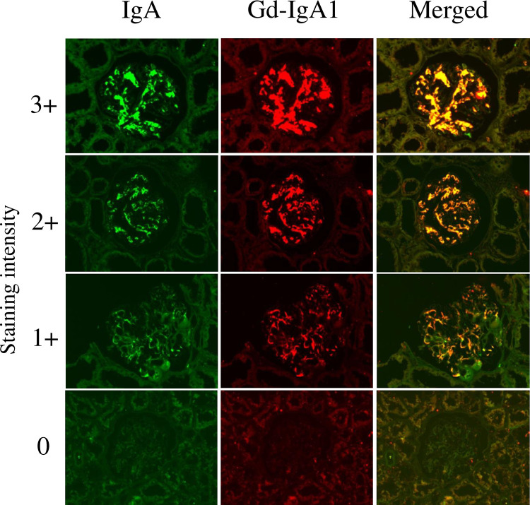
The glomerular staining intensity of IgA and Gd-IgA1 was defined by the intensity of 0 to +3 as shown in Fig 1. IgA and Gd-IgA1 were mainly positive in the mesangial region, and IgA and Gd-IgA1 had the same distribution. There was a significant association between the glomerular staining intensity of IgA and Gd-IgA1 using regression analysis (R = 0.787, P < 0.001).

Fig 1. Glomerular staining intensity of IgA and galactose-deficient IgA1.

Fig 1

Double staining with anti-IgA polyclonal antibody and Gd-IgA1 (KM55) monoclonal antibody was performed on biopsy specimens. Representative images of glomerular IgA and Gd-IgA1 (KM55) in patients with 0, 1+, 2+, and 3+ staining intensity. Seventy-four patients were divided into four groups according to the staining intensity of IgA: group 0 (n = 4), group 1+ (n = 46), group 2+ (n = 12), and group 3+ (n = 12). These patients were also divided into four groups according to the staining intensity of Gd-IgA1: group 0 (n = 4), group 1+ (n = 35), group 2+ (n = 23), and group 3+ (n = 12). Fluorescein-conjugated polyclonal rabbit anti-human IgA antibody (DAKO Japan Inc., Kyoto, Japan), anti-human Gd-IgA1 antibody (KM55) (Immuno-Biological Laboratories Co., Ltd, Gunma, Japan), and Alexa Fluor 555-conjugated goat anti-rat IgG antibody (Life Technologies) was used for immunofluorescence staining. Original magnification: ×200. Gd-IgA1, galactose-deficient IgA1.

Plasma Gd-IgA1 concentrations were measured in 33 of 74 patients. There was no significant association between the glomerular staining intensity of Gd-IgA1 and plasma Gd-IgA1 concentrations (n = 33) (S1 Fig).

Association between glomerular staining intensity of IgA and Gd-IgA1, and the rate of cnm-positive S. mutans in the oral cavity

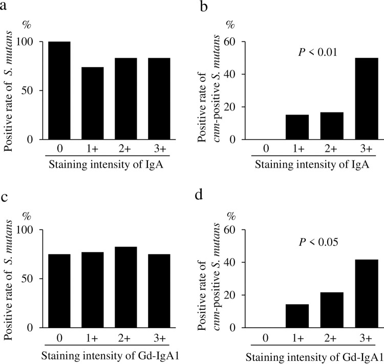
There was no significant association between the glomerular staining intensity of IgA and the positive rate of S. mutans (Fig 2A). However, there was a significant association between the glomerular staining intensity of IgA and the positive rate of cnm-positive S. mutans (Fig 2B). There was also no significant association between the glomerular staining intensity of Gd-IgA1 and positive rate of S. mutans (Fig 2C). However, there was a significant association between the glomerular staining intensity of Gd-IgA1 and the positive rate of cnm-positive S. mutans (Fig 2D).

Fig 2. Association between glomerular staining intensity of IgA and galactose-deficient IgA1 (Gd-IgA1) and the rate of cnm-positive S. mutans in the oral cavity.

Fig 2

Comparison of the glomerular staining intensity of IgA and the positive rate of S. mutans in the oral cavity (a). Comparison of the glomerular staining intensity of IgA and the positive rate of cnm-positive S. mutans in the oral cavity (b). Comparison of the glomerular staining intensity of Gd-IgA1 (KM55) and the positive rate of S. mutans in the oral cavity (c). Comparison of the glomerular staining intensity of Gd-IgA1 (KM55) and the positive rate of cnm-positive S. mutans in the oral cavity (d). Data were analyzed using the Cochran–Armitage trend test. P < 0.05 was considered statistically significant.

There was no significant association between the glomerular staining intensity of IgA and the positive rate of C. rectus (S2A Fig) or between the glomerular staining intensity of Gd-IgA1 and positive rate of C. rectus (S2B Fig). Furthermore, there was no significant association between the glomerular staining intensity of IgA and the positive rate of P. gingivalis (S2C Fig) or between the glomerular staining intensity of Gd-IgA1 and the positive rate of P. gingivalis (S2D Fig).

Comparison of the cnm-positive S. mutans and cnm-negative S. mutans groups

The percentage of the staining intensity of IgA: 3+ was significantly higher in the cnm-positive S. mutans group than in the cnm-negative S. mutans group (Table 1). The percentage of the staining intensity of Gd-IgA1: 3+ was also significantly higher in the cnm-positive S. mutans group than in the cnm-negative S. mutans group (Table 1). No significant differences were found in age, sex, height, body weight, body mass index, systolic blood pressure, diastolic blood pressure, serum creatinine concentrations, eGFR, serum C3 concentrations, serum IgA concentrations, or urinary protein concentrations between the groups (Table 1). The percentage of urinary sediment ≥100 RBCs/high-power field was higher in the cnm-positive S. mutans group than in the cnm-negative S. mutans group, but this was not significant (Table 1). No significant differences were found in the mesangial hypercellularity score, endocapillary hypercellularity score, segmental glomerulosclerosis score, tubular atrophy/interstitial fibrosis score, or the crescent score of the Oxford classification between the groups (Table 2).

Table 1. Comparison between the cnm-positive and cnm-negative S. mutans groups of patients.

Characteristics cnm-negative S. mutans (n = 59) cnm-positive S. mutans (n = 15) P value
Percentage of staining intensity of IgA: 3+ (%) 10.2 40 0.0051
Percentage of staining intensity of Gd-IgA1: 3+ (%) 11.9 33.3 0.0440
Age (years) 44.9 ± 15.3 45.1 ± 13.3 0.9662
Sex (M/F) 27/32 9/6 0.3246
Height (cm) 162.6 ± 8.6 164.3 ± 7.5 0.4834
Body weight (kg) 62.5 ± 13.4 64.0 ± 11.3 0.6966
BMI (kg/m2) 23.6 ± 4.5 23.7 ± 3.7 0.9293
Systolic blood pressure (mmHg) 125.0 ± 21.3 121.5 ± 17.6 0.5582
Diastolic blood pressure (mmHg) 69.2 ± 11.6 68.4 ± 8.0 0.8046
Serum creatinine (mg/dl) 1.0 ± 0.7 1.0 ± 0.5 0.932
eGFR (ml/minute/1.73 m2) 67.3 ± 26.4 67.4 ± 22.6 0.9926
Serum C3 (mg/dl) 108.2 ± 16.1 107.0 ± 18.7 0.8029
Serum IgA (mg/dl) 348.3 ± 158.3 358.5 ± 164.6 0.8255
Urinary protein (g/gCr) 1.4 ± 0.8 0.8 ± 0.7 0.1857
Percentage of urinary sediment ≥ 100 RBCs/HPF 8.5 26.7 0.0543

Values are %, number, or mean ± standard deviation. Bold values indicate statistical significance at P < 0.05. BMI: body mass index, eGFR: estimated glomerular filtration rate.

Table 2. Analysis of renal biopsy specimens using the Oxford classification.

Characteristic cnm-negative S. mutans cnm-positive S. mutans P value
(n = 59) (n = 15)
Positive mesangial hypercellularity score (%) 72.9 86.7 0.2665
Positive endocapillary hypercellularity score (%) 50.8 46.7 0.7725
Positive segmental glomerulosclerosis score (%) 66.1 73.3 0.5932
Tubular atrophy/interstitial fibrosis score ≥1+ (%) 43.9 46.7 0.8565
Crescent score ≥1+ (%) 40.7 33.3 0.6029

Discussion

As far as we know, this is the first study to demonstrate an association between cnm-positive S. mutans in the oral cavity and glomerular Gd-IgA1. We discovered that there was a significant association between the positive rate of cnm-positive S. mutans in the oral cavity and glomerular staining intensity of IgA or Gd-IgA1. Although various factors are considered to be associated with Gd-IgA1, these data indicate that cnm-positive S. mutans in the oral cavity may be one of the factors associated with the development of IgA nephropathy via Gd-IgA1.

Recent clinical studies have demonstrated that cnm-positive S. mutans in the oral cavity is associated with IgAN [2022, 26]. One study showed that the positive rate of cnm-positive S. mutans in the oral cavity was significantly higher in the IgAN group than in the healthy control group (32.1% vs. 14.0%) [20]. However, the positive rate of S. mutans was similar between the IgAN and healthy control groups [20]. Another study suggested that cnm-positive S. mutans in the oral cavity and the dental caries status were associated with urinary protein in patients with IgAN [21]. The Cnm protein in the tonsils may be associated with the severity of IgAN [22]. The report demonstrated the following: (1) the positive Cnm protein area/total tonsillar area rate was significantly higher in patients with IgAN than in the control (chronic tonsillitis) group; (2) Cnm protein existed in the tonsils, not in the glomerulus, in patients with IgAN; and (3) Cnm protein in the tonsils was associated with urinary protein in patients with IgAN [22]. These results suggest that IgAN is aggravated by immune reactions in the tonsils via an unknown mechanism induced by Cnm protein [22]. In rodent models, intravenous administration of cnm-positive S. mutans induced transient IgAN-like lesions [23] and severe dental caries induced by cnm-positive S. mutans caused IgAN-like glomerulonephritis [24]. As mentioned above, we found an association between cnm-positive S. mutans and IgAN. However, we could not identify how cnm-positive S. mutans induced IgAN.

Gd-IgA1 has been demonstrated as a key effector molecule in the pathogenesis of IgAN [69]. We showed an association between the presence of cnm-positive S. mutans and glomerular Gd-IgA1 in this study. This study indicates that cnm-positive S. mutans induced Gd-IgA1 production in the process of causing IgAN, suggesting that the most upstream factor for Gd-IgA1 production is oral bacterial infection, such as cnm-positive S. mutans. We hypothesize that repeated immune reactions involving IgA induced by infection such as cnm-positive S. mutans in the mucosal immune tissues of the oral cavity (e.g., the tonsils) may cause Gd-IgA1 [21]. We believe that this result is important in considering the pathogenic mechanism of IgAN. We do not have enough evidence to explain how cnm-positive S. mutans affects the induction of Gd-IgA1 at this stage and need to elucidate these mechanisms in a further study.

We also investigated the positive rate of C. rectus and P. gingivalis in the oral cavity because these bacteria are reported to be potentially associated with IgA nephropathy [19, 26, 27]. We could not confirm the association between these bacteria and Gd-IgA1 in this research.

Our study showed that the percentage of urinary sediment ≥100 RBCs/high-power field was higher in the cnm-positive S. mutans group than in the cnm-negative S. mutans group. Although this finding did not reach statistical significance, it suggests that cnm-positive S. mutans is associated with the hematuria that is the cardinal symptom of IgAN.

Unfortunately, because the exact mechanism of IgAN is not yet understood, there are no specific disease-targeted therapies for IgAN [5]. Our present findings are consistent with the new concept of an oral–kidney association and could establish a new treatment for IgAN [21]. Considering that S. mutans is a pathogen that causes dental caries, reducing it in the oral cavity to prevent dental caries may improve the prognosis of IgAN [27]. Further studies are needed to elucidate the effects of oral care as a treatment of IgAN.

There are some limitations in this study. First, although we demonstrated that cnm-positive S. mutans in the oral cavity was associated with Gd-IgA1 in patients with IgAN, how cnm-positive S. mutans contributes to Gd-IgA1 needs to be elucidated. Second, given that the only S. mutans protein we examined was Cnm, the possibility that other S. mutans proteins are associated with Gd-IgA1 cannot be excluded. Third, the sample size was small, and all patients were of the same ethnicity and from a single facility. Further studies in larger numbers of ethnically diverse patients from multiple facilities are needed to confirm our present findings.

In conclusion, cnm-positive S. mutans in the oral cavity is associated with Gd-IgA1 in glomeruli in patients with IgAN. This finding suggests that cnm-positive S. mutans is involved in the pathogenicity of IgAN through induction of Gd-IgA1.

Supporting information

S1 Fig. Comparison of the glomerular staining intensity of (Gd-IgA1) (KM55) and plasma Gd-IgA1 (KM55) concentrations.

There was no significant association between the glomerular staining intensity of Gd-IgA1 and the plasma Gd-IgA1 concentration. The data were examined for statistical significance using a simple regression analysis. Gd-IgA1: galactose-deficient IgA1.

(TIF)

S2 Fig. No association between glomerular staining intensity of Gd-IgA1 and the positive rate of C. rectus or P. gingivalis in the oral cavity.

Comparison of the glomerular staining intensity of IgA and the positive rate of Campylobacter rectus in the oral cavity (a). Comparison of the glomerular staining intensity of Gd-IgA1 (KM55) and the positive rate of C. rectus in the oral cavity (b). Comparison of the glomerular staining intensity of IgA and the positive rate of Porphyromonas gingivalis in the oral cavity (c). Comparison of the glomerular staining intensity of Gd-IgA1 (KM55) and the positive rate of P. gingivalis in the oral cavity (d). The data were examined for statistical significance using the Cochran–Armitage trend test. P < 0.05 was considered statistically significant.

(TIF)

Acknowledgments

Ethical statement

This study protocol fully adhered to the Declaration of Helsinki (64th WMA General Assembly, Fortaleza, Brazil, 2013). The protocol was approved by the Ethics Committee of Seirei Hamamatsu General Hospital (approval no. 3646), Osaka University Graduate School of Dentistry (approval no. H29-E9), and Okayama University Graduate School of Medicine (approval no. 1704–036). All patients were informed of the study protocol, and provided written informed consent prior to participating in the study.

Data Availability

All relevant data are within the manuscript and its Supporting Information files.

Funding Statement

This work was supported by Japan Society for the Promotion of Science (grant numbers 19K10098, Dr. Taro Misaki, 21H03149, Dr Kazuhiko Nakano and 21K08242, Dr Yasuyuki Nagasawa). The sponsors or funders did not play any role in the study design, data collection and analysis, decision to publish, or preparation of the manuscript.

References

  • 1.D’Amico G. The commonest glomerulonephritis in the world: IgA nephropathy. Q J Med. 1987;64(245):709–27. Epub 1987/09/01. . [PubMed] [Google Scholar]
  • 2.Julian BA, Waldo FB, Rifai A, Mestecky J. IgA nephropathy, the most common glomerulonephritis worldwide. A neglected disease in the United States? Am J Med. 1988;84(1):129–32. Epub 1988/01/01. doi: 10.1016/0002-9343(88)90019-8 . [DOI] [PubMed] [Google Scholar]
  • 3.Sugiyama H, Yokoyama H, Sato H, Saito T, Kohda Y, Nishi S, et al. Japan Renal Biopsy Registry and Japan Kidney Disease Registry: Committee Report for 2009 and 2010. Clin Exp Nephrol. 2013;17(2):155–73. Epub 2013/02/07. doi: 10.1007/s10157-012-0746-8 . [DOI] [PubMed] [Google Scholar]
  • 4.Conley ME, Cooper MD, Michael AF. Selective deposition of immunoglobulin A1 in immunoglobulin A nephropathy, anaphylactoid purpura nephritis, and systemic lupus erythematosus. J Clin Invest. 1980;66(6):1432–6. Epub 1980/12/01. doi: 10.1172/JCI109998 ; PubMed Central PMCID: PMC371631. [DOI] [PMC free article] [PubMed] [Google Scholar]
  • 5.Wyatt RJ, Julian BA. IgA nephropathy. N Engl J Med. 2013;368(25):2402–14. Epub 2013/06/21. doi: 10.1056/NEJMra1206793 . [DOI] [PubMed] [Google Scholar]
  • 6.Barratt J, Feehally J. IgA nephropathy. J Am Soc Nephrol. 2005;16(7):2088–97. Epub 2005/06/03. doi: 10.1681/ASN.2005020134 . [DOI] [PubMed] [Google Scholar]
  • 7.Moldoveanu Z, Wyatt RJ, Lee JY, Tomana M, Julian BA, Mestecky J, et al. Patients with IgA nephropathy have increased serum galactose-deficient IgA1 levels. Kidney Int. 2007;71(11):1148–54. Epub 2007/03/08. doi: 10.1038/sj.ki.5002185 . [DOI] [PubMed] [Google Scholar]
  • 8.Suzuki H, Moldoveanu Z, Hall S, Brown R, Vu HL, Novak L, et al. IgA1-secreting cell lines from patients with IgA nephropathy produce aberrantly glycosylated IgA1. J Clin Invest. 2008;118(2):629–39. Epub 2008/01/04. doi: 10.1172/JCI33189 ; PubMed Central PMCID: PMC2157566. [DOI] [PMC free article] [PubMed] [Google Scholar]
  • 9.Berthoux F, Suzuki H, Thibaudin L, Yanagawa H, Maillard N, Mariat C, et al. Autoantibodies targeting galactose-deficient IgA1 associate with progression of IgA nephropathy. J Am Soc Nephrol. 2012;23(9):1579–87. Epub 2012/08/21. doi: 10.1681/ASN.2012010053 ; PubMed Central PMCID: PMC3431415. [DOI] [PMC free article] [PubMed] [Google Scholar]
  • 10.Yasutake J, Suzuki Y, Suzuki H, Hiura N, Yanagawa H, Makita Y, et al. Novel lectin-independent approach to detect galactose-deficient IgA1 in IgA nephropathy. Nephrol Dial Transplant. 2015;30(8):1315–21. Epub 2015/06/26. doi: 10.1093/ndt/gfv221 ; PubMed Central PMCID: PMC4513896. [DOI] [PMC free article] [PubMed] [Google Scholar]
  • 11.Suzuki H, Yasutake J, Makita Y, Tanbo Y, Yamasaki K, Sofue T, et al. IgA nephropathy and IgA vasculitis with nephritis have a shared feature involving galactose-deficient IgA1-oriented pathogenesis. Kidney Int. 2018;93(3):700–5. Epub 2018/01/14. doi: 10.1016/j.kint.2017.10.019 . [DOI] [PubMed] [Google Scholar]
  • 12.Donadio JV, Grande JP. IgA nephropathy. N Engl J Med. 2002;347(10):738–48. Epub 2002/09/06. doi: 10.1056/NEJMra020109 . [DOI] [PubMed] [Google Scholar]
  • 13.Suzuki S, Nakatomi Y, Sato H, Tsukada H, Arakawa M. Haemophilus parainfluenzae antigen and antibody in renal biopsy samples and serum of patients with IgA nephropathy. Lancet. 1994;343(8888):12–6. Epub 1994/01/01. doi: 10.1016/s0140-6736(94)90875-3 . [DOI] [PubMed] [Google Scholar]
  • 14.Kusano K, Tokunaga O, Ando T, Inokuchi A. Helicobacter pylori in the palatine tonsils of patients with IgA nephropathy compared with those of patients with recurrent pharyngotonsillitis. Hum Pathol. 2007;38(12):1788–97. Epub 2007/08/24. doi: 10.1016/j.humpath.2007.04.012 . [DOI] [PubMed] [Google Scholar]
  • 15.Iwama H, Horikoshi S, Shirato I, Tomino Y. Epstein-Barr virus detection in kidney biopsy specimens correlates with glomerular mesangial injury. Am J Kidney Dis. 1998;32(5):785–93. Epub 1998/11/20. doi: 10.1016/s0272-6386(98)70134-9 . [DOI] [PubMed] [Google Scholar]
  • 16.Koyama A, Sharmin S, Sakurai H, Shimizu Y, Hirayama K, Usui J, et al. Staphylococcus aureus cell envelope antigen is a new candidate for the induction of IgA nephropathy. Kidney Int. 2004;66(1):121–32. Epub 2004/06/18. doi: 10.1111/j.1523-1755.2004.00714.x . [DOI] [PubMed] [Google Scholar]
  • 17.Rollino C, Vischini G, Coppo R. IgA nephropathy and infections. J Nephrol. 2016. Epub 2016/01/24. doi: 10.1007/s40620-016-0265-x . [DOI] [PubMed] [Google Scholar]
  • 18.Nagasawa Y, Iio K, Fukuda S, Date Y, Iwatani H, Yamamoto R, et al. Periodontal disease bacteria specific to tonsil in IgA nephropathy patients predicts the remission by the treatment. PLoS One. 2014;9(1):e81636. Epub 2014/02/04. doi: 10.1371/journal.pone.0081636 ; PubMed Central PMCID: PMC3904818. [DOI] [PMC free article] [PubMed] [Google Scholar]
  • 19.Nagasawa Y, Nomura R, Misaki T, Ito S, Naka S, Wato K, et al. Relationship between IgA Nephropathy and Porphyromonas gingivalis; Red Complex of Periodontopathic Bacterial Species. Int J Mol Sci. 2021;22(23). Epub 2021/12/11. doi: 10.3390/ijms222313022 ; PubMed Central PMCID: PMC8657970. [DOI] [PMC free article] [PubMed] [Google Scholar]
  • 20.Misaki T, Naka S, Kuroda K, Nomura R, Shiooka T, Naito Y, et al. Distribution of Streptococcus mutans strains with collagen-binding proteins in the oral cavity of IgA nephropathy patients. Clin Exp Nephrol. 2015;19(5):844–50. Epub 2014/12/11. doi: 10.1007/s10157-014-1072-0 . [DOI] [PubMed] [Google Scholar]
  • 21.Misaki T, Naka S, Hatakeyama R, Fukunaga A, Nomura R, Isozaki T, et al. Presence of Streptococcus mutans strains harbouring the cnm gene correlates with dental caries status and IgA nephropathy conditions. Sci Rep. 2016;6:36455. Epub 2016/11/05. doi: 10.1038/srep36455 ; PubMed Central PMCID: PMC5095553. [DOI] [PMC free article] [PubMed] [Google Scholar]
  • 22.Ito S, Misaki T, Naka S, Wato K, Nagasawa Y, Nomura R, et al. Specific strains of Streptococcus mutans, a pathogen of dental caries, in the tonsils, are associated with IgA nephropathy. Sci Rep. 2019;9(1):20130. Epub 2019/12/29. doi: 10.1038/s41598-019-56679-2 ; PubMed Central PMCID: PMC6934739. [DOI] [PMC free article] [PubMed] [Google Scholar]
  • 23.Naka S, Wato K, Misaki T, Ito S, Nagasawa Y, Nomura R, et al. Intravenous administration of Streptococcus mutans induces IgA nephropathy-like lesions. Clin Exp Nephrol. 2020;24(12):1122–31. Epub 2020/09/11. doi: 10.1007/s10157-020-01961-1 ; PubMed Central PMCID: PMC7599197. [DOI] [PMC free article] [PubMed] [Google Scholar]
  • 24.Naka S, Wato K, Misaki T, Ito S, Matsuoka D, Nagasawa Y, et al. Streptococcus mutans induces IgA nephropathy-like glomerulonephritis in rats with severe dental caries. Sci Rep. 2021;11(1):5784. Epub 2021/03/13. doi: 10.1038/s41598-021-85196-4 ; PubMed Central PMCID: PMC7952735. [DOI] [PMC free article] [PubMed] [Google Scholar]
  • 25.Nagasawa Y, Misaki T, Ito S, Naka S, Wato K, Nomura R, et al. Title IgA Nephropathy and Oral Bacterial Species Related to Dental Caries and Periodontitis. Int J Mol Sci. 2022;23(2). Epub 2022/01/22. doi: 10.3390/ijms23020725 ; PubMed Central PMCID: PMC8775524. [DOI] [PMC free article] [PubMed] [Google Scholar]
  • 26.Misaki T, Naka S, Wato K, Hatakeyama R, Nagasawa Y, Ito S, et al. Campylobacter rectus in the Oral Cavity Correlates with Proteinuria in Immunoglobulin A Nephropathy Patients. Nephron. 2018;139(2):143–9. Epub 2018/02/13. doi: 10.1159/000487103 . [DOI] [PubMed] [Google Scholar]
  • 27.Misaki T, Naka S, Nagasawa Y, Matsuoka D, Ito S, Nomura R, et al. Simultaneous Presence of Campylobacter rectus and Cnm-Positive Streptococcus mutans in the Oral Cavity Is Associated with Renal Dysfunction in IgA Nephropathy Patients: 5-Year Follow-Up Analysis. Nephron. 2022:1–10. Epub 2022/08/24. doi: 10.1159/000525511 . [DOI] [PubMed] [Google Scholar]
  • 28.Selwitz RH, Ismail AI, Pitts NB. Dental caries. Lancet. 2007;369(9555):51–9. Epub 2007/01/09. doi: 10.1016/S0140-6736(07)60031-2 . [DOI] [PubMed] [Google Scholar]
  • 29.Naka S, Matsuoka D, Goto K, Misaki T, Nagasawa Y, Ito S, et al. Cnm of Streptococcus mutans is important for cell surface structure and membrane permeability. Front Cell Infect Microbiol. 2022;12:994014. Epub 2022/10/01. doi: 10.3389/fcimb.2022.994014 ; PubMed Central PMCID: PMC9513430. [DOI] [PMC free article] [PubMed] [Google Scholar]
  • 30.Sato Y, Okamoto K, Kagami A, Yamamoto Y, Igarashi T, Kizaki H. Streptococcus mutans strains harboring collagen-binding adhesin. J Dent Res. 2004;83(7):534–9. Epub 2004/06/26. doi: 10.1177/154405910408300705 . [DOI] [PubMed] [Google Scholar]
  • 31.Nomura R, Naka S, Nemoto H, Otsugu M, Nakamura S, Ooshima T, et al. Potential high virulence for infective endocarditis in Streptococcus mutans strains with collagen-binding proteins but lacking PA expression. Arch Oral Biol. 2013;58(11):1627–34. Epub 2013/10/12. doi: 10.1016/j.archoralbio.2013.06.008 . [DOI] [PubMed] [Google Scholar]
  • 32.Kojima A, Nakano K, Wada K, Takahashi H, Katayama K, Yoneda M, et al. Infection of specific strains of Streptococcus mutans, oral bacteria, confers a risk of ulcerative colitis. Sci Rep. 2012;2:332. Epub 2012/03/28. doi: 10.1038/srep00332 ; PubMed Central PMCID: PMC3312205. [DOI] [PMC free article] [PubMed] [Google Scholar]
  • 33.Nakano K, Hokamura K, Taniguchi N, Wada K, Kudo C, Nomura R, et al. The collagen-binding protein of Streptococcus mutans is involved in haemorrhagic stroke. Nat Commun. 2011;2:485. Epub 2011/09/29. doi: 10.1038/ncomms1491 ; PubMed Central PMCID: PMC3220351. [DOI] [PMC free article] [PubMed] [Google Scholar]
  • 34.Tonomura S, Ihara M, Kawano T, Tanaka T, Okuno Y, Saito S, et al. Intracerebral hemorrhage and deep microbleeds associated with cnm-positive Streptococcus mutans; a hospital cohort study. Sci Rep. 2016;6:20074. Epub 2016/02/06. doi: 10.1038/srep20074 ; PubMed Central PMCID: PMC4742798. [DOI] [PMC free article] [PubMed] [Google Scholar]
  • 35.Watanabe I, Kuriyama N, Miyatani F, Nomura R, Naka S, Nakano K, et al. Oral Cnm-positive Streptococcus Mutans Expressing Collagen Binding Activity is a Risk Factor for Cerebral Microbleeds and Cognitive Impairment. Sci Rep. 2016;6:38561. Epub 2016/12/10. doi: 10.1038/srep38561 ; PubMed Central PMCID: PMC5146923. [DOI] [PMC free article] [PubMed] [Google Scholar]
  • 36.Naka S, Nomura R, Takashima Y, Okawa R, Ooshima T, Nakano K. A specific Streptococcus mutans strain aggravates non-alcoholic fatty liver disease. Oral Dis. 2014;20(7):700–6. Epub 2014/11/02. doi: 10.1111/odi.12191 . [DOI] [PubMed] [Google Scholar]
  • 37.Naka S, Hatakeyama R, Takashima Y, Matsumoto-Nakano M, Nomura R, Nakano K. Contributions of Streptococcus mutans Cnm and PA antigens to aggravation of non-alcoholic steatohepatitis in mice. Sci Rep. 2016;6:36886. Epub 2016/11/12. doi: 10.1038/srep36886 ; PubMed Central PMCID: PMC5105074. [DOI] [PMC free article] [PubMed] [Google Scholar]
  • 38.Nomura R, Nagasawa Y, Misaki T, Ito S, Naka S, Okunaka M, et al. Distribution of periodontopathic bacterial species between saliva and tonsils. Odontology. 2022. Epub 2022/12/17. doi: 10.1007/s10266-022-00776-8 . [DOI] [PubMed] [Google Scholar]
  • 39.Working Group of the International Ig ANN, the Renal Pathology S, Cattran DC, Coppo R, Cook HT, Feehally J, et al. The Oxford classification of IgA nephropathy: rationale, clinicopathological correlations, and classification. Kidney Int. 2009;76(5):534–45. Epub 2009/07/03. doi: 10.1038/ki.2009.243 . [DOI] [PubMed] [Google Scholar]
  • 40.Working Group of the International Ig ANN, the Renal Pathology S, Roberts IS, Cook HT, Troyanov S, Alpers CE, et al. The Oxford classification of IgA nephropathy: pathology definitions, correlations, and reproducibility. Kidney Int. 2009;76(5):546–56. Epub 2009/07/03. doi: 10.1038/ki.2009.168 . [DOI] [PubMed] [Google Scholar]
  • 41.Trimarchi H, Barratt J, Cattran DC, Cook HT, Coppo R, Haas M, et al. Oxford Classification of IgA nephropathy 2016: an update from the IgA Nephropathy Classification Working Group. Kidney Int. 2017;91(5):1014–21. Epub 2017/03/28. doi: 10.1016/j.kint.2017.02.003 . [DOI] [PubMed] [Google Scholar]

Decision Letter 0

Maria Leonor S Oliveira

19 Dec 2022

PONE-D-22-21218

cnm -positive Streptococcus mutans is associated with galactose-deficient IgA in patients with IgA nephropathy

PLOS ONE

Dear Dr. Misaki,

Thank you for submitting your manuscript to PLOS ONE. After careful consideration, we feel that it has merit but does not fully meet PLOS ONE’s publication criteria as it currently stands. Therefore, we invite you to submit a revised version of the manuscript that addresses the points raised during the review process.

==============================

Specifically, we request you to revise the conclusions, in order to better reflect the results obtained in the study, as indicated by reviewer #1.

In addition, all the comments and corrections indicated by both reviewers need to be considered.

==============================

Please submit your revised manuscript by Feb 02 2023 11:59PM. If you will need more time than this to complete your revisions, please reply to this message or contact the journal office at plosone@plos.org. When you're ready to submit your revision, log on to https://www.editorialmanager.com/pone/ and select the 'Submissions Needing Revision' folder to locate your manuscript file.

Please include the following items when submitting your revised manuscript:

  • A rebuttal letter that responds to each point raised by the academic editor and reviewer(s). You should upload this letter as a separate file labeled 'Response to Reviewers'.

  • A marked-up copy of your manuscript that highlights changes made to the original version. You should upload this as a separate file labeled 'Revised Manuscript with Track Changes'.

  • An unmarked version of your revised paper without tracked changes. You should upload this as a separate file labeled 'Manuscript'.

If you would like to make changes to your financial disclosure, please include your updated statement in your cover letter. Guidelines for resubmitting your figure files are available below the reviewer comments at the end of this letter.

If applicable, we recommend that you deposit your laboratory protocols in protocols.io to enhance the reproducibility of your results. Protocols.io assigns your protocol its own identifier (DOI) so that it can be cited independently in the future. For instructions see: https://journals.plos.org/plosone/s/submission-guidelines#loc-laboratory-protocols. Additionally, PLOS ONE offers an option for publishing peer-reviewed Lab Protocol articles, which describe protocols hosted on protocols.io. Read more information on sharing protocols at https://plos.org/protocols?utm_medium=editorial-email&utm_source=authorletters&utm_campaign=protocols.

We look forward to receiving your revised manuscript.

Kind regards,

Maria Leonor S Oliveira, PhD

Academic Editor

PLOS ONE

Journal requirements:

When submitting your revision, we need you to address these additional requirements.

1. Please ensure that your manuscript meets PLOS ONE's style requirements, including those for file naming. The PLOS ONE style templates can be found at

https://journals.plos.org/plosone/s/file?id=wjVg/PLOSOne_formatting_sample_main_body.pdf and

https://journals.plos.org/plosone/s/fileid=ba62/PLOSOne_formatting_sample_title_authors_affiliations.pdf.

2. Please include your tables as part of your main manuscript and remove the individual files. Please note that supplementary tables (should remain/ be uploaded) as separate "supporting information" files.

3. Thank you for submitting the above manuscript to PLOS ONE. During our internal evaluation of the manuscript, we found significant text overlap between your submission and previous work in the Methods and Discussion sections. We would like to make you aware that copying extracts from previous publications, especially outside the methods section, word-for-word is unacceptable. In addition, the reproduction of text from published reports has implications for the copyright that may apply to the publications. Please revise the manuscript to rephrase the duplicated text, cite your sources, and provide details as to how the current manuscript advances on previous work. Please note that further consideration is dependent on the submission of a manuscript that addresses these concerns about the overlap in text with published work. We will carefully review your manuscript upon resubmission and further consideration of the manuscript is dependent on the text overlap being addressed in full. Please ensure that your revision is thorough as failure to address the concerns to our satisfaction may result in your submission not being considered further.

4. You indicated that you had ethical approval for your study. Please clarify whether minors (patients below the age of 18 years) were included in your study. If yes, in your Methods section, please ensure you have also stated whether you obtained consent from parents or guardians of the minors included in the study or whether the research ethics committee or IRB specifically waived the need for their consent.

5. Thank you for stating the following in the Acknowledgments Section of your manuscript:

“This work was supported by JSPS KAKENHI [grant numbers 19K10098, 21H03149 and 21K08242].”

We note that you have provided funding information that is not currently declared in your Funding Statement. However, funding information should not appear in the Acknowledgments section or other areas of your manuscript. We will only publish funding information present in the Funding Statement section of the online submission form.

Please remove any funding-related text from the manuscript and let us know how you would like to update your Funding Statement. Currently, your Funding Statement reads as follows:

“This work was supported by JSPS KAKENHI [grant numbers 19K10098 (TM), 21H03149 (KN) and 21K08242 (YN)].

URL of each funder website: https://www.jsps.go.jp/english/index.html

Did the sponsors or funders play any role in the study design, data collection and analysis, decision to publish, or preparation of the manuscript? NO.”

Please include your amended statements within your cover letter; we will change the online submission form on your behalf.

6. We note that you have stated that you will provide repository information for your data at acceptance. Should your manuscript be accepted for publication, we will hold it until you provide the relevant accession numbers or DOIs necessary to access your data. If you wish to make changes to your Data Availability statement, please describe these changes in your cover letter and we will update your Data Availability statement to reflect the information you provide.

[Note: HTML markup is below. Please do not edit.]

Reviewers' comments:

Reviewer's Responses to Questions

Comments to the Author

1. Is the manuscript technically sound, and do the data support the conclusions?

The manuscript must describe a technically sound piece of scientific research with data that supports the conclusions. Experiments must have been conducted rigorously, with appropriate controls, replication, and sample sizes. The conclusions must be drawn appropriately based on the data presented.

Reviewer #1: Partly

Reviewer #2: Yes

**********

2. Has the statistical analysis been performed appropriately and rigorously?

Reviewer #1: Yes

Reviewer #2: I Don't Know

**********

3. Have the authors made all data underlying the findings in their manuscript fully available?

The PLOS Data policy requires authors to make all data underlying the findings described in their manuscript fully available without restriction, with rare exception (please refer to the Data Availability Statement in the manuscript PDF file). The data should be provided as part of the manuscript or its supporting information, or deposited to a public repository. For example, in addition to summary statistics, the data points behind means, medians and variance measures should be available. If there are restrictions on publicly sharing data—e.g. participant privacy or use of data from a third party—those must be specified.

Reviewer #1: Yes

Reviewer #2: No

**********

4. Is the manuscript presented in an intelligible fashion and written in standard English?

PLOS ONE does not copyedit accepted manuscripts, so the language in submitted articles must be clear, correct, and unambiguous. Any typographical or grammatical errors should be corrected at revision, so please note any specific errors here.

Reviewer #1: Yes

Reviewer #2: Yes

**********

5. Review Comments to the Author

Please use the space provided to explain your answers to the questions above. You may also include additional comments for the author, including concerns about dual publication, research ethics, or publication ethics. (Please upload your review as an attachment if it exceeds 20,000 characters)

Reviewer #1: The paper is written in an intelligible way and describes adequately the data obtained by the authors. There are some minor questions related to grammar and clearness of some statements:

line 51: The whole fragment is confuse, the authors should rewrite this line to improve clarity.

line 94: Replace "pathogenesis" for "pathogen"

line 172: The authors should define what TMB solution is.

Although the paper describes clearly the objectives and some findings, the authors were not able to draw a line of evidences which would delineate an horizon to support their hypothesis. Just to be clearer, in lines 230-232, the authors state: "These data indicated that cnm-positive S. mutans was clearly associated with the development of IgA nephropathy via Gd-IgA1."

It is possible that specific Gd-IgA1 directed to Cnm protein would be involved in IgA nephropathy (IgAN) establishment or recrudescence. However, the authors merely showed that IgAN recipients infected with Cnm-positive S. mutans also presented Gd-IgA1 in renal milieu. It represents, undoubtedly, a promising finding which, on the other hand, is not enough to support the hypothesis per se. Therefore, the issues raised in this review are not related to the quality of experiments or the way they were presented, but how the conclusions were drawn in connection with a correlation, which in turn not, necessarily, implicates causality. As a consequence, it seems inadequate to affirm categorically that the presence of Cnm protein is the reason of IgaN status and that the mechanism involves uniquely Gd-IgA1 production in the kidneys, as seen in the fragment mentioned above (lines 230-232).

Co-infection with other bacteria in multispecies biofilms and other physiological indicators, different from those analyzed here, could influence the appearence of IgA. Other factors somewhat ignored cannot be discarded in this context, such as the possibility that other S. mutans proteins could have a role in the pathology. In summary, the authors should have collected other data to support their hypothesis.

In lines 217-219, the authors state: "The percentage of urinary sediment ≥100 RBCs/high-power field was higher in the cnm-positive S. mutans group than in cnm-negative S. mutans group, but this was not significant (Table 1)". The p value of this indicator was very close to the significance level chosen in the analysis. For this reason, the authors should have drawn some inference about this indicator. Above all, statistics indicates tendencies even when the numerical indicator is not strictly between the limits established previously. In this case, this particular indicator should be considered, at least, as a clue to a more complete scenario involving the production of Gd-IgA1, the establishment of IgAN and the presence of S. mutans producing Cnm protein.

Based on these issues, I do not consider this paper suitable for publication in PLoS One. Significant revision, including additional data, are required to justify further consideration of this manuscript in a new submission.

Reviewer #2: The work is interesting and original, the authors have experience in this area. I have several comments regarding the manuscript:

Materials and Methods

In general, I think it is necessary to check if all information is described.

1. Histological studies (line131-132): What are the fluorochromes used?

2. Statistical analysis: I'm not an expert in statistics but it seems to me that it's not the best to do statistical analysis from results that are in percentage (Fig 2).

Results

1. Fig 1. I would like to see images of glomerular staining intensity slices at a lower magnification. The objective would be to have a better view of how frequent this glomerular staining intensity is in renal biopsy samples.

2. Fig 2. The data shown are in percentage and as mentioned earlier (M&M) are significant between groups.

3. Figure Legends: Legends are incomplete. Fig1 is missing data such as number of samples per group, antibodies and fluorochromes used. Fig2 is missing the significance value.

4. Tables: Table 1. Data with significance from data represented in percentage. Table 2. It does not say if the data are in percentage. You could put the number of patients by characteristics analyzed.

Discussion

1. In the second paragraph, change the sentence of line 233.

Supplementary Figure

The supplementary figure must be submitted the same quality as the other figures in the paper. The legend is incomplete.

**********

6. PLOS authors have the option to publish the peer review history of their article (what does this mean?). If published, this will include your full peer review and any attached files.

If you choose “no”, your identity will remain anonymous but your review may still be made public.

Do you want your identity to be public for this peer review? For information about this choice, including consent withdrawal, please see our Privacy Policy.

Reviewer #1: No

Reviewer #2: No

**********

[NOTE: If reviewer comments were submitted as an attachment file, they will be attached to this email and accessible via the submission site. Please log into your account, locate the manuscript record, and check for the action link "View Attachments". If this link does not appear, there are no attachment files.]

While revising your submission, please upload your figure files to the Preflight Analysis and Conversion Engine (PACE) digital diagnostic tool, https://pacev2.apexcovantage.com/. PACE helps ensure that figures meet PLOS requirements. To use PACE, you must first register as a user. Registration is free. Then, login and navigate to the UPLOAD tab, where you will find detailed instructions on how to use the tool. If you encounter any issues or have any questions when using PACE, please email PLOS at figures@plos.org. Please note that Supporting Information files do not need this step.

PLoS One. 2023 Mar 2;18(3):e0282367. doi: 10.1371/journal.pone.0282367.r002

Author response to Decision Letter 0


10 Jan 2023

Journal requirements:

When submitting your revision, we need you to address these additional requirements.

1. Please ensure that your manuscript meets PLOS ONE's style requirements, including those for file naming. The PLOS ONE style templates can be found at …

Response: We thank the editorial staff of PLOS ONE for pointing this out. We have formatted the manuscript according to the instructions provided by PLOS ONE.

2. Please include your tables as part of your main manuscript and remove the individual files. Please note that supplementary tables (should remain/ be uploaded) as separate "supporting information" files.

Response: We appreciate this advice. We have now included our tables in the main manuscript and removed the individual files.

3. Thank you for submitting the above manuscript to PLOS ONE. During our internal evaluation of the manuscript, we found significant text overlap between your submission and previous work in the Methods and Discussion sections. We would like to make you aware that copying extracts from previous publications, especially outside the methods section, word-for-word is unacceptable. In addition, the reproduction of text from published reports has implications for the copyright that may apply to the publications. Please revise the manuscript to rephrase the duplicated text, cite your sources, and provide details as to how the current manuscript advances on previous work. Please note that further consideration is dependent on the submission of a manuscript that addresses these concerns about the overlap in text with published work. We will carefully review your manuscript upon resubmission and further consideration of the manuscript is dependent on the text overlap being addressed in full. Please ensure that your revision is thorough as failure to address the concerns to our satisfaction may result in your submission not being considered further.

Response: We are grateful for these comments. We have revised our manuscript so that it does not overlap with any text from previous publications. The sections in green font are revised text. In the Methods section, the parts of our experimental protocol that were similar to those in previous experiments have been acknowledged and referenced.

4. You indicated that you had ethical approval for your study. Please clarify whether minors (patients below the age of 18 years) were included in your study. If yes, in your Methods section, please ensure you have also stated whether you obtained consent from parents or guardians of the minors included in the study or whether the research ethics committee or IRB specifically waived the need for their consent.

Response: We confirm that our study did not include patients younger than 18 years. We have added the following explanatory text in the Methods section:

“All patients were over 18 years of age.” (lines 102–103)

5. Thank you for stating the following in the Acknowledgments Section of your manuscript:

“This work was supported by JSPS KAKENHI [grant numbers 19K10098, 21H03149 and 21K08242].”

We note that you have provided funding information that is not currently declared in your Funding Statement. However, funding information should not appear in the Acknowledgments section or other areas of your manuscript. We will only publish funding information present in the Funding Statement section of the online submission form.

Please remove any funding-related text from the manuscript and let us know how you would like to update your Funding Statement. Currently, your Funding Statement reads as follows:

“This work was supported by JSPS KAKENHI [grant numbers 19K10098 (TM), 21H03149 (KN) and 21K08242 (YN)].

URL of each funder website: https://www.jsps.go.jp/english/index.html

Did the sponsors or funders play any role in the study design, data collection and analysis, decision to publish, or preparation of the manuscript? NO.”

Please include your amended statements within your cover letter; we will change the online submission form on your behalf.

Response: We apologize for this error and have deleted the funding information in the Acknowledgment section. The Funding statement should read as follows: “This work was supported by JSPS KAKENHI (grant numbers 19K10098 [TM], 21H03149 [KN] and 21K08242 [YN]). The sponsors or funders did not play any role in the study design, data collection and analysis, decision to publish, or preparation of the manuscript.”

6. We note that you have stated that you will provide repository information for your data at acceptance. Should your manuscript be accepted for publication, we will hold it until you provide the relevant accession numbers or DOIs necessary to access your data. If you wish to make changes to your Data Availability statement, please describe these changes in your cover letter and we will update your Data Availability statement to reflect the information you provide.

Response: We are grateful for these comments. Although we could open up the data, we wish to change our Data Availability statement to “No”.

Reviewers' comments:

Reviewer's Responses to Questions

Comments to the Author

3. Have the authors made all data underlying the findings in their manuscript fully available?

The PLOS Data policy requires authors to make all data underlying the findings described in their manuscript fully available without restriction, with rare exception (please refer to the Data Availability Statement in the manuscript PDF file). The data should be provided as part of the manuscript or its supporting information, or deposited to a public repository. For example, in addition to summary statistics, the data points behind means, medians and variance measures should be available. If there are restrictions on publicly sharing data—e.g. participant privacy or use of data from a third party—those must be specified.

Reviewer #1: Yes

Reviewer #2: No

Response: We understand the response of Reviewer 2 to be related to his/her question: “Statistical analysis: I'm not an expert in statistics but it seems to me that it's not the best to do statistical analysis from results that are in percentage (Fig 2).”

We did consider various statistical methods for analysis of our study data. However, the results for the bacteria evaluated in this study were either positive or negative and there was no other method that could be used to evaluate the positivity rate. Therefore, we believe it is appropriate to express the positivity rate as a percentage.

5. Review Comments to the Author

Reviewer #1: The paper is written in an intelligible way and describes adequately the data obtained by the authors. There are some minor questions related to grammar and clearness of some statements:

line 51: The whole fragment is confuse, the authors should rewrite this line to improve clarity.

Response: We apologize for the lack of clarity of the abovementioned text. We have revised it to read as follows:

”The presence of S. mutans and cnm-positive S. mutans was evaluated in saliva samples of 74 patients with IgAN or IgA vasculitis by polymerase chain reaction.” (lines 43–45)

line 94: Replace "pathogenesis" for "pathogen"

Response: We apologize for this typographical error and have corrected it (line 87).

line 172: The authors should define what TMB solution is.

Response: We have expanded “TMB” to “tetramethylbenzidine” (line 160).

Although the paper describes clearly the objectives and some findings, the authors were not able to draw a line of evidences which would delineate an horizon to support their hypothesis. Just to be clearer, in lines 230-232, the authors state: "These data indicated that cnm-positive S. mutans was clearly associated with the development of IgA nephropathy via Gd-IgA1."

It is possible that specific Gd-IgA1 directed to Cnm protein would be involved in IgA nephropathy (IgAN) establishment or recrudescence. However, the authors merely showed that IgAN recipients infected with Cnm-positive S. mutans also presented Gd-IgA1 in renal milieu. It represents, undoubtedly, a promising finding which, on the other hand, is not enough to support the hypothesis per se. Therefore, the issues raised in this review are not related to the quality of experiments or the way they were presented, but how the conclusions were drawn in connection with a correlation, which in turn not, necessarily, implicates causality. As a consequence, it seems inadequate to affirm categorically that the presence of Cnm protein is the reason of IgAN status and that the mechanism involves uniquely Gd-IgA1 production in the kidneys, as seen in the fragment mentioned above (lines 230-232).

Co-infection with other bacteria in multispecies biofilms and other physiological indicators, different from those analyzed here, could influence the appearance of IgA. Other factors somewhat ignored cannot be discarded in this context, such as the possibility that other S. mutans proteins could have a role in the pathology. In summary, the authors should have collected other data to support their hypothesis.

Response: We thank the reviewer for these valuable observations and agree with them.

In this study, we demonstrated an association between the presence of cnm-positive S. mutans in the oral cavity and glomerular Gd-IgA1. However, we acknowledged that other factors could be involved in the pathogenesis of Gd-IgA1.

We have performed an additional experiment to determine the P. gingivalis and C. rectus positivity rates in the oral cavity in view of the findings of previous studies performed by our group that suggest these bacteria may be associated with IgA nephropathy (Nagasawa Y et al., Int J Mol Sci. 2021;22(23), Misaki T et al., Nephron, 2018;139(2):143-9). We have added the findings of this additional experiment as S2 Fig a-d. We found no association between these two bacteria and Gd-IgA1. We have added text in the Methods section (lines 117–118), Results section (lines 195–200), Discussion section (lines 258–261) and the Fig legends in the Supportive information S2 that provides the details of this additional experiment.

We have also included the following explanatory text in the Discussion section:

“Although various factors are considered to be associated with Gd-IgA1, these data indicate that cnm-positive S. mutans in the oral cavity may be one of the factors associated with the development of IgA nephropathy via Gd-IgA1.” (lines 224–227)

We have also added the following text in the paragraph addressing the limitations of the study in the Discussion section:

“Second, given that the only S. mutans protein we examined was Cnm, the possibility that other S. mutans proteins are associated with Gd-IgA1 cannot be excluded.” (lines 275–277).

In lines 217-219, the authors state: "The percentage of urinary sediment ≥100 RBCs/high-power field was higher in the cnm-positive S. mutans group than in cnm-negative S. mutans group, but this was not significant (Table 1)". The p value of this indicator was very close to the significance level chosen in the analysis. For this reason, the authors should have drawn some inference about this indicator. Above all, statistics indicates tendencies even when the numerical indicator is not strictly between the limits established previously. In this case, this particular indicator should be considered, at least, as a clue to a more complete scenario involving the production of Gd-IgA1, the establishment of IgAN and the presence of S. mutans producing Cnm protein.

Based on these issues, I do not consider this paper suitable for publication in PLoS One. Significant revision, including additional data, are required to justify further consideration of this manuscript in a new submission.

Response: We are very grateful to the reviewer for these helpful comments. We were hesitant to discuss non-significant events, so did not do so. However, given our view that cnm-positive S. mutans exacerbates IgAN, we consider that the trend of increased hematuria in the cnm-positive S. mutans group is an important finding. Accordingly, we have added the following text in the Discussion section:

“Our study showed that the percentage of urinary sediment ≥100 RBCs/high-power field was higher in the cnm-positive S. mutans group than in the cnm-negative S. mutans group. Although this finding did not reach statistical significance, it suggests that cnm-positive S. mutans is associated with the hematuria which is the one symptom of the IgAN.” (lines 262–266)

Reviewer #2: The work is interesting and original, the authors have experience in this area. I have several comments regarding the manuscript:

Materials and Methods

In general, I think it is necessary to check if all information is described.

1. Histological studies (line131-132): What are the fluorochromes used?

Response: We appreciate the reviewer’s prompt and have added the name of the IF antibody used as follows:

“ For the immunofluorescence analysis, frozen sections were subjected to fluorescence by fluorescein-conjugated goat IgG fraction to human IgG (F110FC, American Qualex, San Clemente, CA, USA), fluorescein-conjugated goat IgG fraction to human IgA (55077, MP Biomedicals, Solon, OH, USA), fluorescein-conjugated goat IgG fraction to human IgM (55153, MP Biomedicals), fluorescein-conjugated goat IgG fraction to human C3 (55167, MP Biomedicals), fluorescein-conjugated rabbit anti human C1q (F0254, DAKO Japan Inc., Kyoto, Japan), and fluorescein-conjugated goat IgG fraction to human fibrinogen (55169, MP Biomedicals). (lines 124–132).

2. Statistical analysis: I'm not an expert in statistics but it seems to me that it's not the best to do statistical analysis from results that are in percentage (Fig 2).

Response: We appreciate this comment regarding our statistical analysis. We considered alternative statistical methods for analysis of our study data. However, the results for the bacteria evaluated in this study were either positive or negative, and there was no other method that could be used to evaluate the positivity rate. Therefore, we consider it appropriate to express the positivity rate as a percentage.

Results

1. Fig 1. I would like to see images of glomerular staining intensity slices at a lower magnification. The objective would be to have a better view of how frequent this glomerular staining intensity is in renal biopsy samples.

Response: We thank the reviewer for this comment and agree with it. We examined multiple glomeruli in each case and confirmed that several glomeruli had similar staining intensity in each case. In clinical practice, it is generally accepted that the diagnosis is made by IF staining of a single glomerulus. We have presented an example of confirmation in multiple glomeruli for the reviewer only (Figure 20221222). Each glomerulus had similar staining intensity.

2. Fig 2. The data shown are in percentage and as mentioned earlier (M&M) are significant between groups.

Response: We are grateful to the reviewer for this observation. As mentioned in our response to Question 2 posed by Reviewer 2, we considered various statistical methods for analysis of our study data. However, the results for the bacteria evaluated in our study were either positive or negative, and there was no alternative method that could be used to evaluate the positivity rate. Therefore, we consider it appropriate to express the positivity rate as a percentage.

3. Figure Legends: Legends are incomplete. Fig1 is missing data such as number of samples per group, antibodies and fluorochromes used. Fig2 is missing the significance value.

Response: Seventy-four patients were divided into four groups according to the staining intensity of IgA: group 0 (n = 4), group 1+ (n = 46), group 2+ (n = 12), and group 3+ (n = 12). These patients were also divided into four groups according to the staining intensity of Gd-IgA1: group 0 (n = 4), group 1+ (n = 35), group 2+ (n = 23), and group 3+ (n = 12). Fluorescein–conjugated polyclonal rabbit anti-human IgA antibody (DAKO Japan Inc., Kyoto, Japan), anti-human Gd-IgA1 antibody (KM55) (Immuno-Biological Laboratories Co., Ltd, Gunma, Japan), and Alexa Fluor 555-conjugated goat anti-rat IgG antibody (Life Technologies) was used for immunofluorescence staining.

We have also added the following text in the legend to Figure 2:

“P < 0.05 was considered statistically significant.”

4. Tables: Table 1. Data with significance from data represented in percentage. Table 2. It does not say if the data are in percentage. You could put the number of patients by characteristics analyzed.

Response: We agree with the reviewer’s suggestion regarding consistency of reporting. We have revised Table 2 so that the data therein are now expressed as percentages, as in Table 1. In view of the difference in the number of patients in the two groups, we think it appropriate to express the data as percentages only. The number of patients in each group can be calculated easily by checking the number of patients in the column headed “Characteristics”.

Discussion

1. In the second paragraph, change the sentence of line 233.

Response: We thank the reviewer for pointing out the awkward text in line 233. We have changed it from “Recently, the results of recent clinical studies have suggested that cnm-positive S. mutans in the oral cavity is associated with IgAN.” to “Recent clinical studies have suggested that cnm-positive S. mutans in the oral cavity is associated with IgAN.” (lines 228–229).

Supplementary Figure

The supplementary figure must be submitted the same quality as the other figures in the paper. The legend is incomplete.

(Response)

We have revised the abovementioned figure, completed its legend, and included it as Supporting Information in the text.

Attachment

Submitted filename: 20230110Response.docx

Decision Letter 1

Maria Leonor S Oliveira

14 Feb 2023

cnm -positive Streptococcus mutans is associated with galactose-deficient IgA in patients with IgA nephropathy

PONE-D-22-21218R1

Dear Dr. Misaki,

We’re pleased to inform you that your manuscript has been judged scientifically suitable for publication and will be formally accepted for publication once it meets all outstanding technical requirements.

Within one week, you’ll receive an e-mail detailing the required amendments. When these have been addressed, you’ll receive a formal acceptance letter and your manuscript will be scheduled for publication.

An invoice for payment will follow shortly after the formal acceptance. To ensure an efficient process, please log into Editorial Manager at http://www.editorialmanager.com/pone/, click the 'Update My Information' link at the top of the page, and double check that your user information is up-to-date. If you have any billing related questions, please contact our Author Billing department directly at authorbilling@plos.org.

If your institution or institutions have a press office, please notify them about your upcoming paper to help maximize its impact. If they’ll be preparing press materials, please inform our press team as soon as possible -- no later than 48 hours after receiving the formal acceptance. Your manuscript will remain under strict press embargo until 2 pm Eastern Time on the date of publication. For more information, please contact onepress@plos.org.

Kind regards,

Maria Leonor S Oliveira, PhD

Academic Editor

PLOS ONE

Additional Editor Comments (optional):

Reviewers' comments:

Reviewer's Responses to Questions

Comments to the Author

1. If the authors have adequately addressed your comments raised in a previous round of review and you feel that this manuscript is now acceptable for publication, you may indicate that here to bypass the “Comments to the Author” section, enter your conflict of interest statement in the “Confidential to Editor” section, and submit your "Accept" recommendation.

Reviewer #2: All comments have been addressed

**********

2. Is the manuscript technically sound, and do the data support the conclusions?

The manuscript must describe a technically sound piece of scientific research with data that supports the conclusions. Experiments must have been conducted rigorously, with appropriate controls, replication, and sample sizes. The conclusions must be drawn appropriately based on the data presented.

Reviewer #2: Yes

**********

3. Has the statistical analysis been performed appropriately and rigorously?

Reviewer #2: I Don't Know

**********

4. Have the authors made all data underlying the findings in their manuscript fully available?

The PLOS Data policy requires authors to make all data underlying the findings described in their manuscript fully available without restriction, with rare exception (please refer to the Data Availability Statement in the manuscript PDF file). The data should be provided as part of the manuscript or its supporting information, or deposited to a public repository. For example, in addition to summary statistics, the data points behind means, medians and variance measures should be available. If there are restrictions on publicly sharing data—e.g. participant privacy or use of data from a third party—those must be specified.

Reviewer #2: Yes

**********

5. Is the manuscript presented in an intelligible fashion and written in standard English?

PLOS ONE does not copyedit accepted manuscripts, so the language in submitted articles must be clear, correct, and unambiguous. Any typographical or grammatical errors should be corrected at revision, so please note any specific errors here.

Reviewer #2: Yes

**********

6. Review Comments to the Author

Please use the space provided to explain your answers to the questions above. You may also include additional comments for the author, including concerns about dual publication, research ethics, or publication ethics. (Please upload your review as an attachment if it exceeds 20,000 characters)

Reviewer #2: (No Response)

**********

7. PLOS authors have the option to publish the peer review history of their article (what does this mean?). If published, this will include your full peer review and any attached files.

If you choose “no”, your identity will remain anonymous but your review may still be made public.

Do you want your identity to be public for this peer review? For information about this choice, including consent withdrawal, please see our Privacy Policy.

Reviewer #2: No

**********

Acceptance letter

Maria Leonor S Oliveira

17 Feb 2023

PONE-D-22-21218R1

cnm-positive Streptococcus mutans is associated with galactose-deficient IgA in patients with IgA nephropathy

Dear Dr. Misaki:

I'm pleased to inform you that your manuscript has been deemed suitable for publication in PLOS ONE. Congratulations! Your manuscript is now with our production department.

If your institution or institutions have a press office, please let them know about your upcoming paper now to help maximize its impact. If they'll be preparing press materials, please inform our press team within the next 48 hours. Your manuscript will remain under strict press embargo until 2 pm Eastern Time on the date of publication. For more information please contact onepress@plos.org.

If we can help with anything else, please email us at plosone@plos.org.

Thank you for submitting your work to PLOS ONE and supporting open access.

Kind regards,

PLOS ONE Editorial Office Staff

on behalf of

Dr. Maria Leonor S Oliveira

Academic Editor

PLOS ONE

Associated Data

    This section collects any data citations, data availability statements, or supplementary materials included in this article.

    Supplementary Materials

    S1 Fig. Comparison of the glomerular staining intensity of (Gd-IgA1) (KM55) and plasma Gd-IgA1 (KM55) concentrations.

    There was no significant association between the glomerular staining intensity of Gd-IgA1 and the plasma Gd-IgA1 concentration. The data were examined for statistical significance using a simple regression analysis. Gd-IgA1: galactose-deficient IgA1.

    (TIF)

    S2 Fig. No association between glomerular staining intensity of Gd-IgA1 and the positive rate of C. rectus or P. gingivalis in the oral cavity.

    Comparison of the glomerular staining intensity of IgA and the positive rate of Campylobacter rectus in the oral cavity (a). Comparison of the glomerular staining intensity of Gd-IgA1 (KM55) and the positive rate of C. rectus in the oral cavity (b). Comparison of the glomerular staining intensity of IgA and the positive rate of Porphyromonas gingivalis in the oral cavity (c). Comparison of the glomerular staining intensity of Gd-IgA1 (KM55) and the positive rate of P. gingivalis in the oral cavity (d). The data were examined for statistical significance using the Cochran–Armitage trend test. P < 0.05 was considered statistically significant.

    (TIF)

    Attachment

    Submitted filename: 20230110Response.docx

    Data Availability Statement

    All relevant data are within the manuscript and its Supporting Information files.


    Articles from PLOS ONE are provided here courtesy of PLOS

    RESOURCES