Abstract
Introduction:
Physeal fractures are a rare entity to occur around the knee. However, they may prove to be dangerous when encountered due to their proximity to the popliteal artery and associated risk of premature physeal closure. A distal femur displaced, SH type I, physeal fracture is very uncommon and is most likely caused by high velocity trauma.
Case Report:
Here is a case of right sided distal femur physeal fracture dislocation in a 15-year-old boy having positional vascular compromise (popliteal vessel involvement) as a result of the fracture displacement. He was immediately planned for open reduction and fixation using multiple K wires due to limb threatening condition. We focus on the potential immediate and remote complications, the treatment modality and the functional outcome of the fracture.
Conclusion:
Due to the potential risk of immediate limb-threatening complication due to vascular compromise, this kind of injury needs emergency fixation. Furthermore, long-term complications like growth disturbances have to be considered and prevented by early definitive treatment.
Keywords: Distal femur, adolescent, physeal fracture, salter-harris, growth plate, vascular compromise, operative treatment
Learning Point of the Article:
Smooth pins/wires are used to stabilize physeal injuries as they are less likely to cause further physeal growth arrest.
Introduction
Trauma to the distal femoral physeal plate is of great concern as it has a major contribution in the longitudinal growth of the femur. The distal femur physis is in turn responsible for the ultimate lower limb growth, and hence complications like limb length discrepancy are a major risk [1]. For physeal injury the gold standard reference used is Salter and Harris classification. Because of its reliable prognostic value, it is used in decision making and planning further line of management [2]. As per literature, Salter–Harris type 1 and 2 injuries usually show a better prognosis at sites other than the distal femur, provided that the initial displacement is adequately reduced. However, similar physeal fractures encountered in the distal femur have increased probability to develop growth related problems. Fortuitously, these fractures are infrequent, with an incidence of only 1% amongst all the physeal injuries [3]. These and are caused mostly by high velocity trauma. Immediate complications may include vascular compromise (popliteal vessel injury), whereas limb-length discrepancy, residual angular deformity (genu valgum, genu varum, and recurvatum), and restricted joint movements have been reported as possible mid and long-term complications of these injuries. Rarely, compartment syndrome can also occur as an early complication.
The ideal treatment plan in case of a displaced physeal fracture includes adequate reduction of the fracture followed by internal fixation using a kwire. While doing so, care must be taken that the growth cartilage is preserved. The prognosis of distal femur physeal fracture dislocation is comparatively better in adolescents than in children [4]. The incidence of complications among undisplaced fractures is less as compared with the displaced physeal fractures.
Case Presentation
A 15-year-old averagely built male, having no significant medical history, presented to the casualty with a history of closed trauma of the right knee following self-fall while playing. The mechanism of injury was hyperextension of the knee joint with a valgus stress. On physical examination, the patient had difficulty to move and was unable to stand on his right lower limb. There was visible swelling over his right knee and distal third of thigh with no skin defect. The attitude of the affected limb was that of abduction and external rotation with visible deformity around the right knee. There was a limb shortening of about 3 cm of the right lower limb as compared to the normal limb. The pulsations of the popliteal, tibialis posterior, and dorsalis pedis arteries were positionally palpable.
An urgent color Doppler of the right lower limb was done which revealed positional variability in the pulsatality of the popliteal artery with compromised lumen and showing triphasic waveform in certain positions only. The anterior and posterior tibial arteries and dorsalis pedis artery also showed positional variability in the pulsatality with biphasic waveform in certain positions. Thus, the limb was maintained in the position where pulsations of the major arteries were felt (Fig. 1a). The capillary refill and toe movements were present. No motor or sensory defect was found. Plain AP and lateral X-ray of the right knee revealed a distal femur physeal fracture (Salter and Harris type I) with complete metaphyseal fragment displacement (Fig. 1b).
Figure 1.
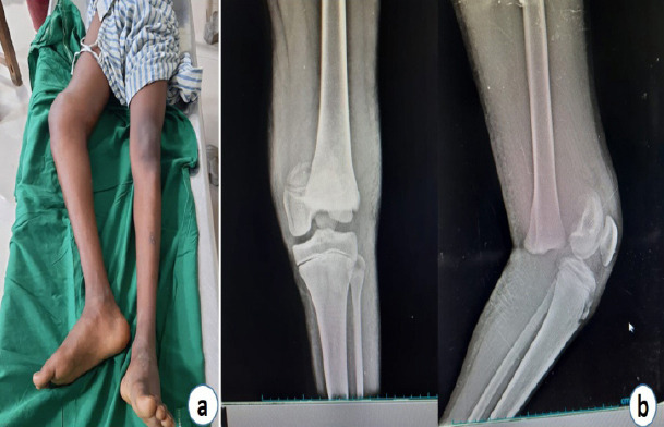
(a) Clinical presentation and (b) radiographs at presentation.
The patient was operated within 12 h after the injury. Under spinal anesthesia, patient was positioned supine. Closed reduction was attempted using a proximal tibial Steinman pin for skeletal traction and manipulation of the right lower limb. After a gentle continuous traction, manipulation was done in the form of downward pressure on the distal epiphysis and an upward pressure on the proximal metaphyseal fragment of the femur. The reduction was not achieved and hence the fracture site was opened up using a Lateral approach. After the fracture site was visualized, the proximal fragment was held using bone holding forceps and the distal fragment fixed using a clamp (Fig. 2), and traction and manipulation was done to achieve anatomic reduction of the fracture site. Reduction was confirmed under fluoroscopy and fixed using three 2.5 mm K-wires in a crossing manner (Figs. 3 and 4), 2 from lateral to medial side and 1 from medial to lateral side. For additional stability, a long leg splint was given anteriorly in 20–30 degrees flexion of the knee.
Figure 2.
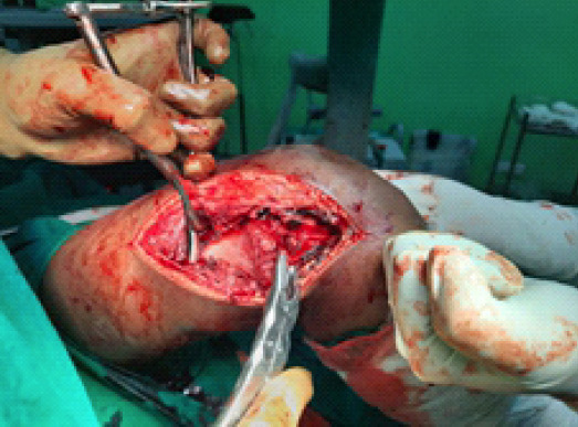
Intra-operative image of the fracture site exposure and manipulation using clamps.
Figure 3.
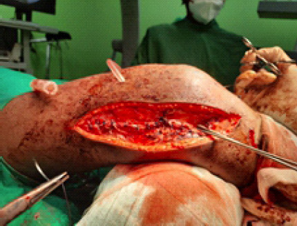
Intra-operative image showing internal fixation with K wires.
Figure 4.
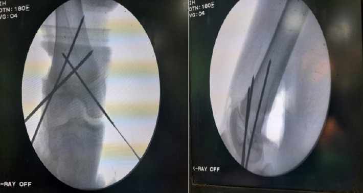
Intra-operative reduction checked under fluoroscope.
Post-operative radiographs revealed anatomic reduction of the physis of the distal femur with restoration of the normal axis of femur (Fig. 5). Post-operative surgical outcome was satisfactory. Close monitoring of the patient was done for the first 24 h postoperatively and no signs of secondary vascular thrombosis were found. Patient was started with hamstring and quadriceps strengthening exercises postoperatively. The slab was kept for 6 weeks, following which the K wires were removed. A review X-ray done showed healing of the fracture (Fig. 6), and partial weight bearing was initiated with the help of walker. On subsequent follow-up, after 4 and 8 months, correct limb axis alignment and no significant post-operative complications were found. After 6 months, the patient had become free of pain and had a complete range of movement in the right knee joint (Fig. 7). There was no significant angular deformity or shortening in the right lower extremity.
Figure 5.
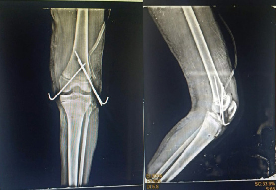
Post-operative radiographs showing the fixation.
Figure 6.
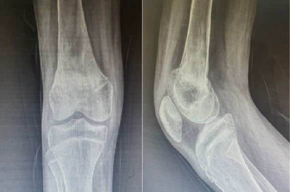
weeks follow-up radiograph showing alignment of the femur after k wire removal.
Figure 7.
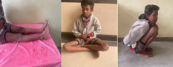
Follow-up clinical images showing patient comfortably able to flex and extend his knees, squat, and sit crossed legs.
Discussion
The distal femoral epiphysis is present since birth [5]. In the first 5 years of life the growth is rapid, after which growth slows down between ages of five to puberty and remains stable at about 2 cm a year, eventually becoming 5 times the initial length. This makes it the fastest growing physis in the human body [6]. At puberty, the physis undergoes active growth, during this period the physis is at its thickest but also the weakest. Closure of physis and growth cessation generally occurs between ages 14 and 16 in girls and 16–18 in boys [1].
Although uncommon, the distal femoral physeal injuries usually occur in adolescents. 80% of all the physeal fractures in distal femur are usually type I and II Salter and Harris fractures [4]. The mechanism of injury most commonly resulting in physeal fracture of the distal femur is a varus or valgus (lateral) stress. In some cases, hyperextension of the knee joint can result in posterior displacement of the metaphyseal fragment [7]. Posterior displacement could lead to outstretching and compression of the posterior neurovascular bundle. Thus, in posteriorly displaced knee injuries the popliteal artery can be damaged. The best strategy for diagnosing vascular compromise occurring as a complication of knee injury is still a topic of debate. Not only physical examination findings, but even sonographic color Doppler study can miss some serious latent vascular injuries like intimal tears of the popliteal artery with possibility of subsequent late thrombosis [8]. Conferring from a study led by Segal et al. [9] systematic angiography had no added advantage in terms of limb salvage in younger individuals with otherwise normal arteries. In such condition, ankle-brachial index can be used as an alternative as it is simple and easily reproducible with reliable specificity and sensitivity for diagnosing arterial injuries [8]. For the mentioned patient, the plan was to go for fracture reduction without unnecessary delay since positional variability of the pulse was present on both physical evaluation and Doppler study. However, the popliteal, tibialis posterior, and dorsalis pedis arterial pulses were closely observed in the first 24 h for early detection of any signs of secondary thrombosis.
There are varying options for treatment of distal femoral physeal fractures. A study was performed by Arkader, in which ten patients were conservatively managed with casting, traction, or both. In this study, it was noticed that initial reduction was lost within the first 2 weeks in seven patients, and there was asymmetrical growth development in nine children (angular deformity, limb length discrepancy, or flexion contracture) in the first 6 months following the injury [10]. Hence, a high incidence of displacement was encountered with closed reduction and casting. A better alternative to this is closed reduction and percutaneous fixation with non-threaded Kirschner wires. This method of fixation across the physis using percutaneous smooth wires is a frequently used method for fracture stabilization of limbs in younger age group, including the lower end of radius, lower end of humerus, distal femur, and the lower end of tibia. This practice is observed to be safer for normal physeal growth; however, there is not enough literature to associate the outcome with accordance to the size of the pin, number of attempts, and pin position. An experimental study performed by Siffert et al. on rabbits in 1956 showed that using K-wires for provisional fixation across the physeal plate of a long bone does not hinder with the longitudinal growth of the bone [11]. The study conducted by Arkader et al. [1] proposed that physeal growth arrest may occur due to placing of pins across the physis. In another animal study done by Janarv et al., [15] it was seen that the physeal drill injury of about 7–9% was required to cause growth disturbance.
The aim of surgery for these injuries is to obtain proper alignment of the fracture fragments as well as the joint, and also to maintain a steady fixation. It must be remembered that physeal growth can be repressed in case the fracture fixation crosses the growth plate [8, 12]. Thus, in such cases plates or threaded screws are not preferred, instead smooth non-threaded pins/wires are used to stabilize these fractures in order to reduce further damage to the physis. A simple fixation generally suffices in case of type I and II Salter Harris fractures, as the normal shape of the bone is restored as growth occurs. However, in case of marked lysis with a fracture dislocation operative management is deemed necessary. Thus, in this case, a cross k-wire fixation was done so as to avoid re-displacement of the fracture.
Physeal injuries involving the distal femur are reported to have high incidence of complications, the most common being premature physeal closure [3, 9, 11]. The occurrence of an early closure of physis is considered to be multifactorial: (1) High energy trauma to the bone in such trauma causes both the germinal zone injury as well as damage to the epiphyseal blood supply, (2) the extent of fracture displacement (3) high chances of shearing injury to the reserve cells of the physis, (4) common occurrence in adolescents, that is, when the physeal plate is about to close, and (5) additional iatrogenic injury taking place due to surgical intervention [3, 4, 11].
The distal femoral physeal plate is accountable for almost 40% of the longitudinal growth of the lower limb. Thus, if its growth potential is hampered due to any partial or complete arrest, it may result in significant angular deformity (genu valgum, genu varum, and recurvatum) or limb length discrepancy. These growth disturbances usually result if the growth plate alignment is not properly obtained at the time of reduction. However, these growth disturbances only get revealed 3–4 months after the initial trauma. In case growth arrest occurs, it can be appreciated on X-ray appearing like sclerotic bounds lying parallelly to the physeal plate. As a result of this, it is extremely important to have an early as well as annual follow-up of the patient until growth cessation is achieved, especially in case of younger ages. Further likelihood of disability can be reduced by early planning of appropriate procedures such as epiphysiodesis or limb lengthening. It is seen that due to a higher physiological load, shortening and deformities of the lower limbs are not as well tolerated as in the upper limbs. Fortunately, as these fractures remain rare, and most likely occur near the end of growth period, long-term consequences are minimized.
In the last follow-up, which was 15 months after the injury, none of these complications were found in our patient. The patient being close to the end of his growing period minimized the chances of potential premature closure of the physeal plate. A normal X-ray picture of the growth plate at this point makes the consequent growth arrest a very unlikely occurrence.
Conclusion
One must know that physeal injuries of the distal femur are rare in occurrence, but can prove to be dangerous due to its proximity to the popliteal vessels and involvement of the fertile growth plate of the lower limb. The potential risks of immediate limb-threatening vascular complications and long-term growth disturbances are to be considered, prevented, and thus need to be treated in an emergency. It is seen that despite initial unavoidable injury to the growth plate due to trauma, anatomical reduction, and fracture fixation is achieved it can prevent re-displacement, which, if it occurs, could result in further injury. Thus, the treatment is usually aimed at minimizing further damage to the growth plate. Cross-pinning with smooth non-threaded K-wires is an effective modality of treatment for physeal injuries. Therefore, stability of the construct is extremely important to avoid re-displacement of the fracture. Finally, patient families need to be counseled regarding the possibility of complications, and long-term follow up, preferably until skeletal maturity is achieved.
Clinical Message.
In posteriorly displaced knee injuries the popliteal artery can be damaged. Hence, one must always be watchful of peripheral pulsations in such injuries. For fixation of physeal injuries, use of smooth k wires is the dictum. Furthermore, to avoid lower limb deformities it is advisable to have a follow-up of the patient until cessation of growth is achieved.
Biography


Footnotes
Conflict of Interest: Nil
Source of Support: Nil
Consent: The authors confirm that informed consent was obtained from the patient for publication of this case report
References
- 1.Arkader A, Warner WC, Jr, Horn BD, Shaw RN, Wells L. Predicting the outcome of physeal fractures of the distal femur. J Pediatr Orthop. 2007;27:703–8. doi: 10.1097/BPO.0b013e3180dca0e5. [DOI] [PubMed] [Google Scholar]
- 2.Ilharreborde B, Raquillet C, Morel E, Fitoussi F, Bensahel H, Penneçot GF, et al. Long-term prognosis of Salter-Harris Type 2 injuries of the distal femoral physis. J Pediatr Orthop B. 2006;15:433–8. doi: 10.1097/01.bpb.0000228384.01690.aa. [DOI] [PubMed] [Google Scholar]
- 3.Chen J, Abel MF, Fox MG. Imaging appearance of entrapped periosteum within a distal femoral Salter-Harris II fracture. Skeletal Radiol. 2015;44:1547–51. doi: 10.1007/s00256-015-2201-x. [DOI] [PubMed] [Google Scholar]
- 4.Edgard-Rosa G, Launay F, Glard Y, Guillaume JM, Jouve JL, Bollini G. Salter and Harris Type-II distal femoral physeal fracture-separations at adolescent age:A new therapeutic approach (preliminary study) Rev Chir Orthop Réparatrice Appar Mot. 2008;94:546–51. doi: 10.1016/j.rco.2008.01.007. [DOI] [PubMed] [Google Scholar]
- 5.Chauvin N, Jaramillo D. Occult distal femoral physeal injury with disruption of the perichondrium. J Comput Assist Tomogr. 2012;36:310–2. doi: 10.1097/RCT.0b013e31825039a6. [DOI] [PubMed] [Google Scholar]
- 6.Sponseller PD, Stanitski CL. Distal femoral epiphyseal fractures. In: Beaty JH, Kasser JR, editors. Rockwood and Wilkins'Fractures in Children. 5th ed. Philadelphia, PA: Lippincott Williams and Wilkins; 2001. pp. 982–1010. [Google Scholar]
- 7.Basener CJ, Mehlman CT, DiPasquale TG. Growth disturbance after distal femoral growth plate fractures in children:A meta-analysis. J Orthop Trauma. 2009;23:663–7. doi: 10.1097/BOT.0b013e3181a4f25b. [DOI] [PubMed] [Google Scholar]
- 8.Cassebaum WH, Patterson AH. Fractures of the distal femoral epiphysis. Clin Orthop Relat Res. 1965;41:79–91. [PubMed] [Google Scholar]
- 9.Segal LS, Shrader MW. Periosteal entrapment in distal femoral physeal fractures:Harbinger for premature physeal arrest?Acta Orthop Belg. 2011;77:684–90. [PubMed] [Google Scholar]
- 10.Graham JM, Gross RH. Distal femoral physeal problem fractures. Clin Orthop Relat Res. 1990;255:51–3. [PubMed] [Google Scholar]
- 11.Dahl WJ, Silva S, Vanderhave KL. Distal femoral physeal fixation:Are smooth pins really safe? J Pediatr Orthop. 2014;34:134–8. doi: 10.1097/BPO.0000000000000083. [DOI] [PubMed] [Google Scholar]
- 12.Garrett BR, Hoffman EB, Carrara H. The effect of percutaneous pin fixation in the treatment of distal femoral physeal fractures. J Bone Joint Surg Br. 2011;93:689–94. doi: 10.1302/0301-620X.93B5.25422. [DOI] [PubMed] [Google Scholar]
- 13.Riseborough EJ, Barrett IR, Shapiro F. Growth disturbances following distal femoral physeal fracture-separations. J Bone Joint Surg Am. 1983;65:885–93. [PubMed] [Google Scholar]
- 14.Lombardo SJ, Harvey JP., Jr Fractures of the distal femoral epiphyses. J Bone Joint Surg Am. 1977;59:742–51. [PubMed] [Google Scholar]
- 15.Janarv PM, Wikstrom B, Hirsch G. The influence of transphyseal drilling and tendon grafting on bone growth:An experimental study in the rabbit. J Peiatric Orthop. 1998;18:149–54. [PubMed] [Google Scholar]
- 16.Aitken AP, Magill HK. Fractures involving the distal femoral epiphyseal cartilage. J Bone Joint Surg Am. 1952;34-A:96–108. [PubMed] [Google Scholar]


