Summary:
Refractory axillary lymphorrhea is a postoperative complication of breast cancer with no established standard treatment. Recently, lymphaticovenular anastomosis (LVA) was used to treat not only lymphedema but also lymphorrhea and lymphocele in the inguinal and pelvic regions. However, only a few reports have been published on the treatment of axillary lymphatic leakage with LVA. This report presents a case of successful treatment of refractory axillary lymphorrhea after breast cancer surgery with LVA. A 68-year-old woman underwent nipple-sparing mastectomy for right breast cancer, axillary lymph node dissection, and immediate subpectoral tissue expander placement. Postoperatively, the patient developed refractory lymphorrhea and subsequent seroma around the tissue expander, and underwent postmastectomy radiation therapy and frequent percutaneous aspiration of the seroma. However, lymphatic leakage persisted, and surgical treatment was planned. Preoperative lymphoscintigraphy showed lymphatic outflow from the right axilla to the space around the tissue expander. There was no dermal backflow in the upper extremities. To reduce lymphatic flow into the axilla, LVA was performed at two sites in the right upper arm. The lymphatic vessels used for anastomosis were 0.35 mm and 0.50 mm in diameter, and each was anastomosed to the vein in an end-to-end fashion. The axillary lymphatic leakage stopped shortly after the operation, and there were no postoperative complications. LVA may be a safe and simple option for the treatment of axillary lymphorrhea.
Lymphatic leakage and seroma formation after axillary lymph node dissection in breast cancer surgery is a common complication. Although various methods of prevention and treatment have been reported, there is no effective treatment for refractory axillary lymphorrhea.1
Lymphaticovenular anastomosis (LVA) is a standard technique for the treatment of secondary lymphedema, and recent reports have demonstrated that LVA was effective for lymphatic cysts and leakage in the inguinal and pelvic regions.2–4 Yet, only a few reports have touched on the efficacy of LVA for axillary lymphorrhea.3,5 This report presents our experience with LVA for refractory axillary lymphorrhea in postoperative breast cancer.
CASE REPORT
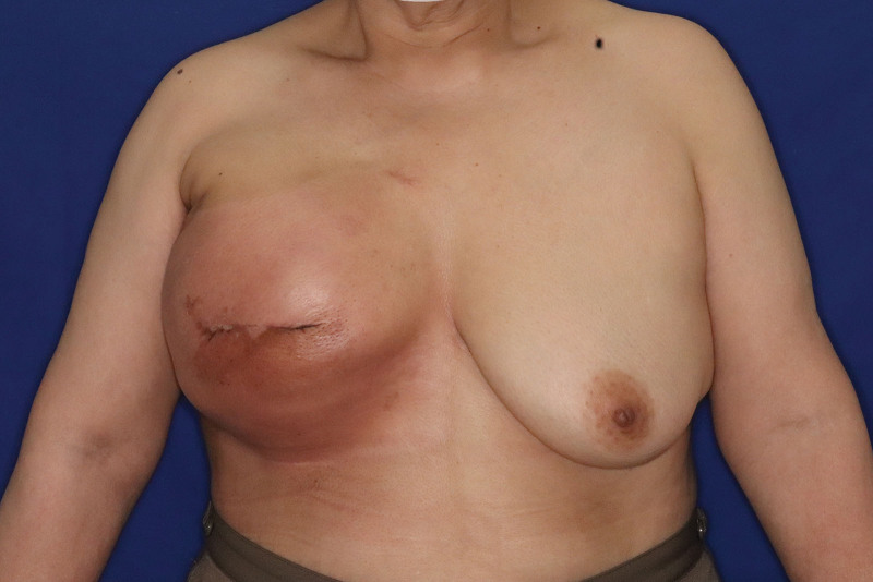
A 68-year-old woman underwent nipple-sparing mastectomy (her nipple was unfortunately resected due to necrosis several days after surgery) and level Ⅰ and Ⅱ axillary lymph node dissection for right breast cancer, and immediate subpectoral tissue expander (TE) breast reconstruction was performed. However, the volume of axillary drainage remained about 200 mL per day even after 20 days postoperatively. Given that further drainage was unlikely to reduce drainage volume, the drain was removed. Subsequently, a seroma formed around the TE, and the patient began to complain of pain due to chest tenderness. Percutaneous puncture was continued twice a week, with a drainage volume per puncture of about 400 mL. Two months postoperatively, postmastectomy radiation therapy (PMRT; 50 Gy/25 Fr) was started. Puncture was continued during PMRT, but the fluid reaccumulated each time. Three months postoperatively, the seroma did not decrease in size (Fig. 1). On lymphoscintigraphy of the upper extremities, there were no obvious areas of dermal backflow, but lymphatic leakage from the axilla to the space around the TE was noted, suggesting the possibility of refractory axillary lymphorrhea (Fig. 2).
Fig. 1.
Preoperative photograph. The seroma was located near the TE, causing tension in the chest skin. The skin also had redness from radiodermatitis.
Fig. 2.
Preoperative lymphoscintigraphy. The lymphatic flow moved up the right arm to the axilla and accumulated around the TE due to lymphorrhea. The arrowhead shows the TE, and the arrow shows the site of lymphorrhea.
We still had several months before the scheduled surgery date for autologous tissue reconstruction, and we needed to stop the lymphatic leakage because of the possibility of TE infection. We considered it unlikely that conservative therapy would improve the lymphatic leakage, so we planned LVA on the upper arm to reduce lymphatic flow into the axillary region. Before surgery, indocyanine green lymphangiography was performed to confirm the location of linear lymphatic vessels in the upper arm. After identifying veins in the vicinity of the lymphatic vessels using ultrasonography, a 2-cm incision was made at these sites, and LVA was performed at two locations. Veins and lymph vessels were anastomosed in an end-to-end fashion, using 12-0 Nylon. The diameters of the lymphatic vessels were 0.35 mm and 0.50 mm. After anastomosis, patency was confirmed by using PDE-neo (Hamamatsu Photonics, Hamakita, Japan). (See Video [online], which shows the intraoperative view after LVA in the upper arm.) We punctured the seroma on postoperative day 2, and no further accumulation was observed. Chest tenderness also improved postoperatively (Fig. 3).
Fig. 3.
Postoperative photograph (1 month after surgery). The tenderness in the chest skin improved postoperatively.
Video 1. shows the intraoperative view after LVA in the upper arm.
Lymphoscintigraphy was performed 2 months after surgery and revealed no lymphatic leakage around the TE (Fig. 4). The patient could continue the breast reconstruction procedure, and she finally underwent autologous tissue reconstruction with DIEP flap.
Fig. 4.
Postoperative lymphoscintigraphy. There was no lymphatic effusion around the TE.
DISCUSSION
In the present case, LVA was performed on the upper arm for refractory axillary lymphorrhea after breast cancer surgery, and the seroma around the TE disappeared. Based on clinical and lymphoscintigraphic findings, the large amount of serous effusion around the TE was most likely due to axillary lymphatic leakage.
Lymphorrhea may be treated with procedures such as ligation of lymphatic vessels, sclerotherapy, and embolization.1,6,7 However, as described by Giacalone et al and Kadota et al, such procedures are likely to induce subsequent lymphedema due to the obstruction of lymphatic vessels.3,4 The best treatment would be to directly repair the damaged lymphatic vessels in the axilla by anastomosing them with intact lymphatic vessels.2 However, because our patient had already undergone TE implantation and PMRT, it was expected to be difficult to find, dissect, and anastomose the lymphatic vessels through a chest skin incision. In addition, we considered that an incision to the chest skin would not only be highly invasive but would also cause postoperative complications that might disrupt the breast reconstruction procedure. Marthan et al performed LVA in addition to capsulotomy for postoperative breast seroma,5 but we decided on an LVA-only approach to reduce lymphatic flow into the axilla to minimize invasiveness. As for the site chosen for LVA, we hypothesized that LVA closer to the axilla in the upper extremity would effectively reduce the flow of lymph fluid into the axillary region. Therefore, we identified linear lymphatic vessels in the upper arm, with a focus on the proximal part of the upper arm, and targeted them for LVA.
It is difficult to determine the extent to which LVA contributed to the disappearance of seroma around the TE. However, because the seroma persisted for more than 3 months after the onset, it was more than likely that the seroma improved with the formation of a bypass around the axillary lymph-leakage spot by LVA, rather than by spontaneous resolution. After the postoperative day 2 puncture, no seroma accumulation was observed, which is compatible with previous reports noting the time required from LVA to resolution of lymphorrhea.2,4 It is also possible that the lymphatic leakage was resolved due to radiotherapy, but recurrent seroma was observed after the puncture near the end of PMRT, suggesting again that LVA likely contributed to the resolution of lymphorrhea.
LVA may reduce the risk of developing lymphedema in the future as a prophylactic treatment.8,9 On the other hand, there is also a possibility that lymphedema may develop due to postoperative obstruction at the site of LVA. Although no obvious lymphedema has developed in our patient as of this writing (at 6 months postoperatively), careful follow-up will be necessary.
We performed LVA as a means to reduce the inflow of lymphatic fluid to the injured site. However, in the present case, surgical treatment might have been avoided with the use of somatostatin and dietary guidance (eg, low-fat diet). As such, conservative therapies should also be considered as treatment options in future cases.10
CONCLUSIONS
LVA allowed for minimally invasive treatment of axillary lymphorrhea, and breast reconstruction was able to continue in this case. We believe our report is significant because, although the efficacy of LVA for lymphatic leakage after axillary lymph node dissection has been reported, case reports describing the detailed treatment course are scarce. LVA would likely be a useful treatment for similar cases. However, further accumulation of cases will be needed to confirm our findings.
Footnotes
Disclosure: The authors have no financial interests to declare in relation to the content of this article.
Related Digital Media are available in the full-text version of the article on www.PRSGlobalOpen.com.
REFERENCES
- 1.van Bemmel AJM, van de Velde CJH, Schmitz RF, et al. Prevention of seroma formation after axillary dissection in breast cancer: a systematic review. Eur J Surg Oncol. 2011;37:829–835. [DOI] [PubMed] [Google Scholar]
- 2.Yamamoto T, Yoshimatsu H, Koshima I. Navigation lymphatic supermicrosurgery for iatrogenic lymphorrhea: supermicrosurgical lymphaticolymphatic anastomosis and lymphaticovenular anastomosis under indocyanine green lymphography navigation. J Plast Reconstr Aesthet Surg. 2014;67:1573–1579. [DOI] [PubMed] [Google Scholar]
- 3.Giacalone G, Yamamoto T, Hayashi A, et al. Lymphatic supermicrosurgery for the treatment of recurrent lymphocele and severe lymphorrhea. Microsurgery. 2019;39:326–331. [DOI] [PubMed] [Google Scholar]
- 4.Kadota H, Shimamoto R, Fukushima S, et al. Lymphaticovenular anastomosis for lymph vessel injury in the pelvis and groin. Microsurgery. 2021;41:421–429. [DOI] [PubMed] [Google Scholar]
- 5.Marthan J, Struk S, Bennis Y, et al. Lymphaticovenous anastomosis: treatment of a persistent breast seroma. Ann Chir Plast Esthet. 2020;65:332–337. [DOI] [PubMed] [Google Scholar]
- 6.Klode J, Klötgen K, Körber A, et al. Polidocanol foam sclerotherapy is a new and effective treatment for post-operative lymphorrhea and lymphocele. J Eur Acad Dermatol Venereol. 2010;24:904–909. [DOI] [PubMed] [Google Scholar]
- 7.Inoue M, Nakatsuka S, Yashiro H, et al. Lymphatic intervention for various types of lymphorrhea: access and treatment. Radiographics. 2016;36:2199–2211. [DOI] [PubMed] [Google Scholar]
- 8.Rodriguez JR, Fuse Y, Yamamoto T. Microsurgical strategies for prophylaxis of cancer-related extremity lymphedema: a comprehensive review of the literature. J Reconstr Microsurg. 2020;36:471–479. [DOI] [PubMed] [Google Scholar]
- 9.Ciudad P, Escandón JM, Bustos VP, et al. Primary prevention of cancer-related lymphedema using preventive lymphatic surgery: systematic review and meta-analysis. Indian J Plast Surg. 2022;55:01818–01025. [DOI] [PMC free article] [PubMed] [Google Scholar]
- 10.Mahmoud SA, Abdel-Elah K, Eldesoky AH, et al. Octreotide can control lymphorrhea after axillary node dissection in mastectomy operations. Breast J. 2007;13:108–109. [DOI] [PubMed] [Google Scholar]


