Abstract
Objectives
This study aims to investigate the expression patterns of mitochondrially encoded cytochrome c oxidase 1 (MT-CO1) in different organs and tissues of MRL/lpr mice aged six and 18 weeks.
Materials and methods
Six-week-old female MRL/lpr mice (n=10) were considered young lupus model mice, and 18-week-old MRL/lpr mice (n=10) were considered old lupus model mice. Additionally, six-week-old (n=10) and 39-week-old (n=10) female Balb/c mice were used as the young and old controls, respectively. The messenger ribonucleic acid (mRNA) and protein expression levels of MT-CO1 in nine organs/tissues were detected via quantitative polymerase chain reaction (qPCR) and Western blot. Malondialdehyde (MDA) levels were determined with thiobarbituric acid colorimetry. The correlation coefficient of MT-CO1 mRNA levels and MDA levels in each organ/tissue at different ages was analyzed by Pearson correlation analysis.
Results
The results showed that most non-immune organs/tissues (heart, lung, liver, kidneys, and intestines) showed increased MT-CO1 expression levels in younger MRL/lpr mice (p<0.05) and decreased MT-CO1 expression in older mice (p<0.05). Expression of MT-CO1 in the lymph nodes was low in younger mice but high in older mice. In other immune organs (spleen and thymus), MT-CO1 expression was low in older MRL/lpr mice. Lower mRNA expression and higher MDA levels were observed in the brains of MRL/lpr mice. However, all MRL/lpr mice showed higher MDA levels than Balb/c mice in every organ no matter younger or older MRL/lpr mice.
Conclusion
Our study results suggest that lymphoid mitochondrial hyperfunction at organ level may be an important intrinsic pathogenesis in systemic lupus erythematosus activity, which may affect mitochondrial dysfunction in non-immune organs.
Keywords: Cytochrome C oxidase subunits 1, malondialdehyde, oxidative stress, systemic lupus erythematosus.
Introduction
Systemic lupus erythematosus (SLE) is a chronic autoimmune disease. The anti-doublestranded deoxyribonucleic acid DNA (dsDNA) antibodies in SLE are important biomarkers, and immune inflammation is the core pathogenesis of SLE. Mitochondrial DNA (mtDNA) is a natural dsDNA and is prone to mutation owing to its susceptibility to reactive oxygen species (ROS). Mitochondrial ROS can facilitate the release of higher levels of cytochrome C by participating in the formation of apoptosome, leading to the decrease of enzyme activities of mitochondrial complexes I, IV and V, which may be associated with swelling of mitochondria and its depolarization.[1] Moreover, mitochondria are closely related to programmed cell death, with mtDNA involved in apoptosis, Neutrophil extracellular traps (NETs) and pyroptosis can induce immune inflammation via the cGAS-Sting pathway.[2] Mitochondrial dysfunction may cause abnormal redox reaction, decreased functioning of biogenesis-related enzymes, increased NETosis, harmful cytokine effects, and aberrant lymphocyte behavior.[3] SLE is closely related to mitochondrial dysfunction and overproduction of ROS.[4]
The mitochondrial respiratory chain (RC) is the site of adenosine triphosphate (ATP) synthesis and ROS generation. The ATP is the most important energetic molecule and participates in all major life activities. The ROS also have important implications in the biological system. Oxidative stress acts as an etiological factor in a wide variety of pathologies. The mitochondrially encoded cytochrome c oxidase 1 (MT-CO1) gene encodes a crucial subunit of the RC complex IV.[5] Decreased complex IV activity is related to increased oxidative stress in MT-CO1 mutants.[6] Our previous research revealed the high mutation rate of MT-CO1 and its low messenger ribonucleic acid (mRNA) and protein levels in blood samples from MRL/lpr mice were related to decreased antioxidant capacity, indicating that the MT-CO1 gene of the mitochondrial RC in MRL/lpr mice is susceptible to ROS-induced oxidative damage.[7] SLE is an autoimmune disease that leads to damage in multiple organs and systems. The mtDNA participates in important physiological and pathological processes of the body; thus, MT-CO1 is likely also involved in the processes of organ damage in SLE. The distribution of mitochondria in various organs and tissues depends on energetic demands and age-related changes. Therefore, in the present study, we aimed to investigate the distinct functional characteristics of MT-CO1 and MDA levels in nine organs/tissues of MRL/lpr mice at different ages and to examine the potential mechanisms of SLE.
Patients and Methods
This experimental study was conducted at the State Key Laboratory of Respiratory Diseases between September 2020 and March 2021. The animal research followed the standard of the Animal Research: Reporting of in vivo Experiments (ARRIVE) guidelines.[8]
Experimental animals and randomization
Twenty female MRL/lpr mice and 20 BALB/c mice were included in the study and randomly divided into four groups: six-week-old female MRL/lpr mice (n=10) were considered young lupus model mice, and 18-weekold mice (n=10) were considered old lupus model mice. Additionally, six-week-old (n=10) and 39-week-old (n=10) female Balb/c mice were used as the young and old controls, respectively. The MRL/lpr mice were provided by Shanghai Shrek Laboratory Animal Co. Ltd (Shanghai, China) (animal-quality certificate number: 44007200001471). The Balb/c mice were purchased from Guangdong Experimental Animal Center (Foshan, China) (animal-quality certificate number: 44007200055054).
After one week of adaptation, each group was raised to the corresponding ages listed above. Each mouse was, then, anesthetized via intraperitoneal injection of 10% chloral hydrate (1 mL/kg), after which the right eyeball was enucleated for blood collection. Ethylenediaminetetraacetic acid (EDTA) anticoagulant tubes were used to collect whole blood samples and, then, each anesthetized mouse was killed via cervical dislocation. Heart, liver, kidney, spleen, lung, intestinal, lymph node, thymus, and brain tissues were collected from each mouse. The specific procedure was as follows: First, the abdominal skin of each mouse was cut off. Then, the heart, lung, thymus, liver, kidney, spleen, and intestine were separated with tweezers. Next, the auxiliary skin of each mouse was removed, and the lymph nodes were dissociated. Finally, the skin was cut away from the head of each mouse, and the brain tissue was subsequently dissected. All of the above steps were performed on ice. One kidney from each mouse was separated and fixed in formaldehyde; the remaining tissue samples were stored at -80°C until further use.
Experimental materials
The following experimental materials were used in this study: radioimmunoprecipitation assay (RIPA) cell lysate (Crystal Color Biological Technology Co. Ltd., Xian, China); enhanced chemiluminescence Western blot detection kit (WBKLS0100; Millipore, Burlington, MA, USA); MT-CO1 antibody (ab203912; Abcam, Cambridge, UK); β-actin Rabbit pAb (30102ES40; Yeasen, Shanghai, China); peroxidase-conjugated goat anti-rabbit immunoglobulin G (H+L) (33101ES6; Yeasen, Shanghai, China); BCA protein quantitative kit (Crystal Color Biological Technology Co. Ltd., Xian, China); Alexa Fluor 594 Goat Anti-mouse lgG (HþL) (A11005, Life Technologies Corp., Carlsbad, CA, USA), bioflake HEp-2 (FA15101010-1, EUROIMMUN, Medical Laboratory Diagnostics AG, Lübeck, Germany), lipid oxidation (malondialdehyde [MDA]) detection kit (Blue Skies Biological Technology Co. Ltd., Guangzhou, China); RNA-easy Isolation Reagent (Vazyme, Nanjing, China); HiScript III RT SuperMix for quantitative polymerase chain reaction (qPCR) (+gDNA wiper) (Vazyme, Nanjing, China); ChamQ Universal SYBR qPCR Master Mix (Vazyme, Nanjing, China); and primers (Sangon Biotech, Shanghai, China). The sequences of the primers were as follows. MT-CO1 primer sequence: forward 5’-TATGTTCTATCAATGGGAGC-3’, reverse: 5’-GTAGTCTGAGTAGCGTCGTG-3’, Nix primer sequence: forward 5’-CTGGAGCACGTTCCTTCCTC-3’, reverse: 5 ’- A C A G T G C G A A C T G C C T C T T G - 3 ’, and housekeeping gene β-actin: forward: 5’-TCGCTGCGCTGGTCGTC-3’, reverse: 5’-GGCCTCGTCACCCACATAGGA-3’.
Cell culture
A mouse glomerular mesangial cell line (MGCs, the MES-13 cells from an SV40 transgenic mouse) was obtained from Guangzhou Scissor Hand Gene Technology Co. Ltd. (icell-m056; Shanghai iCELL Biotechnology Co., Ltd, Shanghai, China) and maintained in a culture medium with a 3:1 mixture of DMEM and Ham’s medium, supplemented with antibiotics (100 μg/mL of penicillin and 100 μg/mL of streptomycin) and 10% fetal bovine serum at 37°C and 5% CO2. Cell experiments were performed between the third and eighth passages; 80% confluent cells were made quiescent by serum-free starvation with 24-h incubation in DMEM before lipopolysaccharide (LPS) treatment (1.5 mg/L). A total of 104 cells per well in a 96-well plate were, then, incubated with and without 1.5 mg/mL of LPS for 24 h.
RNA isolation and qPCR analysis
Total RNA from each group of tissues and MGCs was extracted using RNA-easy Isolation Reagent. The RNA was transcribed into single-stranded DNA using a HiScript-III RT SuperMix for qPCR with complementary DNA (cDNA) as a template. The PCR for MT-CO1 or Nix was performed using a ChamQ Universal SYBR qPCR Master Mix for qPCR. Then, mRNA expression was determined via qPCR. The cDNA generated from each sample was amplified using specific primers. The mRNA levels of the endogenous housekeeping gene, β-actin, were used to normalize the expression levels. Relative abundances of genes were calculated using the 2-ΔΔCT method. The qPCR conditions were as follows: 40 cycles of 65°C for 3 min, 95°C for 10 sec, and 60°C for 20 sec.
Detection of lipid oxidation (MDA)
Tissues were homogenized with RIPA cell lysate and centrifuged at 10,000 to 12,000 g for 10 min to obtain the supernatant from each sample. Protein concentrations in each sample were measured according to the instructions of the BCA protein-concentration assay kit, and MDA levels in each sample were measured according to the instructions of the Biyuntian lipid oxidation (MDA) assay kit. The values of each of these parameters were determined using spectrophotometry, a standard curve was established using standard solutions, and the measured values in each sample were calculated with reference to the standard curve. After calculating the MDA content in the sample solution, the MDA content (μmoL/mg protein) in the initial sample was expressed as the protein content per unit weight.
Western blotting
Total protein from each of the tissues and MGCs was extracted with RIPA, and the protein concentrations were quantitatively detected using a BCA kit. Equal amounts of protein were separated using 10% sodium dodecyl sulfate-polyacrylamide gel electrophoresis, followed by transference to a polyvinylidene fluoride membrane. The membrane was blocked with 3% bovine serum albumin and probed with primary antibody overnight at 4°C and, then, reacted with 1:10,000 diluted secondary antibody at room temperature for 2 h. The electrochemical luminescence method and ImageJ software (National Institutes of Health, Bethesda, MD, USA) were used to read the gray values of each protein band and analyze MT-CO1 protein expression levels.
Statistical analysis
Statistical analysis was performed using the IBM SPSS for Windows version 25.0 software (IBM Corp., Armonk, NY, USA). Descriptive data were expressed in mean ± standard deviation (SD). The Shapiro-Wilk statistical test was used to check the normality of the data. Analysis of variance (ANOVA) test was used to assess the significance of differences between the means. If one or all of the assumptions are not met, the Mann-Whitney U test was used for two independent groups, the Wilcoxon test was used for two conjugate groups, and the Kruskal-Wallis test is used for more than two independent groups. The correlation between MT-CO1 mRNA and MDA concentration was analyzed using the Pearson correlation test. A p value of <0.05 was considered statistically significant.
Results
Expression patterns of MT-CO1 mRNA and protein in MGCs
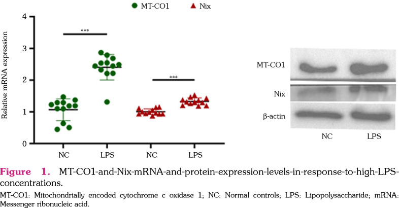
The mRNA and protein expression of MT-CO1 and Nix were all significantly induced by high LPS concentrations (1.5 mg/L) at 24 h (MT-CO1, t=8.778, p<0.001; Nix, t=7.953, p<0.001) (Figure 1).
Figure 1. MT-CO1-and-Nix-mRNA-and-protein-expression-levels-in-response-to-high-LPSconcentrations. MT-CO1: Mitochondrially encoded cytochrome c oxidase 1; NC: Normal controls; LPS: Lipopolysaccharide; mRNA: Messenger ribonucleic acid.
Expression patterns of MT-CO1 mRNA in nine organs and tissues
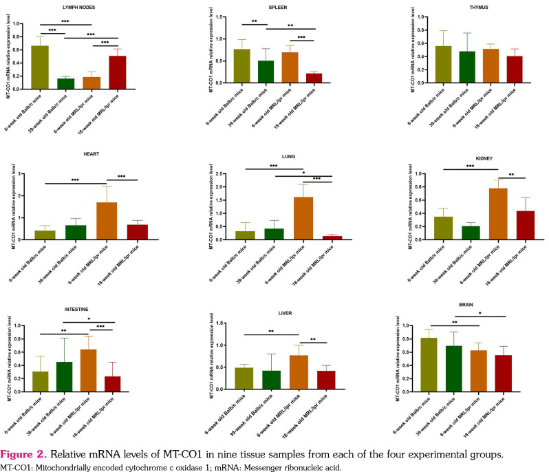
We evaluated changes in MT-CO1 mRNA expression in nine tissues for the four groups of mice. As shown in Figure 2, in immune organs, MT-CO1 mRNA expression levels in the lymph nodes were significantly decreased in six-week-old MRL/lpr mice compared to six-week-old Balb/c mice. In the spleen and thymus, the differences in MT-CO1 mRNA expression levels were both non-significant between six-week-old MRL/lpr mice and six-week-old Balb/c mice. However, in 18-week-old MRL/lpr mice, MT-CO1 mRNA expressions levels were significantly increased in the lymph nodes but decreased in the spleen and thymus, in comparison with 39-week-old Balb/c and six-week-old MRL/lpr mice. In five different non-immune organs, the MT-CO1 mRNA expression features were divided into two categories. One category was that in which the MT-CO1 mRNA expression levels in the lung, heart, kidney, and intestine were significantly increased in six-week-old MRL/lpr mice compared to six-week-old Balb/c mice; however, in 18-week-old MRL/lpr mice, these levels were significantly decreased, as compared to six-weekold MRL/lpr mice. The other category was that in which the MT-CO1 mRNA expression levels in the brain were significantly decreased in sixweek-old MRL/lpr mice compared to six-weekold Balb/c mice and significantly decreased in 18-week-old MRL/lpr mice compared to 39-week-old Balb/c mice; however, there were no significant differences between six-week-old and 18-week-old MRL/lpr mice.
Figure 2. Relative mRNA levels of MT-CO1 in nine tissue samples from each of the four experimental groups. MT-CO1: Mitochondrially encoded cytochrome c oxidase 1; mRNA: Messenger ribonucleic acid.
Expression patterns of MDA levels in nine organs/tissues
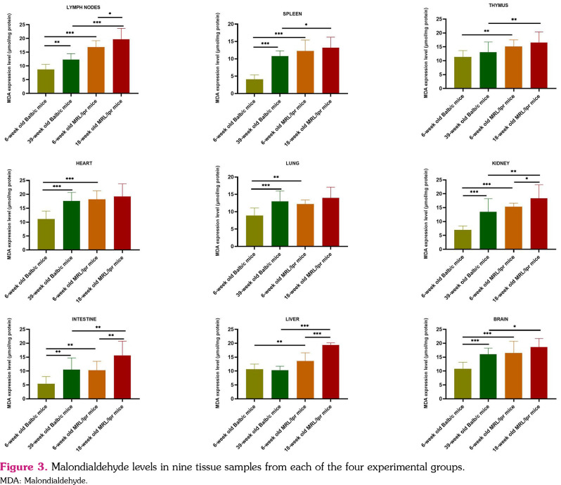
We also measured the changes in MDA levels in nine tissues among the four different groups of mice. As shown in Figure 3, the levels of MDA in these four groups were all increased in six-week-old MRL/lpr mice as compared to six-week-old Balb/c mice and in 18-week-old MRL/lpr mice compared to 39-week-old Balb/c mice. No significant difference was observed in the MDA levels between six-week-old MRL/lpr mice and 18-week-old MRL/lpr mice. These results indicated that, in most organs of the SLE model mice, MDA was highly expressed at younger ages and remained at similar, harmfully high levels at older ages.
Figure 3. Malondialdehyde levels in nine tissue samples from each of the four experimental groups. MDA: Malondialdehyde.
Expression patterns of MT-CO1 protein in nine organs/tissues
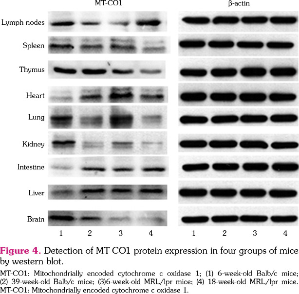
As shown in Figure 4, the expression levels of MT-CO1 protein were similar to those of mRNA. The MT-CO1 protein expression was first downregulated at younger ages and later upregulated at older ages in the lymph nodes, whereas these remained at similar levels in young mice but were finally downregulated in older mice in the spleen and thymus. On the contrary, in five different non-immune organs, the expression levels of MT-CO1 protein were first upregulated at younger ages and then downregulated at older ages in the lung, heart, kidney, and intestine; however, low expression levels were maintained in the brain of MRL/lpr mice, regardless of age.
Figure 4. Detection of MT-CO1 protein expression in four groups of mice by western blot. MT-CO1: Mitochondrially encoded cytochrome c oxidase 1; (1) 6-week-old Balb/c mice; (2) 39-week-old Balb/c mice; (3)6-week-old MRL/lpr mice; (4) 18-week-old MRL/lpr mice. MT-CO1: Mitochondrially encoded cytochrome c oxidase 1.
Pearson correlation analysis of MT-CO1 mRNA levels and MDA levels in each tissue at different ages
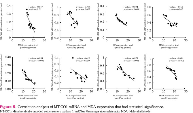
The Pearson correlation coefficients are shown in Figure 5 and Table 1. In general, there was a negative correlation between MT-CO1 expression and MDA levels in MRL/lpr mice, particularly in the spleen and brain of six-week-old and 18-week-old MRL/lpr mice, the lymph nodes of six-week-old MRL/lpr mice, and the thymus, lung, and kidney of 18-week-old MRL/lpr mice.
Figure 5. Correlation-analysis-of-MT-CO1-mRNA-and-MDA-expression-that-had-statistical-significance. MT-CO1: Mitochondrially encoded cytochrome c oxidase 1; mRNA: Messenger ribonucleic acid; MDA: Malondialdehyde.
Table 1. Pearson correlation coefficients between MT-CO1 mRNA expression and MDA concentration.
| Lymph nodes | Spleen | Thymus | Heart | Lung | Kidney | Intestine | Brain | Liver | |
| p | p | p | p | p | p | p | p | p | |
| 6-week old Balb/c mice | -0.515 | -0.223 | -0.230 | 0.418 | 0.258 | -0.032 | -0.617 | -0.318 | 0.288 |
| 39-week old Balb/c mice | -0.396 | -0.319 | 0.226 | 0.013 | -0.349 | -0.448 | 0.117 | -0.512 | -0.491 |
| 6-week old MRL/Ipr mice | -0.817** | -0.734** | -0.032 | -0.292 | 0.040 | -0.324 | -0.065 | -0.878** | 0.105 |
| 18-week old MRUIpr mice | -0.076 | -0.894** | -0.752** | -0.226 | -0.854** | -0.626* | 0.455 | -0.864** | 0.019 |
| MT-CO1: Mitochondrially encoded cytochrome c oxidase 1; MDA: Malondialdehyde; * 0.05; ** 0.01. | |||||||||
Discussion
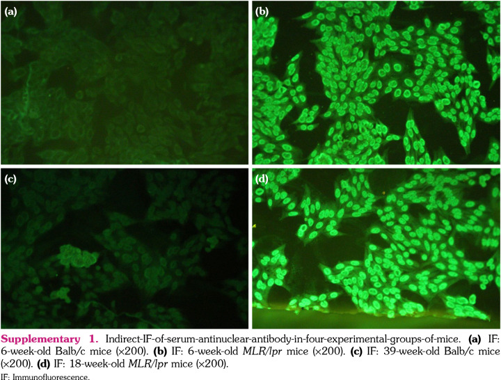
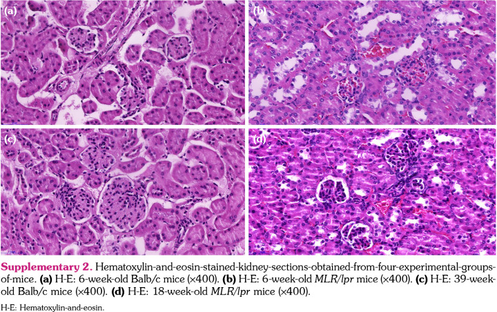
Systemic lupus erythematosus is an autoimmune disease characterized by congenital and acquired immune disorders with multiple organ damage. In our study, the result of serum antinuclear antibody (Supplementary 1) and pathological changes in the kidney (Supplementary 2) were both positive in 6-week-old and 18-week-old MRL/lpr mice, indicating that MRL/lpr mouse is a mature animal model of SLE. Previous studies have shown that oxidative stress is associated with SLE disease activity and has both negative and positive impacts on the disease.[9] Our results showed that ROS were increased in all SLE mouse model tissues, which indicated that the pathological changes in SLE mice resulted in an increase of oxidative stress, inevitably leading to the enhancement of immune inflammation. Our results also showed that the expression of MT-CO1 was obviously different. On the one hand, MT-CO1 expression was significantly different in different organs at the same disease stage under the same conditions of elevated ROS, with downregulation and upregulation of MT-CO1, forming a sharp contrast. On the other hand, the expression of MT-CO1 in most organs was also significantly different at different disease stages under conditions of elevated ROS, with downregulation and upregulation, also forming a sharp contrast. Our study results suggest that the relationship between ROS and mitochondrial RC genes is not simply a vicious cycle of mutual damage, as described in aging theory. It has been found that ROS can not only be considered a damage factor, but also a beneficial biomolecule at low concentrations, which can stimulate the organism to have completely different biological effects or even effects that are opposite to oxidative damage.[10]
Recent studies have shown that ROS undergo a transformation from molecules that cause damage to signal pathways that regulate normal physiological and biological responses. To give a well-known example, physical exercise is a strategy to increase ROS, thereby activating musculoskeletal strength via beneficial pathways.[11] The ROS are also believed to play a dual role in plant biology.[12] Many cellular processes such as differentiation, proliferation, and apoptosis require redox signals. Redox signals are also crucial in the control of various diseases, including tumorigenesis and autoimmune diseases.[13] Whether a normal reaction or an abnormal reaction, ROS affect the redox biology in the body. Therefore, it is not only the ROS that have the dual role of inducing oxidative stress damage and redox signaling, but the mitochondria also have the dual role of ROS production and response to reactive oxygen-mediated changes. Recent studies have also shown that the relationship between mitochondria and ROS is more complicated than previously thought. Hydrogen peroxide (H2O2) is an important secondary messenger for coordinating oxidative metabolism and cellular physiological changes. Mitochondria act as a ‘reactive oxygen stabilizing device’, buffering cellular H2O2 levels in mammalian cells. The production of mitochondrial H2O2 seems to be more sensitive to changes in mitochondrial energetics than the consumption pathway.[14] Mitochondria can act as regulators of ROS and can adapt to the mitochondrial ROS environment. Unlike the traditional concept of oxidative damage caused by ROS, the signal regulation of ROS has a more favorable effect on mitochondrial biogenesis and explains why MT-CO1 expression was increased in most non-immune major organs of young MRL/lpr mice in our study.
Previous studies have also shown that, in most non-immune major organs, activating the DNA repair pathway to maintain mitochondrial genome stability requires physiological levels of intracellular ROS.[15] Our study results suggest that appropriate levels of ROS induced by early SLE activity may act as a stimulus to increase gene expression in the mitochondrial RC.
In the current study, the expression of MT-CO1 in the lymph nodes of six-week-old MRL/lpr mice was lower than that of normal mice, but the expression of MT-CO1 in most non-immune organs was higher than that of normal mice. The MRL/lpr mice usually begin to develop symptoms at six weeks of age,[16] and immune complex deposition occurs at eight weeks.[17] Although ROS were increased in all tissues, there were significant differences in the effects of ROS on different tissues. Lymph nodes are the peripheral immune organs in SLE activity and directly affected with immune inflammation. However, as a tissue with high oxygen consumption and poor antioxidant capacity, brain tissue is the most severely affected by oxidative damage. The ROS and mitochondrial RC genes damage each other, resulting in a significant decrease in the expression of MT-CO1. In most non-immune organs with high oxygen consumption, including the heart and liver, the expression of MT-CO1 did not only not decrease, but was significantly increased in young MRL/lpr mice in our study. These results suggest that, in the early stages of SLE, ROS in most important organs of the body may serve as biologically active molecules to activate DNA repair functions and promote the expression of mitochondrial RC genes. Cell mitochondrial function is in a compensatory state, and the oxidative damage to major organs is not obvious.
What we are most concerned about is that the expression of MT-CO1 in the lymph nodes of 18-week-old MRL/lpr mice was not only higher than that of normal mice, but was also significantly higher than that of young MRL/lpr mice. The lifespan of MRL/lpr mice ends at 20 to 22 weeks (a normal mouse lifespan is 2.5 years, about 130 weeks). The 18-week MRL/lpr mice were approaching death.[16] In 18-week MRL/lpr mice, MT-CO1 expression was decreased in all other non-immune organs but increased only in the peripheral lymph nodes, and the lymph nodes of older MRL/lpr mice may be systemically hyperplastic, which is also found in clinical practice. Lymphocytes and their dysfunction are considered to be an important pathogenesis in SLE.[18] Our study results demonstrated that during disease progression in MRL/lpr mice, the lymphocytes in enlarged lymph nodes are functional, and the lymphatic system is also hyperfunction at overall organ level, that is, when the expression of mitochondrial RC genes in all other tissues decreases, it increases in the lymph nodes. Mitochondria are important energy generators and well-functioning organelles of the cell. The inevitable result of the active functioning of immunocompetent cells is immune hyperfunction, which damages other non-immune organs and develops progressively, leading to shortened lifespan and death in MRL/lpr mice (which live only a quarter the lifespan of normal mice). Indeed, prior to the use of glucocorticoids in the treatment of SLE, its clinicopathological characteristics were aggravated and lethal disease.[19] The natural development of SLE in humans is very similar to the pathological process in MRL/lpr mice.
From the perspective of disease development, there is still a sharp contrast between lymph nodes and non-immune organs. The expression of MT-CO1 in the lymph nodes was decreased at six weeks and increased in compensation at 18 weeks in our mouse model. The dynamic change trend was from decreased decompensation in the early stage to compensatory increase in mitochondrial hyperfunction in the late stage. On the contrary, in most non-immune organs, the expression of MT-CO1 was increased at six weeks and decreased at 18 weeks. The dynamic change trend was from early functional compensation to decreased decompensation in the late stage. In the past, there was a concern about the reduction of mitochondrial RC gene expression owing to oxidative damage of ROS during aging.[6,20] In all non-immune major organs of old MRL/lpr mice, excessive ROS also damage the structure and function of the mitochondria. Mitochondrial RC genes are susceptible to oxidative damage and eventually cause immune inflammation in SLE. Our previous studies also showed that the expression of MT-CO1 in peripheral blood mononuclear cells of young MRL/lpr mice was decreased and was closely related to ROS.[7] In this study, however, we pay close attention to the fact that the mitochondrial RC gene MT-CO1 had both a compensatory increase in most non-immune organs in the early stage and in the lymph nodes in the late stage of SLE; however, in the same environment with elevated ROS, MT-CO1 expression decreased in most non-immune organs in the late stage and in the lymph nodes in the early stage of SLE. These study results indicate that the condition of ROS is not the only factor that affects the expression of mitochondrial RC gene MT-CO1.
In mitochondria, certain other mechanisms have similar effects. In response to mitochondrial damage, mitophagy may be an important mechanism for mitochondrial quality control and may explain the increased MT-CO1 mRNA levels in young MRL/lpr mice. Excessive ROS can trigger mitophagy, which serves to remove damaged mitochondria and reduce the level of ROS, to maintain stable mitochondrial function in the cell.[21,22] Therefore, mitophagy can maintain cellular homeostasis under oxidative stress and protect cells from damage owing to cytotoxic ROS, thereby reducing the damage to mitochondrial RC genes.
Our in vitro experiments also confirmed that the expression of both the mitochondrial gene MT-CO1 and the mitochondrial autophagy gene Nix in renal mesangial cells was increased under stimulation with LPS for 24h, indicating that the quality control of the mitochondrial autophagy function can promote the expression of the mitochondrial RC gene in the early stage of disease, which is conducive to maintaining stability of the internal cellular environment. Autophagy may contribute to reducing oxidative damage by engulfing and degrading oxidized substances. The tight interactions between ROS and autophagy are reflected in two aspects: the induction of autophagy by oxidative stress and the reduction of ROS by autophagy.[23]
In all non-immune organs, the heart, in particular, has a potent effect on the quality control of mitochondria in the organs. It is evident that there is a good quality control system in the heart tissue to ensure its normal function.[24] As the tissue with cells that have the highest metabolic rate, the myocardium has the highest production of ATP and high production of ROS. In the heart, ROS undergo rapid dismutation by mitochondrial superoxide dismutase to H2O2, exiting the mitochondrion to the cytoplasm and serving as important redox signals for antioxidant protection, which trigger cellular and mitochondria repair mechanisms.[25] When ROS damage the mitochondria, resulting in a decreased mitochondrial membrane potential and plasma membrane depolarization, phosphatidylethanolamine exposure serves as an autophagic signal and induces mitophagy to engulf the injured mitochondria and maintain myocardium cell homeostasis.[26] Apart from the heart tissue, the liver tissue also has a compensatory mechanism. Owing to abundant antioxidative enzymes such as superoxide dismutase, catalases, and glutathione peroxidases-as well as antioxidative molecules such as vitamin E, ascorbic acid, and glutathionethe liver tissue can maintain low ROS levels and circumvent ROS-induced oxidative damage.[27] Interestingly, a study has shown that pulmonary mtDNA is 50 times more sensitive to oxidative damage than nuclear DNA,[28] indicating that high sensitivity to oxygen partial pressure may increase the amount of mtDNA for adapting to oxidative damage in the lung tissue. This process, along with the complementary and closely interrelated process of mitophagy, protects and preserves well-functioning mtDNA and mitochondria. Our results revealed that ROS might act as an oxidation signal rather than an oxidative damage factor in most non-immune organs of young MRL/lpr mice, adapting to early SLE activity with a compensatory reaction by enhancing MT-CO1 expression.
We are particularly concerned with the fact that mitochondrial DNA, including the mitochondrial RC gene, is dsDNA. Anti-dsDNA antibody is recognized as the most important SLE-specific landmark antibody. Until now, there has not been any direct evidence to demonstrate that mtDNA is the antigen of anti-dsDNA antibody. Indeed, the natural dsDNA of the mitochondrion evolved from α-proteobacteria engulfed by parasitic eukaryotic cells billions of years ago.[29] The origin of mitochondrial DNA is bacterial DNA; therefore, it is reasonable that mtDNA has heterogeneity or antigenicity in the body. A large number of studies have shown that ROS can affect the replication, transcription, and modification of mtDNA, inducing the mutation of mtDNA.[30] Obviously, abnormal ROS, ATP, and mtDNA could form a vicious feedback cycle, which would increase the antigenicity of mitochondria and might induce anti-dsDNA antibody. Meanwhile, ATP serves as the main energy supply for life activities, and its insufficiency can be affected by mitochondrial gene dysfunction, inevitably leading to cell and organ dysfunction.[31] The dysfunction of the mitochondrial RC is closely related to the production of ATP and ROS. Thus, MT-CO1 decompensated decline could be an important pathological mechanism in multiple organ damage in SLE. As mtDNA is ds-DNA and ROS can aggravate mtDNA mutation, ROS can inevitably lead to enhanced mtDNA antigenicity, we suspect that abnormal MT-CO1 in MRL/lpr mice may be related to the mechanism of antiDNA antibodies formation in SLE.
In conclusion, the mRNA levels of MT-CO1 exhibited tissue-specific characteristics and were correlated with the level of ROS in MRL/lpr mice. During the early stage of SLE, the expression of MT-CO1 in most non-immune organs was compensatorily increased. Given that MT-CO1 is closely related to the production ATP and ROS, which are bioactive molecules that are very important to cell and organ functions, we conclude that abnormal MT-CO1 in MRL/lpr mice could be a potential pathological mechanism and could represent an effective therapeutic target of SLE. Early intervention of MT-CO1 could improve SLE activity and the formation of anti-dsDNA antibodies in patients with SLE.
Supplementary 1. Indirect-IF-of-serum-antinuclear-antibody-in-four-experimental-groups-of-mice. (a) IF: 6-week-old Balb/c mice (x200). (b) IF: 6-week-old MLR/lpr mice (x200). (c) IF: 39-week-old Balb/c mice (x200). (d) IF: 18-week-old MLR/lpr mice (x200). IF: Immunofluorescence.
Supplementary 2. Hematoxylin-and-eosin-stained-kidney-sections-obtained-from-four-experimental-groupsof-mice. (a) H-E: 6-week-old Balb/c mice (x400). (b) H-E: 6-week-old MLR/lpr mice (x400). (c) H-E: 39-weekold Balb/c mice (x400). (d) H-E: 18-week-old MLR/lpr mice (x400). H-E: Hematoxylin-and-eosin.
Footnotes
Conflict of Interest: The authors declared no conflicts of interest with respect to the authorship and/or publication of this article.
Author Contributions: Wrote the manuscript: X.H., P.Y.; Designed experiments: X.S., S.Z.; Performed experiments: Y.D., C.H.; Analyzed experiments: X.Z., S.L.; Modified manuscript: X.C., D.L.
Financial Disclosure: This work was supported by the National Natural Science Foundation of China (grant no. 81673983 and 82074172) and the Zhongnanshan Medical Foundation of Guangdong Province (ZNSA-2020013).
References
- 1.Leishangthem BD, Sharma A, Bhatnagar A. Role of altered mitochondria functions in the pathogenesis of systemic lupus erythematosus. Lupus. 2016;25:272–281. doi: 10.1177/0961203315605370. [DOI] [PubMed] [Google Scholar]
- 2.Riley JS, Tait SW. Mitochondrial DNA in inflammation and immunity. e49799EMBO Rep. 2020;21 doi: 10.15252/embr.201949799. [DOI] [PMC free article] [PubMed] [Google Scholar]
- 3.Lee HT, Wu TH, Lin CS, Lee CS, Wei YH, Tsai CY, et al. The pathogenesis of systemic lupus erythematosus - From the viewpoint of oxidative stress and mitochondrial dysfunction. Mitochondrion. 2016;30:1–7. doi: 10.1016/j.mito.2016.05.007. [DOI] [PubMed] [Google Scholar]
- 4.López-López L, Nieves-Plaza M, Castro Mdel R, Font YM, Torres-Ramos CA, Vilá LM, et al. Mitochondrial DNA damage is associated with damage accrual and disease duration in patients with systemic lupus erythematosus. Lupus. 2014;23:1133–1141. doi: 10.1177/0961203314537697. [DOI] [PMC free article] [PubMed] [Google Scholar]
- 5.Kadenbach B, Hüttemann M. The subunit composition and function of mammalian cytochrome c oxidase. Mitochondrion. 2015;24:64–76. doi: 10.1016/j.mito.2015.07.002. [DOI] [PubMed] [Google Scholar]
- 6.Shen X, Han G, Li S, Song Y, Shen H, Zhai Y, et al. Association between the T6459C point mutation of the mitochondrial MT-CO1 gene and susceptibility to sepsis among Chinese Han people. J Cell Mol Med. 2018;22:5257–5264. doi: 10.1111/jcmm.13746. [DOI] [PMC free article] [PubMed] [Google Scholar]
- 7.Zhang S, Deng Y, Huang X, Li N, Fan H, Xiang B, et al. The expression characteristics of cytochrome C oxidase subunit I in mitochondrial of MRL/lpr lupus mice. Clin Exp Rheumatol. 2021;39:44–51. doi: 10.55563/clinexprheumatol/qd9bag. [DOI] [PubMed] [Google Scholar]
- 8.Percie du Sert N, Hurst V, Ahluwalia A, Alam S, Avey MT, Baker M, al al. The ARRIVE guidelines 2. 0: Updated guidelines for reporting animal research. e3000410PLoS Biol. 2020;18 doi: 10.1371/journal.pbio.3000410. [DOI] [PMC free article] [PubMed] [Google Scholar]
- 9.Lightfoot YL, Blanco LP, Kaplan MJ. Metabolic abnormalities and oxidative stress in lupus. Curr Opin Rheumatol. 2017;29:442–449. doi: 10.1097/BOR.0000000000000413. [DOI] [PMC free article] [PubMed] [Google Scholar]
- 10.Valko M, Leibfritz D, Moncol J, Cronin MT, Mazur M, Telser J. Free radicals and antioxidants in normal physiological functions and human disease. Int J Biochem Cell Biol. 2007;39:44–84. doi: 10.1016/j.biocel.2006.07.001. [DOI] [PubMed] [Google Scholar]
- 11.Powers SK, Deminice R, Ozdemir M, Yoshihara T, Bomkamp MP, Hyatt H. Exercise-induced oxidative stress: Friend or foe. J Sport Health Sci. 2020;9:415–425. doi: 10.1016/j.jshs.2020.04.001. [DOI] [PMC free article] [PubMed] [Google Scholar]
- 12.Mittler R. ROS are good. Trends Plant Sci. 2017;22:11–19. doi: 10.1016/j.tplants.2016.08.002. [DOI] [PubMed] [Google Scholar]
- 13.Sanz MJ, Alcaraz MJ. Pharmacological modulation of redox signaling pathways in disease. Free Radic Biol Med. 2020;157:1–2. doi: 10.1016/j.freeradbiomed.2020.04.003. [DOI] [PubMed] [Google Scholar]
- 14.Munro D, Treberg JR. A radical shift in perspective: Mitochondria as regulators of reactive oxygen species. J Exp Biol. 2017;220:1170–1180. doi: 10.1242/jeb.132142. [DOI] [PubMed] [Google Scholar]
- 15.Zhao M, Wang Y, Li L, Liu S, Wang C, Yuan Y, et al. Mitochondrial ROS promote mitochondrial dysfunction and inflammation in ischemic acute kidney injury by disrupting TFAM-mediated mtDNA maintenance. Theranostics. 2021;11:1845–1863. doi: 10.7150/thno.50905. [DOI] [PMC free article] [PubMed] [Google Scholar]
- 16.Rodriguez W, Mold C, Marnell LL, Hutt J, Silverman GJ, Tran D, et al. Prevention and reversal of nephritis in MRL/lpr mice with a single injection of C-reactive protein. Arthritis Rheum. 2006;54:325–335. doi: 10.1002/art.21556. [DOI] [PubMed] [Google Scholar]
- 17.Tang B, Matsuda T, Akira S, Nagata N, Ikehara S, Hirano T, et al. Age-associated increase in interleukin 6 in MRL/lpr mice. Int Immunol. 1991;3:273–278. doi: 10.1093/intimm/3.3.273. [DOI] [PubMed] [Google Scholar]
- 18.Hoffman RW. T cells in the pathogenesis of systemic lupus erythematosus. D1369-78Front Biosci. 2001;6 doi: 10.2741/hoffman. [DOI] [PubMed] [Google Scholar]
- 19.Zhao X, Zhang J, Liang Y, Li J, Ding S, Wang Y, et al. Advances in drug therapy for systemic lupus erythematosus. Curr Med Chem. 2021;28:1251–1268. doi: 10.2174/0929867327666200625150408. [DOI] [PubMed] [Google Scholar]
- 20.Quan Y, Xin Y, Tian G, Zhou J, Liu X. Mitochondrial ROS-modulated mtDNA: A potential target for cardiac aging. Oxid Med Cell Longev. 2020;2020:9423593–9423593. doi: 10.1155/2020/9423593. [DOI] [PMC free article] [PubMed] [Google Scholar]
- 21.Yang SK, Zhang HR, Shi SP, Zhu YQ, Song N, Dai Q, et al. The role of mitochondria in systemic lupus erythematosus: A glimpse of various pathogenetic mechanisms. Curr Med Chem. 2020;27:3346–3361. doi: 10.2174/0929867326666181126165139. [DOI] [PubMed] [Google Scholar]
- 22.Wincup C, Radziszewska A. Abnormal mitochondrial physiology in the pathogenesis of systemic lupus erythematosus. Rheum Dis Clin North Am. 2021;47:427–439. doi: 10.1016/j.rdc.2021.05.001. [DOI] [PubMed] [Google Scholar]
- 23.Li L, Tan J, Miao Y, Lei P, Zhang Q. ROS and autophagy: Interactions and molecular regulatory mechanisms. Cell Mol Neurobiol. 2015;35:615–621. doi: 10.1007/s10571-015-0166-x. [DOI] [PMC free article] [PubMed] [Google Scholar]
- 24.Campos JC, Bozi LH, Bechara LR, Lima VM, Ferreira JC. Mitochondrial quality control in cardiac diseases. Front Physiol. 2016;7:479–479. doi: 10.3389/fphys.2016.00479. [DOI] [PMC free article] [PubMed] [Google Scholar]
- 25.Bartz RR, Suliman HB, Piantadosi CA. Redox mechanisms of cardiomyocyte mitochondrial protection. Front Physiol. 2015;6:291–291. doi: 10.3389/fphys.2015.00291. [DOI] [PMC free article] [PubMed] [Google Scholar]
- 26.Jang S, Chapa-Dubocq XR, Tyurina YY, St Croix CM, Kapralov AA, Tyurin VA, et al. Elucidating the contribution of mitochondrial glutathione to ferroptosis in cardiomyocytes. Redox Biol. 2021;45:102021–102021. doi: 10.1016/j.redox.2021.102021. [DOI] [PMC free article] [PubMed] [Google Scholar]
- 27.Cichoż-Lach H, Michalak A. Oxidative stress as a crucial factor in liver diseases. World J Gastroenterol. 2014;20:8082–8091. doi: 10.3748/wjg.v20.i25.8082. [DOI] [PMC free article] [PubMed] [Google Scholar]
- 28.Schumacker PT, Gillespie MN, Nakahira K, Choi AM, Crouser ED, Piantadosi CA, et al. Mitochondria in lung biology and pathology: More than just a powerhouse. L962-74Am J Physiol Lung Cell Mol Physiol. 2014;306 doi: 10.1152/ajplung.00073.2014. [DOI] [PMC free article] [PubMed] [Google Scholar]
- 29.Dyall SD, Brown MT, Johnson PJ. Ancient invasions: From endosymbionts to organelles. Science. 2004;304:253–257. doi: 10.1126/science.1094884. [DOI] [PubMed] [Google Scholar]
- 30.Pan Q, Chen Y, Wang S, Xu YZ, Liu HF. Commentary on “The Role of Mitochondria in Systemic Lupus Erythematosus: A Glimpse of Various Pathogenetic Mechanisms” by Prof. Yang et al., Department of Nephrology and Rheumatology, The Third Xiangya Hospital, Central South University, Changsha, Hunan, China, Curr. Med. Chem., 2020, 27(20), 3346- 3361. Curr Med Chem. 2021;28(10):2077–2079. doi: 10.2174/092986732810210416082734. [DOI] [PubMed] [Google Scholar]
- 31.Sheeran FL, Pepe S. In: Mitochondrial Dynamics in Cardiovascular Medicine. Santulli G, editor. Cham: Springer International Publishing; 2017. Mitochondrial Bioenergetics and Dysfunction in Failing Heart; pp. 65–80. [DOI] [PubMed] [Google Scholar]


