
Fig. 1 |. Cryo-EM structure of apo and pemetrexed-bound PCFT.
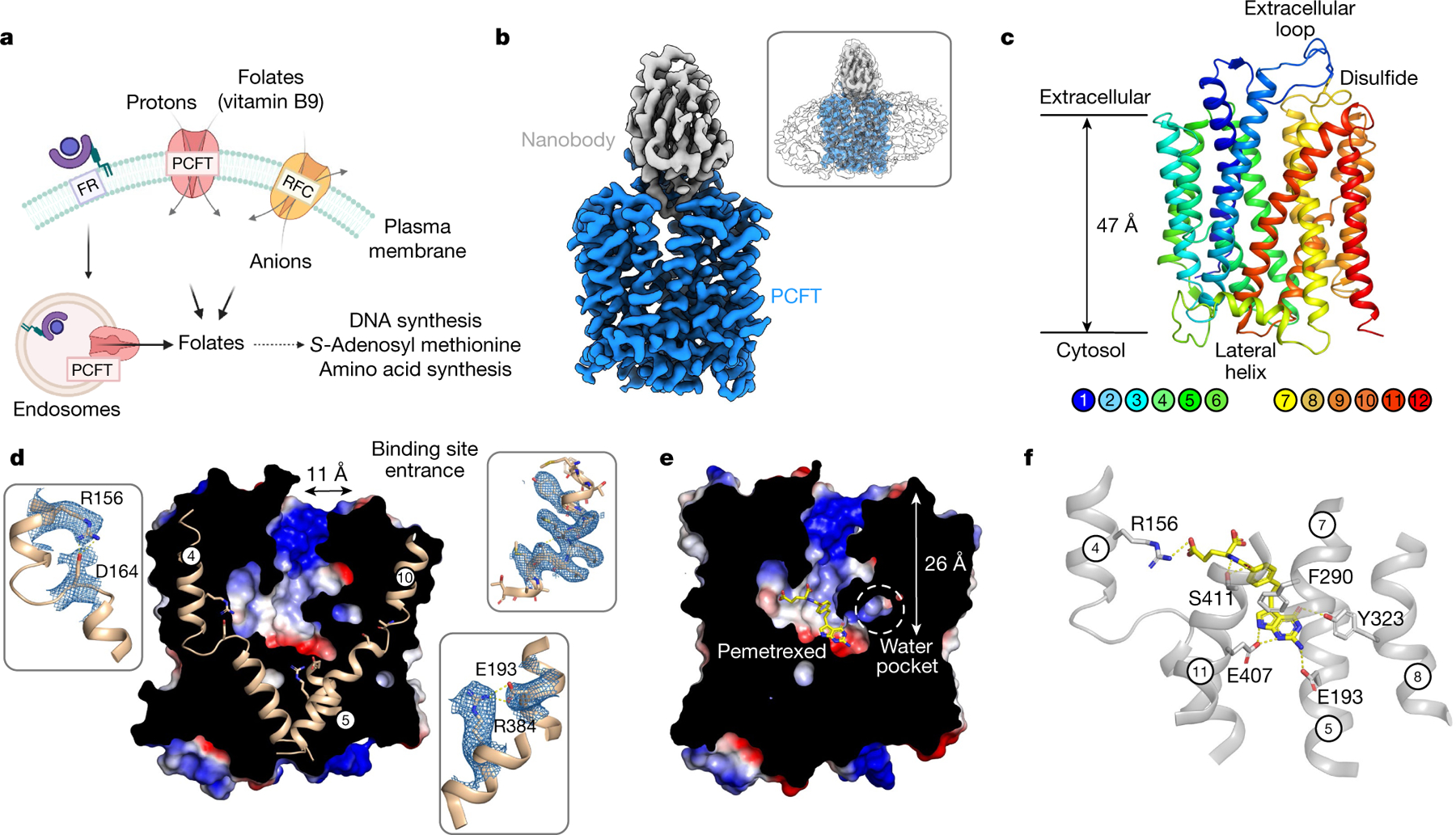
a, Three distinct mechanisms for folate transport exist in mammalian cells. The folate receptor (FR) internalizes extracellular folates within endosomes, whereas PCFT and RFC function to shuttle folates across the plasma membrane. b, Cryo-EM density of the PCFT–nanobody complex contoured at a threshold level of 1.0. Inset, superposition with lower-contoured (threshold level of 0.22) volume to display the detergent micelle. c, Cartoon representation of PCFT. d, Electrostatic surface representation of PCFT, highlighting key structural features. Inset, cryo-EM density for represented side chains (5σ). e, Electrostatic surface representation of PCFT, highlighting the central folate-binding site, enlarged water pocket and narrow vestibule entrance. The bound pemetrexed molecule is shown as sticks. f, Binding site, showing the main polar interactions formed with pemetrexed. Key residues interacting with the drug are shown as sticks and hydrogen bonds are represented as dashed lines.