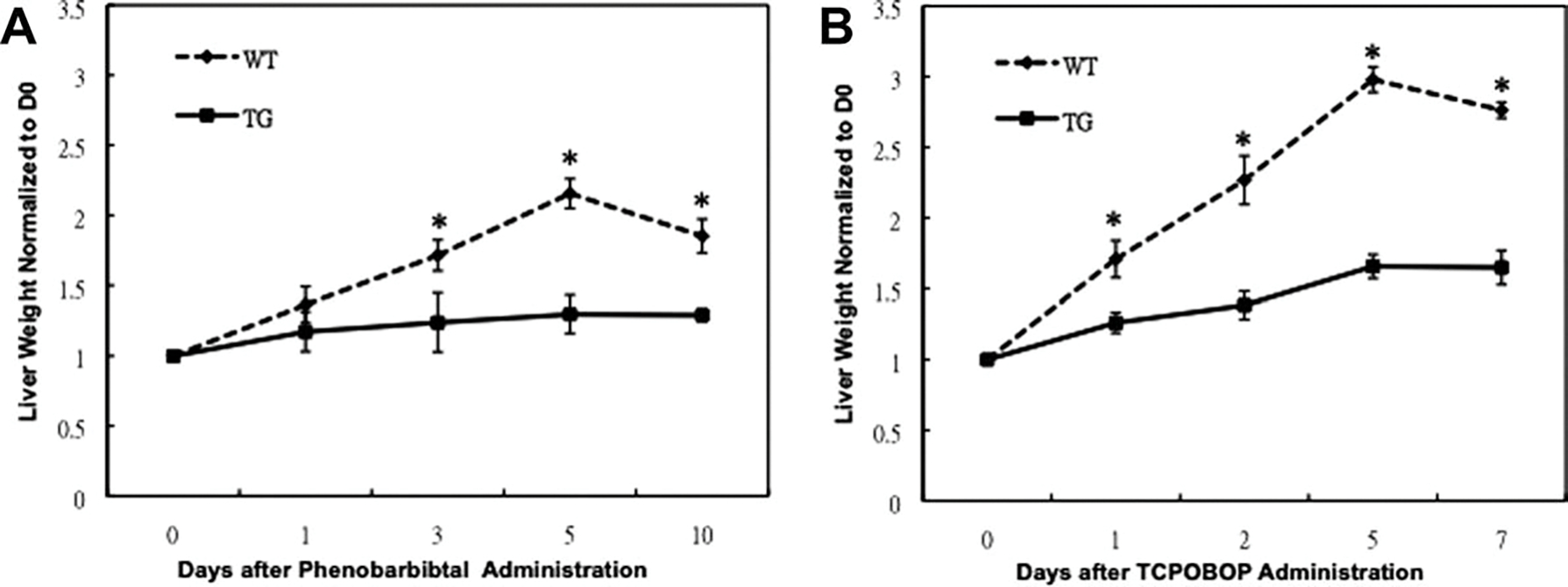
Fig. 2.

Suppression of hepatocyte proliferation after PB and TCPOBOP administration in GPC3 TG mice. Immunohistochemical staining for Ki67 (brown), a proliferation marker, in paraffin sections of WT and TG mouse livers at days 3 and 10 after PB administration (A) and at days 2 and 7 after TCPOBOP administration (D). Percentage of Ki67-positive nuclei in hepatocyte was counted in low-power field (200×) in 15 random sections from at least three different WT and TG mice. A significant decrease in Ki-67-positive hepatocyte nuclei was observed in TG mice at day 3 after PB administration (B) and at day 2 after TCPOBOP administration (E). Western blotting analysis of PCNA. Pooled liver samples from at least three mice were used for protein analysis after PB (C) and TCPOBOP (F) administration. Ponceau staining was used as loading control for nuclear lysates.
